


Home / Training / Manuals / Atlas of breast cancer early detection / Learning
.png)
Click on the pictures to magnify and display the legends

Click on this icon to display a case study
Atlas of breast cancer early detection
Filter by language: English / РусскийBreast imaging Mammography interpretation Mammography lexicon Mass |
Mass is a finding seen to be persistent in two different mammographic projections and is described by its shape, margins, and density. Shape The shape of the mass can be described as:
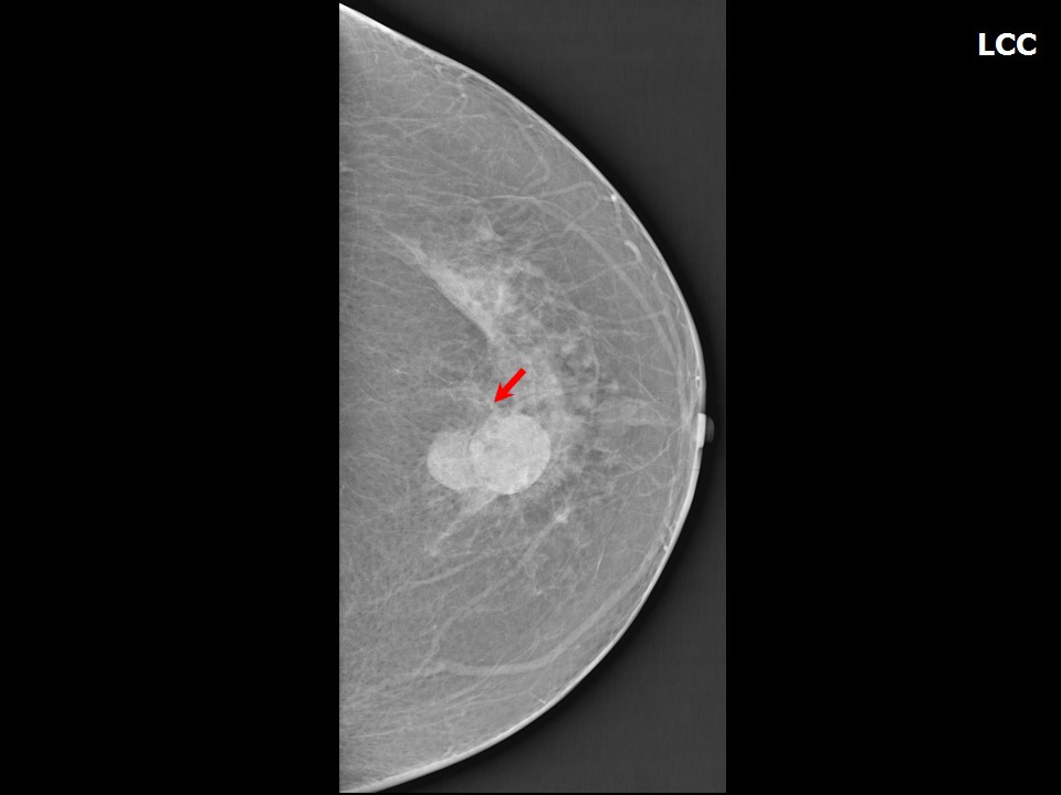
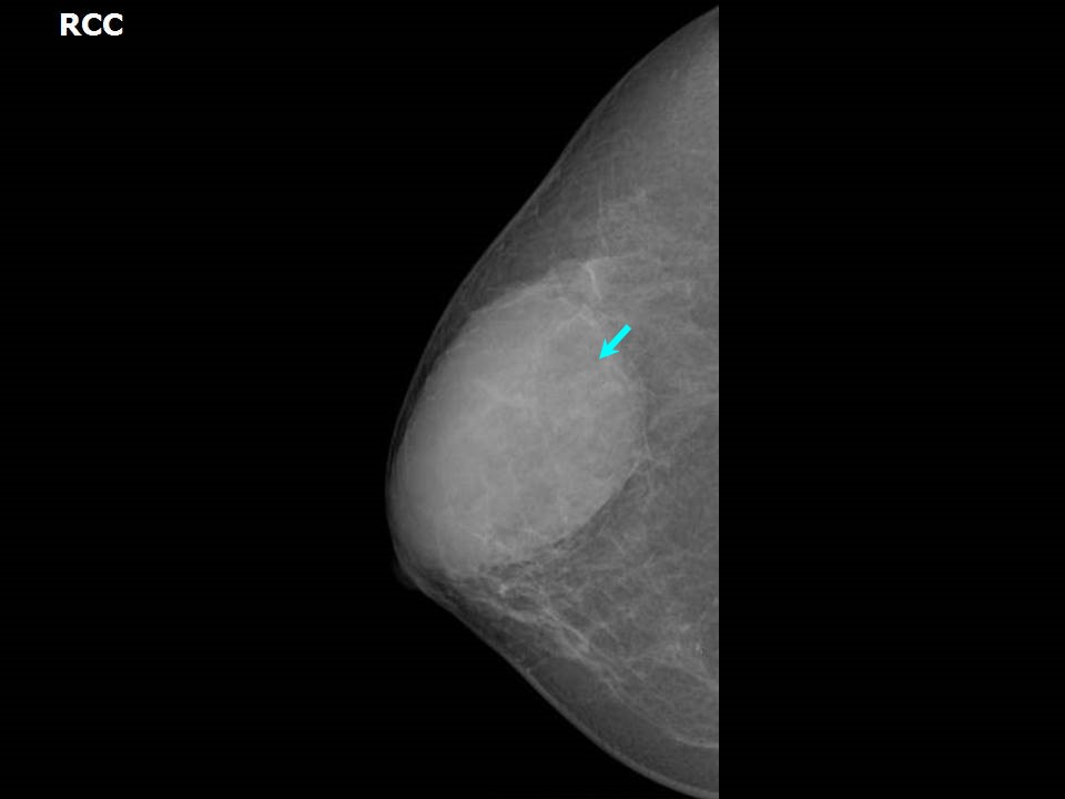
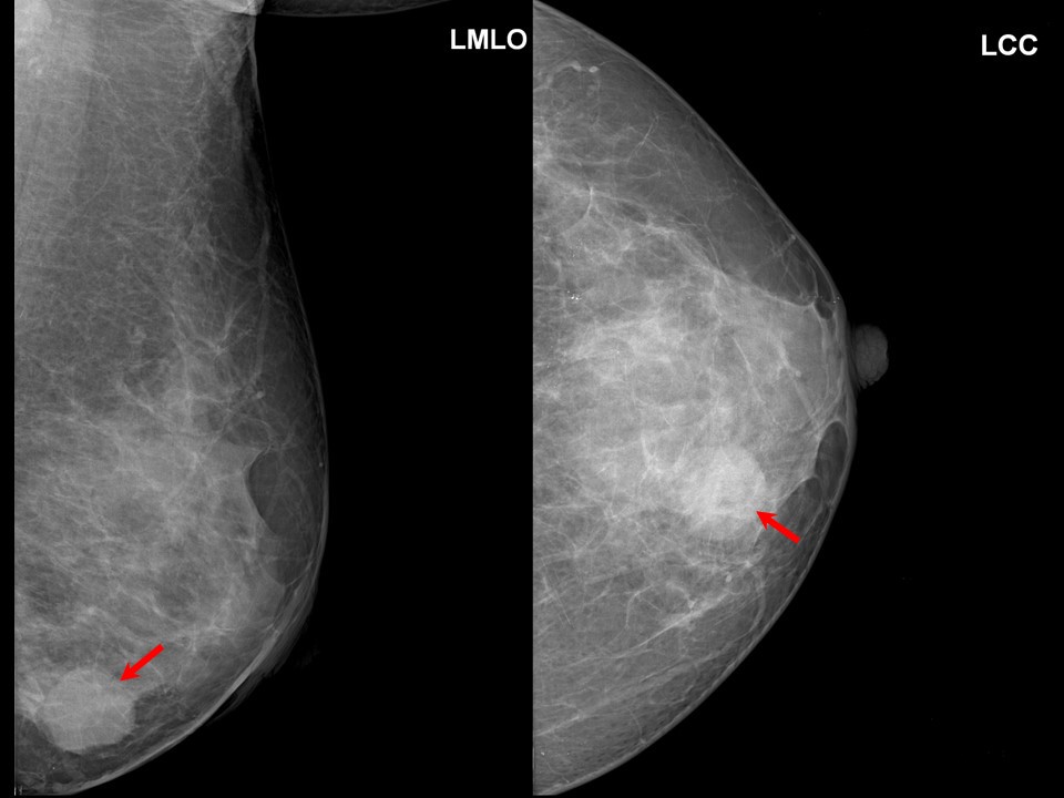
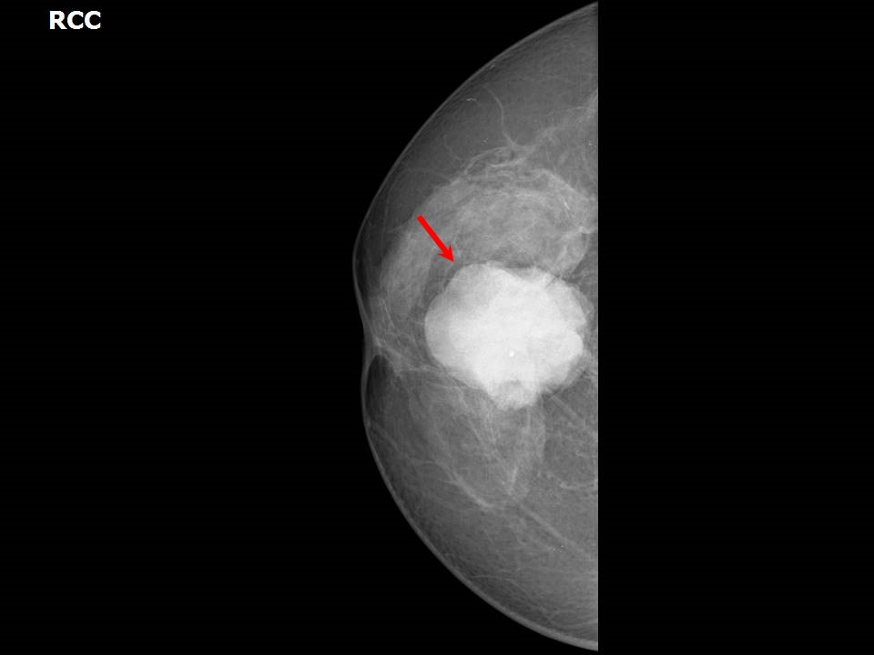
Round shape Round shape is usually suggestive of a benign lesion. However, a well-demarcated cancer, such as intracystic papillary carcinoma, mucinous carcinoma, or medullary carcinoma, may be round in shape. Differential diagnoses include benign proliferative lesion, encapsulated papillary carcinoma , and simple cyst .Other differential diagnoses include complicated cyst , lymphoma, phyllodes, metastasis ; invasive breast carcinoma , and organised seroma .
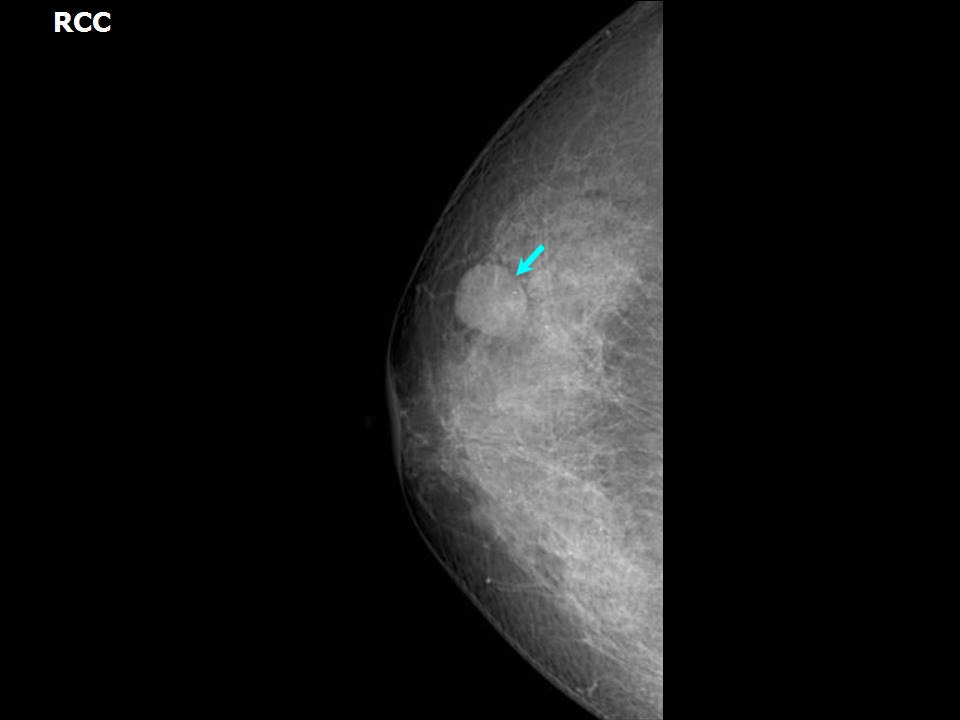
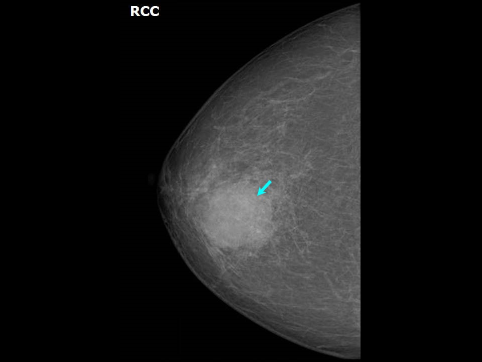
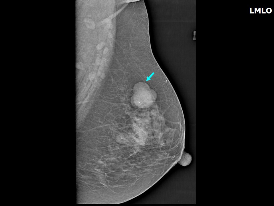
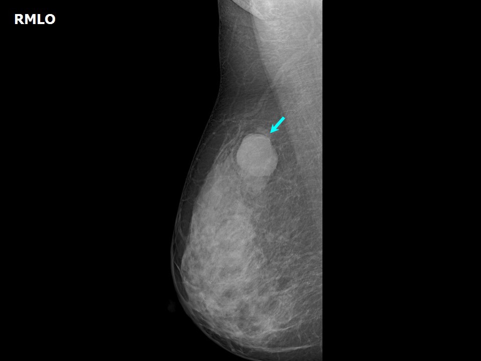
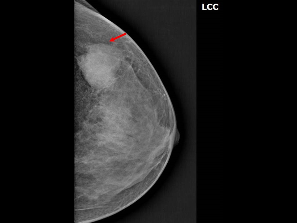
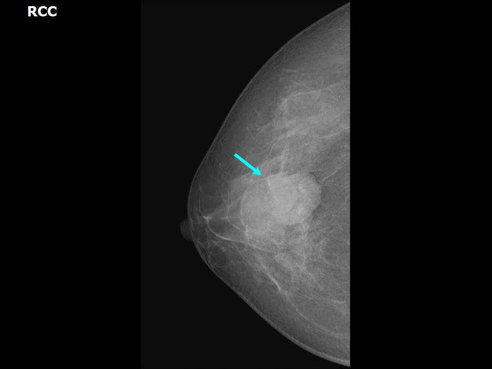
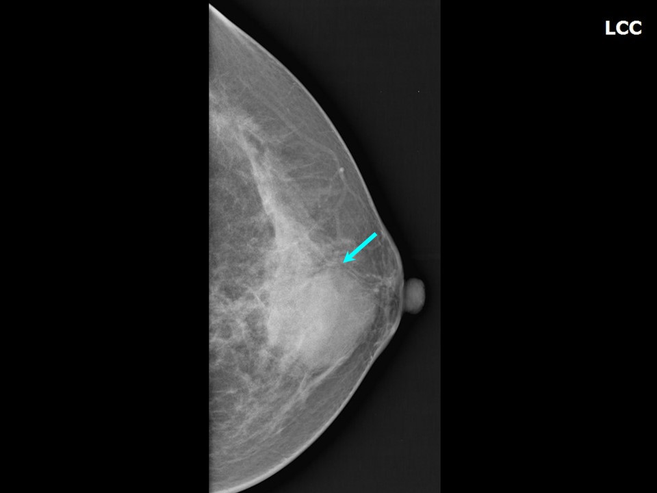
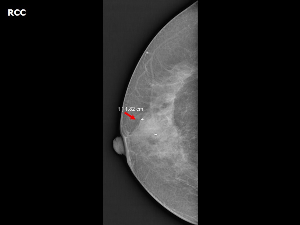
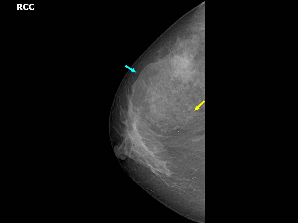
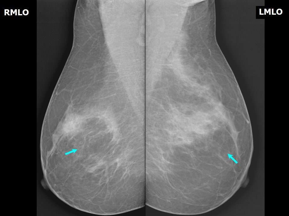
Oval shape Oval shape is usually suggestive of a benign lesion. However, a well-demarcated cancer, such as mucinous carcinoma or metastasis, may be oval in shape. Differential diagnoses include fibroadenoma , fibroadenoma with two smooth lobulations and benign calcification , and medullary carcinoma .Other differential diagnoses include simple cyst , complicated cyst , intramammary node , invasive breast carcinoma , mucinous carcinoma, phyllodes, lymphoma, and metastasis .
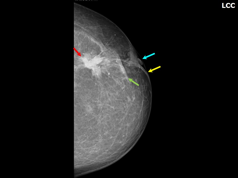
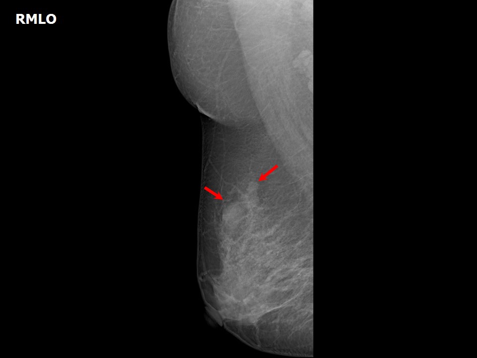
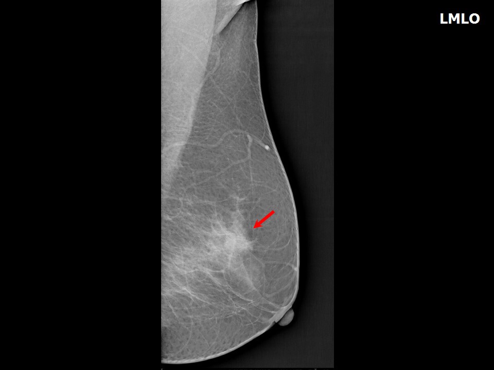
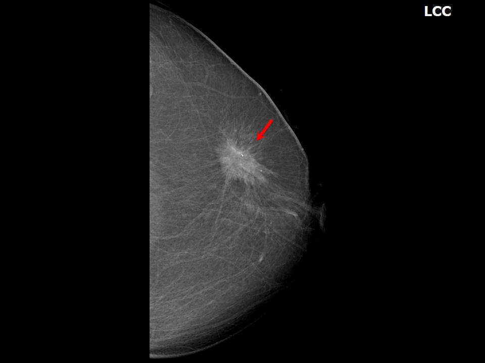
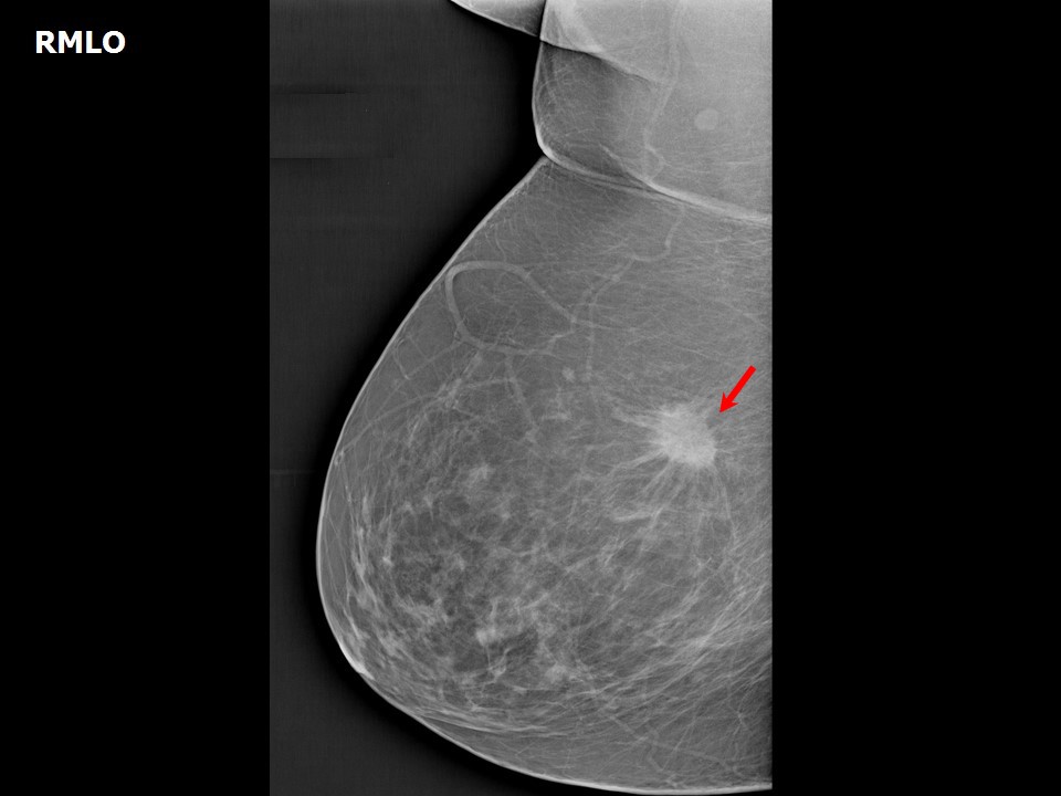
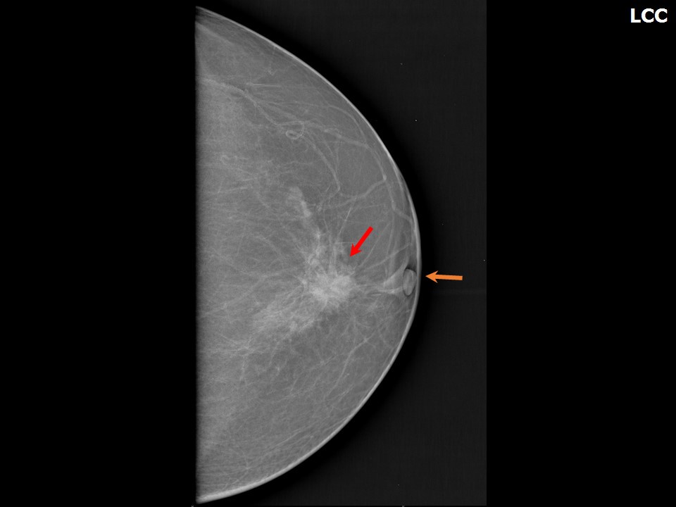
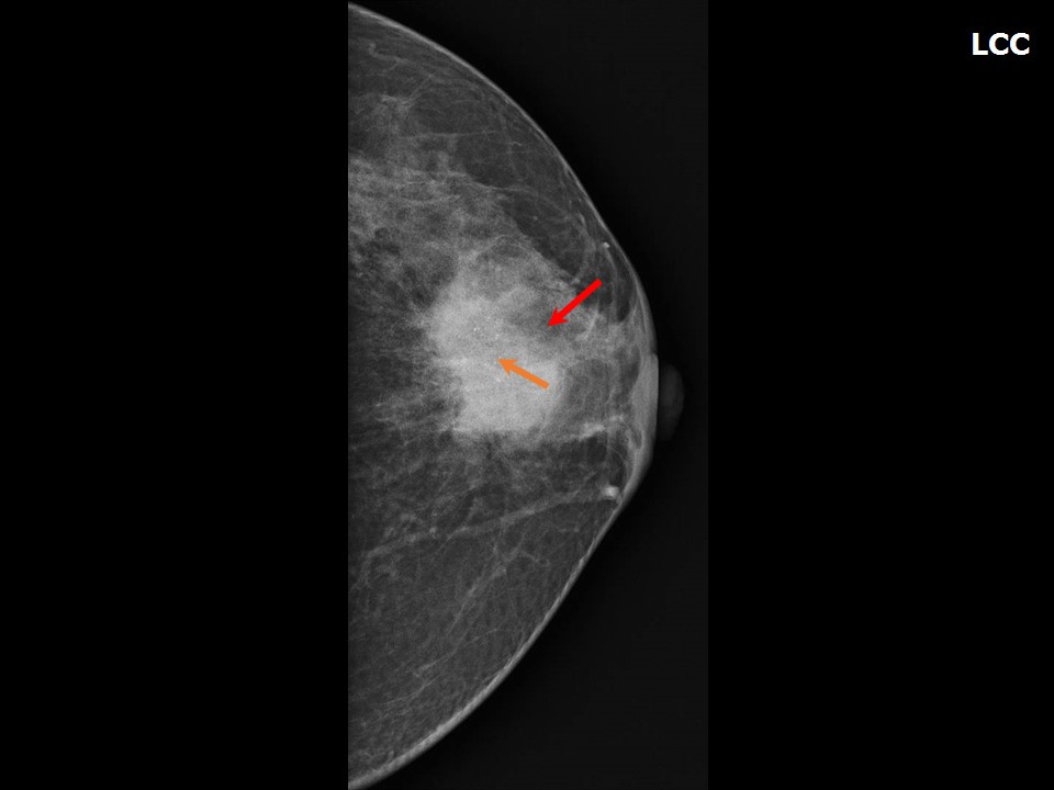
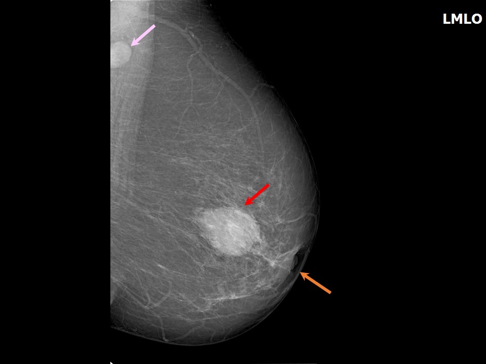
Irregular shape Irregular shape is suspicious for malignancy . Lesions of irregular shape may also be seen in women with a history of surgery as a result of, for example, scar tissue or seroma collection.Margins The margins of a mass indicate its demarcation from the adjacent normal breast parenchyma. They may be categorized as:
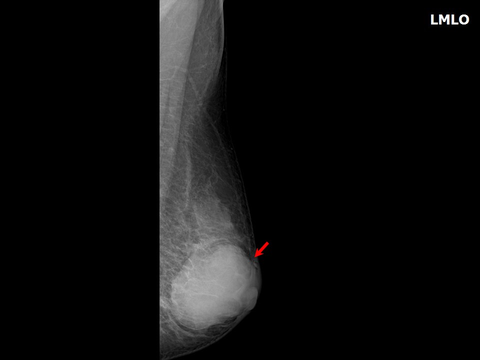
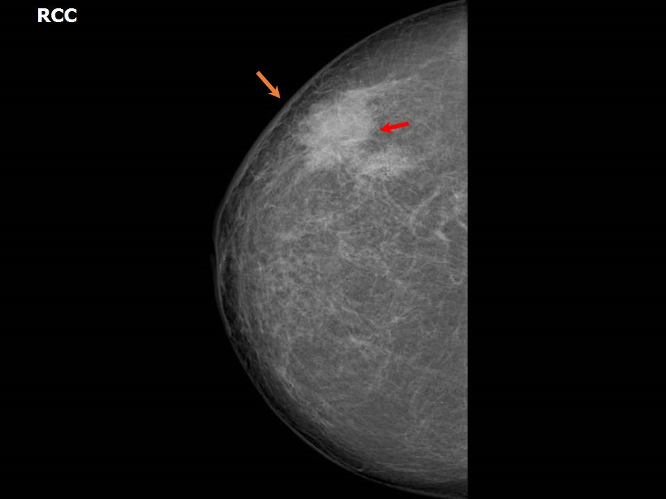
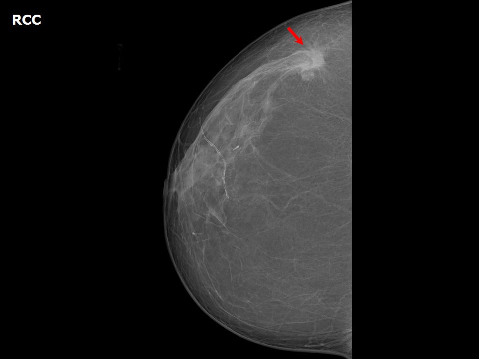
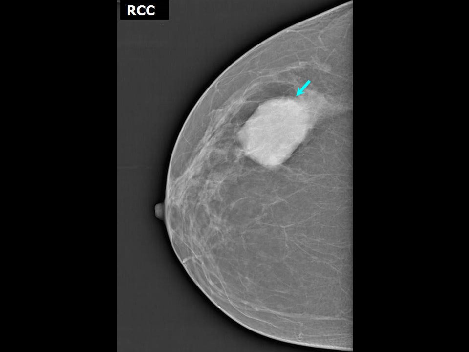
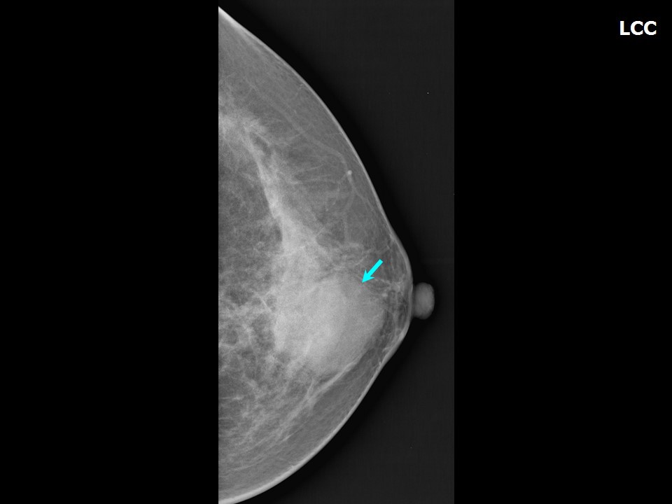
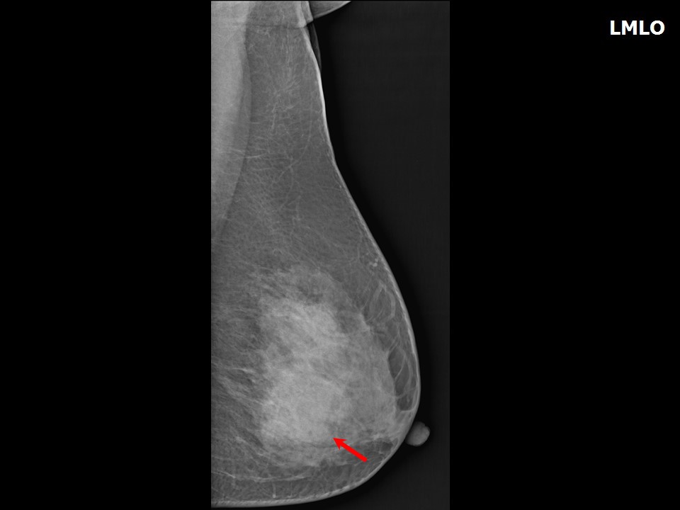
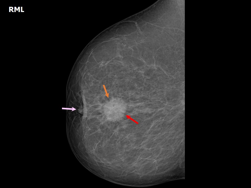
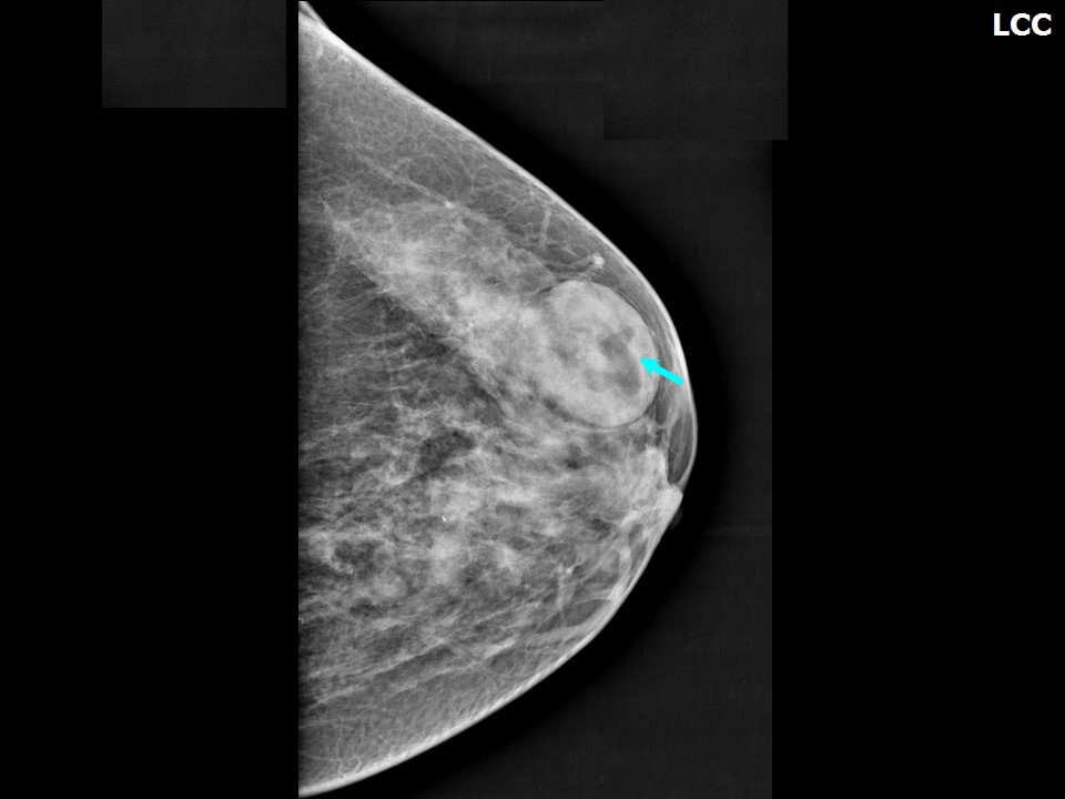
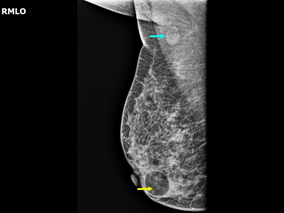
Circumscribed margin This is more likely a feature of a possible benign mass. Differential diagnoses include fibroadenoma , invasive breast carcinoma , and simple cyst .Obscured margin The margin may be obscured because of the superimposition of fibroglandular tissue over the mass. It may be a completely or partially obscured margin. Differential diagnoses include multifocal medullary carcinoma , simple cyst , and invasive breast carcinoma.Microlobulated margin A microlobulated margin is suspicious for breast carcinoma . Other differential diagnoses include DCIS or fibrocystic change.Indistinct margin An indistinct margin is suspicious for malignancy. Indistinct indicates a lack of clear demarcation from the surrounding breast parenchyma, and leads to the possibility of infiltration. Differential diagnoses include invasive carcinoma , papillary carcinoma , fat necrosis , metaplastic carcinoma , and fibrocystic changes .Spiculated margin A spiculated margin is highly suggestive of malignancy. Differential diagnoses include postoperative scar and fat necrosis.Density The density of a mass seen on mammography is compared with the adjacent fibroglandular tissue and is categorized as:
High density A lesion of high density is highly suggestive of breast carcinoma .Equal density As a stand-alone feature, equal density is inconclusive to differentiate between a benign or malignant mass. The margins of the mass, any calcifications, and axillary nodes need to be seen. Differential diagnoses include fibroadenoma , simple cyst , and intraductal papillary carcinoma .Low density Low density is a more likely feature of a benign lesion. Differential diagnoses include benign proliferative lesion , fat necrosis , and hamartoma .Fat-containing A mass visualized on mammography may be categorized as probable malignant or probable benign based on the combination of various features. A round or oval circumscribed mass of equal or low or fat-containing density is more likely to be a benign mass. An irregular not circumscribed mass of high density is more likely to be malignant mass. |


Click on the pictures to magnify and display the legends
Click on this icon to display a case study
25 avenue Tony Garnier CS 90627 69366, LYON CEDEX 07 France - Tel: +33 (0)4 72 73 84 85
© IARC 2026 - Terms of use - Privacy Policy.
© IARC 2026 - Terms of use - Privacy Policy.