


Home / Training / Manuals / Atlas of breast cancer early detection / Cases
Atlas of breast cancer early detection
Filter by language: English / Русский
Go back to the list of case studies
.png) Click on the pictures to magnify and display the legends
Click on the pictures to magnify and display the legends
| Case number: | 020 |
| Age: | 60 |
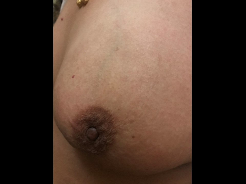
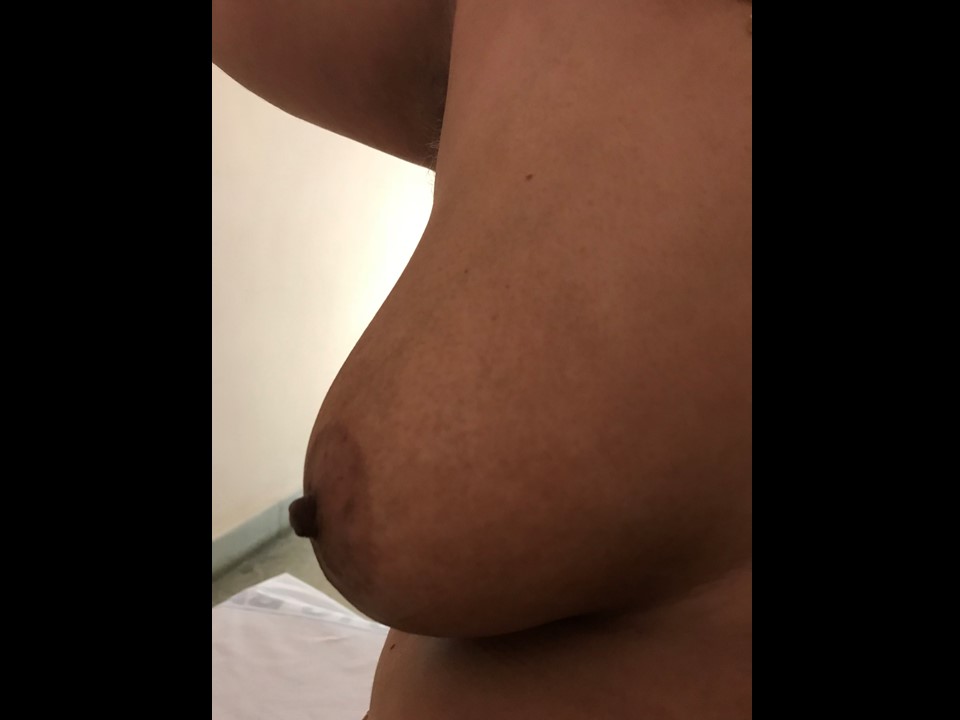
| Clinical presentation: | Postmenopausal woman presented with left nipple retraction noticed a month ago. |
 |  |
 |
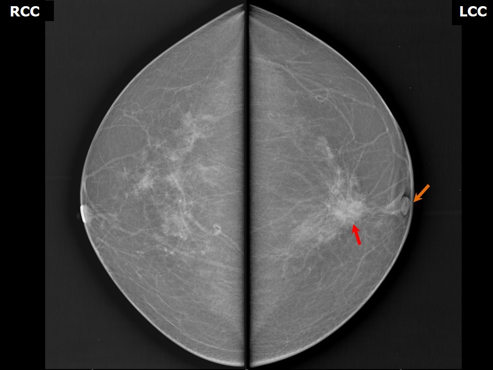
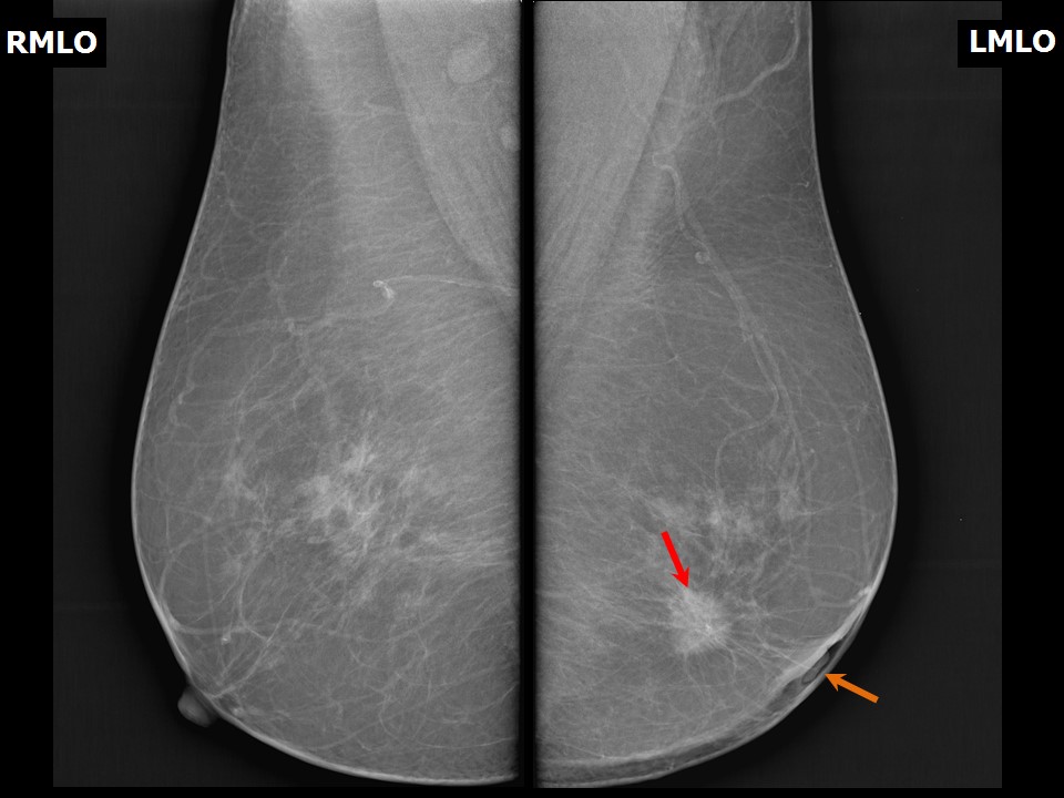
Mammography:
 |  |
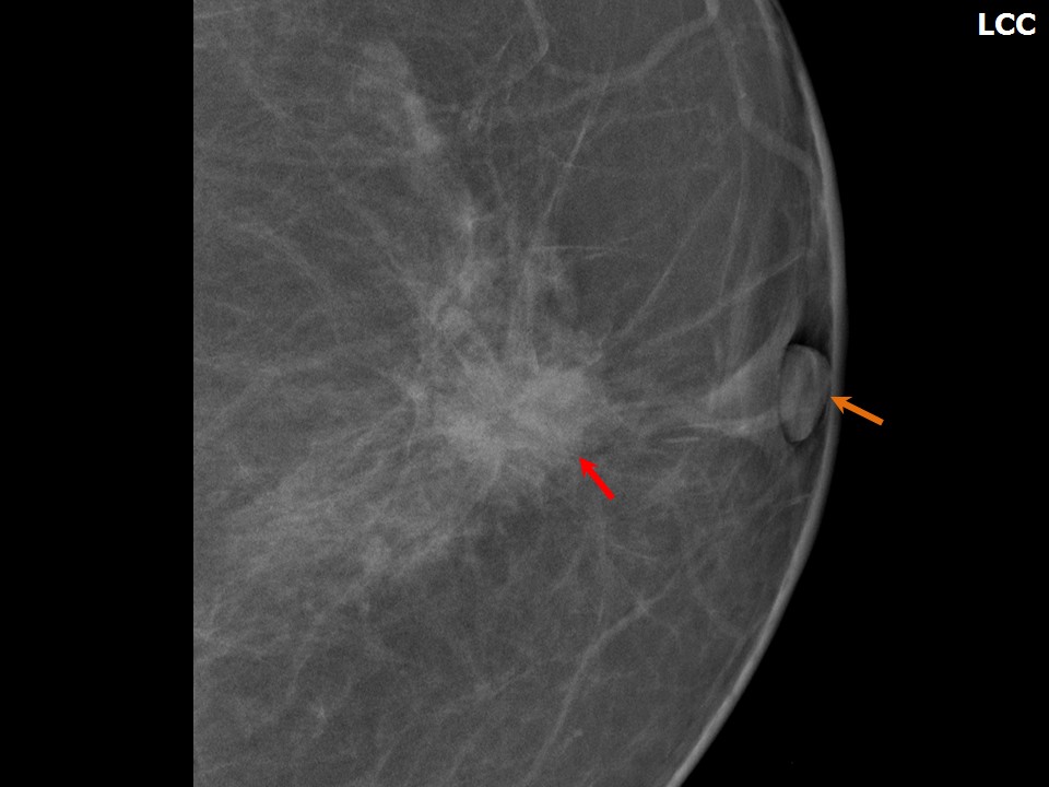 |
| Breast composition: | ACR category a (the breasts are almost entirely fatty) | Mammography features: |
| ‣ Location of the lesion: | Left breast, central portion of the breast, central zone at 6 oclock, middle third |
| ‣ Mass: | |
| • Number: | 1 |
| • Size: | 2.0 × 1.4 cm |
| • Shape: | Irregular |
| • Margins: | Spiculated |
| • Density: | High |
| ‣ Calcifications: | |
| • Typically benign: | None |
| • Suspicious: | None |
| • Distribution: | None |
| ‣ Architectural distortion: | None |
| ‣ Asymmetry: | None |
| ‣ Intramammary node: | None |
| ‣ Skin lesion: | None |
| ‣ Solitary dilated duct: | None |
| ‣ Associated features: | Retracted nipple and skin thickening |
Ultrasound:
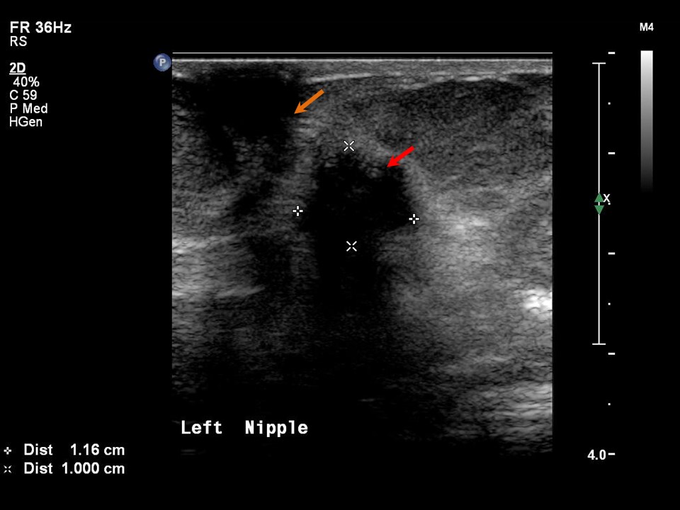 | 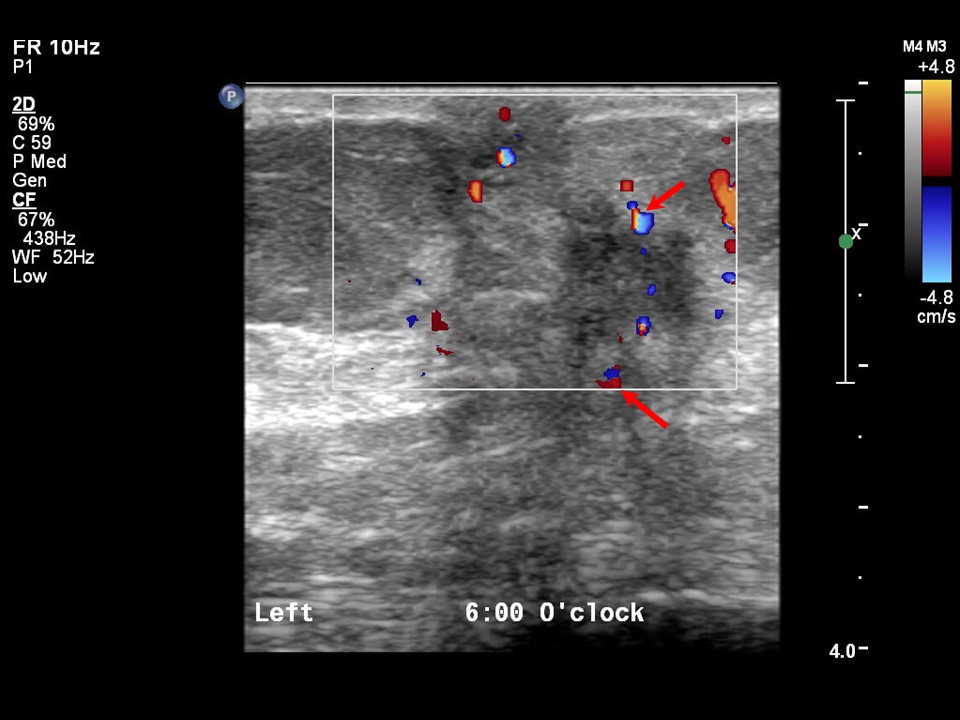 |
| Ultrasound features: Left breast, central portion of the breast at 6 o'clock | |
| ‣ Mass | |
| • Location: | Left breast, central portion of the breast at 6 o'clock |
| • Number: | 1 |
| • Size: | 1.2 × 1.0 cm |
| • Shape: | Irregular |
| • Orientation: | Not parallel |
| • Margins: | spiculated |
| • Echo pattern: | Hypoechoic |
| • Posterior features: | Strong posterior shadowing |
| ‣ Calcifications: | None |
| ‣ Associated features: | Nipple retraction, skin thickening |
| ‣ Special cases: | None |
BI-RADS:
BI-RADS Category: 5 (highly suggestive of malignancy)Further assessment:
Further assessment advised: Referral for cytologyCytology:
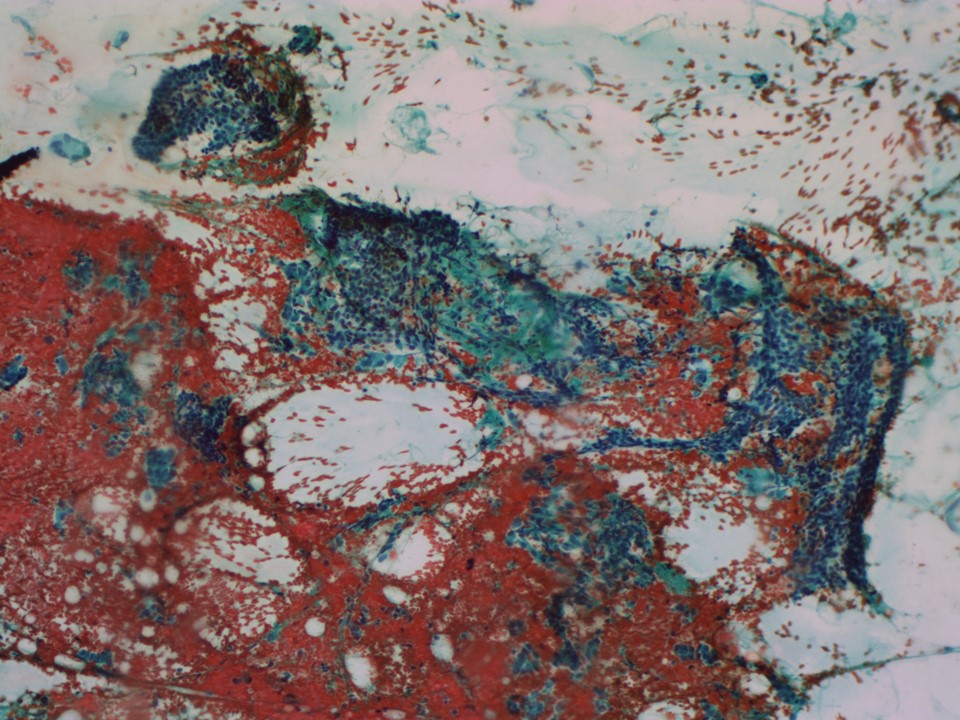 |
| Cytology features: | |
| ‣ Type of sample: | FNAC |
| ‣ Site of biopsy: | |
| • Laterality: | Left |
| • Quadrant: | Retroareolar region |
| • Localization technique: | Ultrasound-guided |
| • Nature of aspirate: | Haemorrhagic |
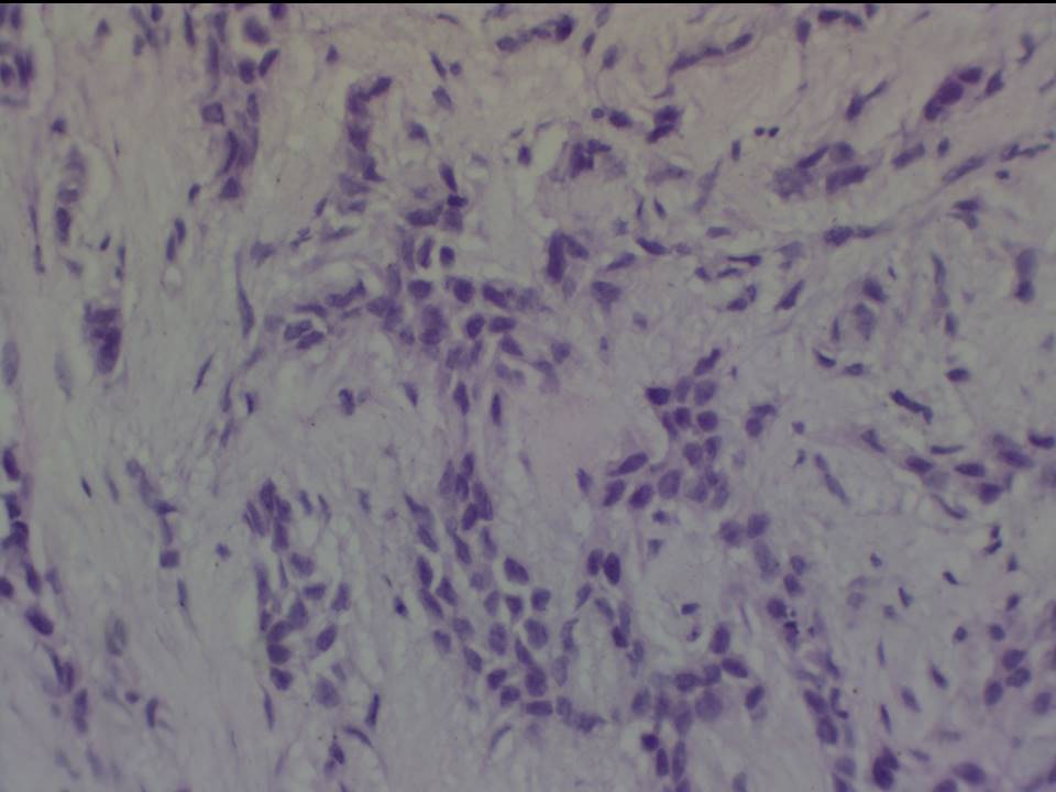
| ‣ Cytological description: | Cellular smear reveals loosely cohesive clusters and a few papillary fragments of ductal epithelial cells along with isolated cells with nuclear atypia. Many ductal epithelial cells show nuclear pleomorphism suspicious for malignancy. Cohesive sheets of benign ductal cells with myoepithelial cells are also seen |
| ‣ Reporting category: | Suspicious, probably in situ or invasive carcinoma |
| ‣ Diagnosis: | Suspicious for in situ or invasive carcinoma |
| ‣ Comments: | None |
Histopathology:
Breast-conserving surgery
 |  |
| Histopathology features: | |
| ‣ Specimen type: | Breast-conserving surgery |
| ‣ Laterality: | Left |
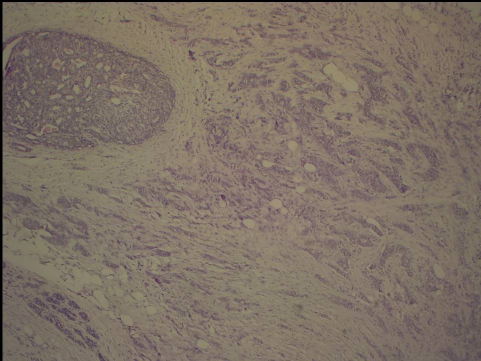
| ‣ Macroscopy: | Lumpectomy specimen (5.5 × 4.0 × 3.0 cm) oriented with long suture laterally and short suture superiorly, with a skin flap (2.5 × 1.0 cm). On serial sectioning a firm, greyish white area (1.0 × 1.0 × 1.5 cm) was identified, located 2.0 cm from skin, 1.5 cm from the posterior margin, 1.6 cm from the superior margin, 1.5 cm from the inferior margin, 1.6 cm from the medial margin, and 2.0 cm from the lateral margin. There are fibrotic areas in the remaining breast parenchyma |
| ‣ Histological type: | Invasive carcinoma of no special type |
| ‣ Histological grade: | Grade 2 (3 + 2 + 2 = 7). |
| ‣ Mitosis: | 15 |
| ‣ Maximum invasive tumour size: | 1.5 cm |
| ‣ Lymph node status: | 0/13 |
| ‣ Peritumoural lymphovascular invasion: | Absent |
| ‣ DCIS/EIC: | DCIS solid and cribriform type of low nuclear grade. EIC is absent |
| ‣ Margins: | All margins were free of tumour |
| ‣ Pathological stage: | pT1cN0 |
| ‣ Biomarkers: | |
| ‣ Comments: | Areas with UDH and ADH and benign papilloma present |
Case summary:
| Postmenopausal woman presented with left nipple retraction. Diagnosed as left breast carcinoma with left nipple retraction, BI-RADS 5 on imaging, as suspicious for malignancy on cytology, and as invasive breast carcinoma of no special type, pT1cN0 on histopathology. |
Learning points:
|