


Home / Training / Manuals / Atlas of breast cancer early detection / Learning
.png)
Click on the pictures to magnify and display the legends

Click on this icon to display a case study
Atlas of breast cancer early detection
Filter by language: English / РусскийAnatomy of the breast Vascular and lymphatic supply |
The breast is supplied by the internal mammary (or thoracic) artery, the intercostal arteries, and the axillary artery, which gives rise to the external mammary or lateral thoracic artery. The veins form a ring around the base of the nipple (circulus venosus). The large veins pass from the circulus venosus to the circumference of the mammary gland and drain into the external mammary vein and then to the axillary vein. Some of the venous channels also drain the internal mammary (thoracic) vein to the subclavian vein.
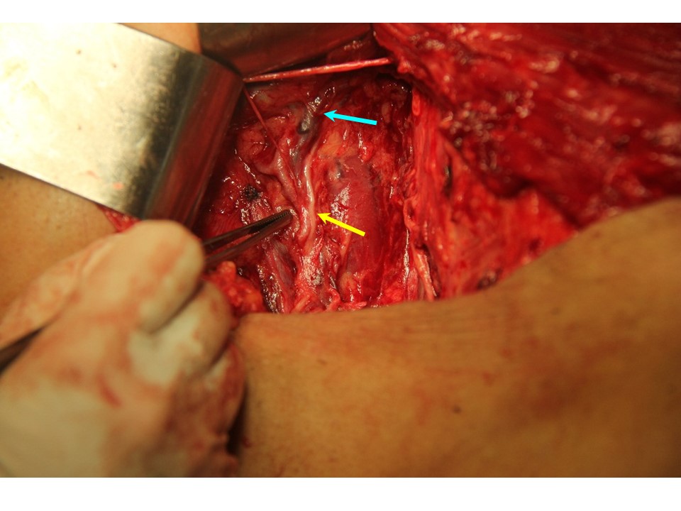
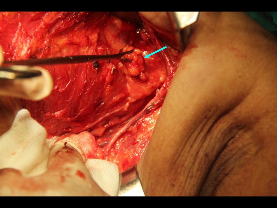
Intraoperative image of axilla The lymphatic drainage of the breast originates from the breast lobules and flows into a subareolar plexus (Sappey plexus). From this plexus, the lymph drains through three main routes:
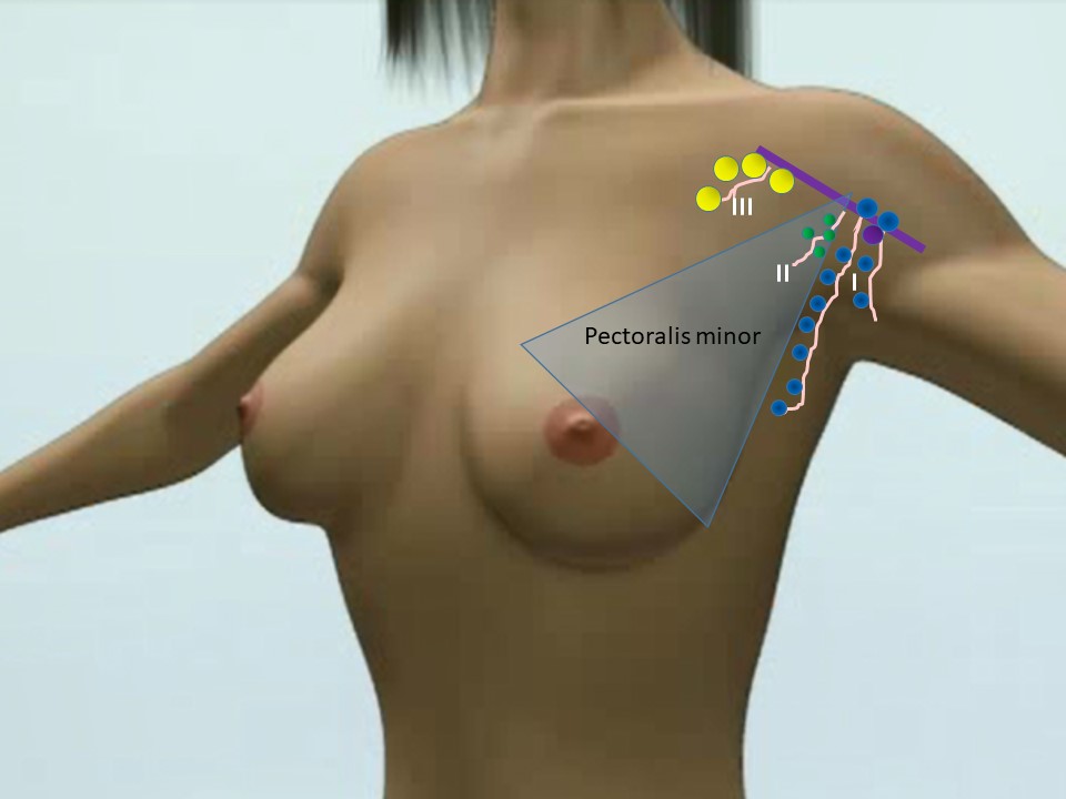
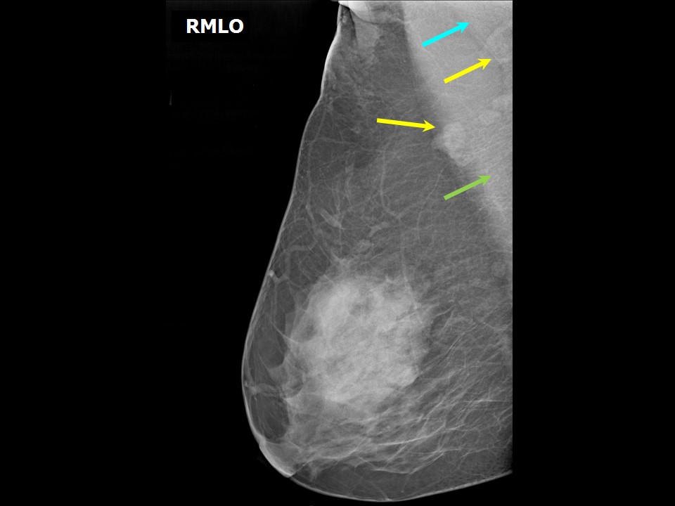
Axillary nodes
|
Click on the pictures to magnify and display the legends
Click on this icon to display a case study
25 avenue Tony Garnier CS 90627 69366, LYON CEDEX 07 France - Tel: +33 (0)4 72 73 84 85
© IARC 2025 - Terms of use - Privacy Policy.
© IARC 2025 - Terms of use - Privacy Policy.