


Home / Training / Manuals / Atlas of breast cancer early detection / Learning
.png)
Click on the pictures to magnify and display the legends

Click on this icon to display a case study
Atlas of breast cancer early detection
Filter by language: English / РусскийAnatomy of the breast Microscopic anatomy |
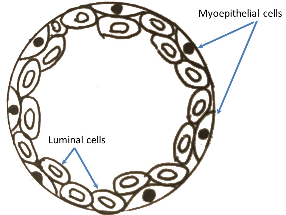
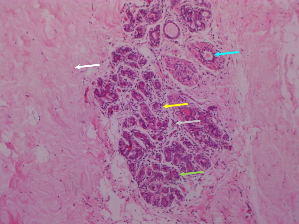
Microscopic anatomy of the epithelial components The TDLU has two layers of epithelium resting on the basement membrane. The inner layer (luminal cells) comprises a single layer of cuboidal cells and may be pseudostratified. The outer layer is made up of myoepithelial cells. Adipose and connective tissue (stromal elements) The glandular elements of the breast are embedded in the fatty (adipose) tissue and connective tissue; together these make up the stroma of the breast. The TDLUs contain and are surrounded by loose specialized connective tissue (intralobular connective tissue), which is different from the stromal connective tissue found in the rest of the breast. The lobes are surrounded by septa formed of the dense connective tissue (interlobular connective tissue). Hollow conical projections of fibrous tissue that arise from these septa are known as the suspensory ligaments of Cooper. The apices of these cones attach the breasts firmly to the superficial fascia and the skin. The base of the breast rests on the pectoral fascia, which is a flat sheet of connective tissue covering the pectoralis major. The ligaments of Cooper are attached to this fascia. Microscopic anatomy of stromal elements Stromal tissue is divided into the interlobular stroma, which surrounds the large ducts and TDLUs, and the intralobular stroma, which surrounds the acini within the TDLUs. The interlobular stroma is denser and more collagenous than the intralobular connective tissue. |

Click on the pictures to magnify and display the legends
Click on this icon to display a case study
25 avenue Tony Garnier CS 90627 69366, LYON CEDEX 07 France - Tel: +33 (0)4 72 73 84 85
© IARC 2025 - Terms of use - Privacy Policy.
© IARC 2025 - Terms of use - Privacy Policy.