


Home / Training / Manuals / Atlas of breast cancer early detection / Learning
.png)
Click on the pictures to magnify and display the legends

Click on this icon to display a case study
Atlas of breast cancer early detection
Filter by language: English / РусскийBreast imaging Mammography technique Mammography procedure Additional mammographic views Lumpectomy specimen mammography |
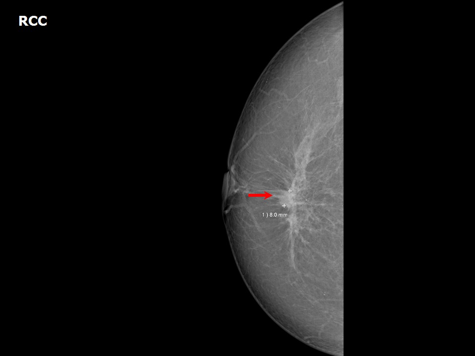
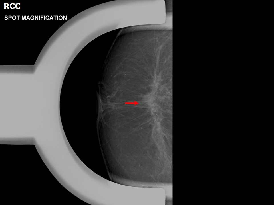
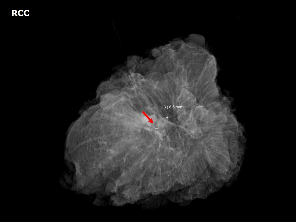
Preclinical cancers or suspicious clusters of microcalcifications detected on screening mammography are further evaluated with image-guided biopsy. If carcinoma in situ or malignancy is detected, most patients undergo wide excision of the lesion as a breast-conserving procedure. Hookwire localization is used for the excision and biopsy of clinically non-palpable breast lesions. Mammography of the lumpectomy specimen is an important intraoperative step to ensure complete excision.
Advantages of specimen mammography
Mammogram right breast shows developing asymmetry Lumpectomy specimen mammogram |
Click on the pictures to magnify and display the legends
Click on this icon to display a case study
25 avenue Tony Garnier CS 90627 69366, LYON CEDEX 07 France - Tel: +33 (0)4 72 73 84 85
© IARC 2025 - Terms of use - Privacy Policy.
© IARC 2025 - Terms of use - Privacy Policy.