


Home / Training / Manuals / Atlas of breast cancer early detection / Learning
.png)
Click on the pictures to magnify and display the legends

Click on this icon to display a case study
Atlas of breast cancer early detection
Filter by language: English / РусскийBreast imaging The evolution of breast imaging |
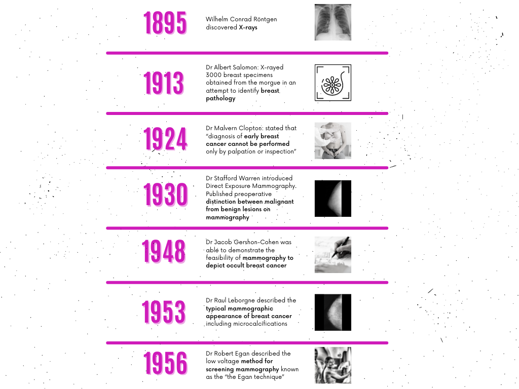
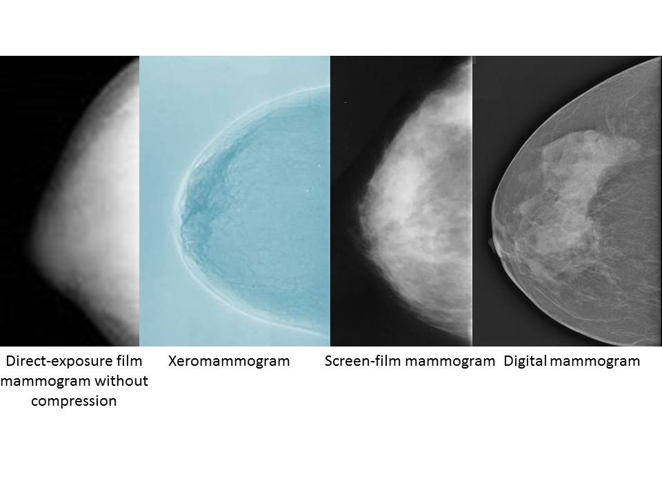
Imaging studies of breast tissue and initiatives to detect breast disease using X-rays were first used in the early twentieth century. There have been significant advances in the technology used in breast imaging since the early days of direct-exposure film mammography in the 1960s. Mammography detects changes in the breast tissue caused by physiological processes or by pathological conditions. These changes can then be characterized depending on the morphological features of the image obtained.
Mammography is a specialized imaging technique that uses X-rays to produce detailed images of the breast structure and to locate and characterize lesions in the breast. Mammography remains the reference standard for the detection of breast abnormalities, particularly those caused by early cancer. By 1985, mammography was established as a sensitive method for breast cancer screening and was the only method recommended for asymptomatic women with an average risk of breast cancer. However, the sensitivity of mammography can be highly variable, ranging from 98% in postmenopausal women with fatty breast parenchyma to 36% in young women with dense breasts. |
Click on the pictures to magnify and display the legends
Click on this icon to display a case study
25 avenue Tony Garnier CS 90627 69366, LYON CEDEX 07 France - Tel: +33 (0)4 72 73 84 85
© IARC 2025 - Terms of use - Privacy Policy.
© IARC 2025 - Terms of use - Privacy Policy.