


Home / Training / Manuals / Atlas of breast cancer early detection / Learning
.png)
Click on the pictures to magnify and display the legends

Click on this icon to display a case study
Atlas of breast cancer early detection
Filter by language: English / РусскийBreast imaging Breast ultrasound Ultrasound lexicon Special cases Cysts |
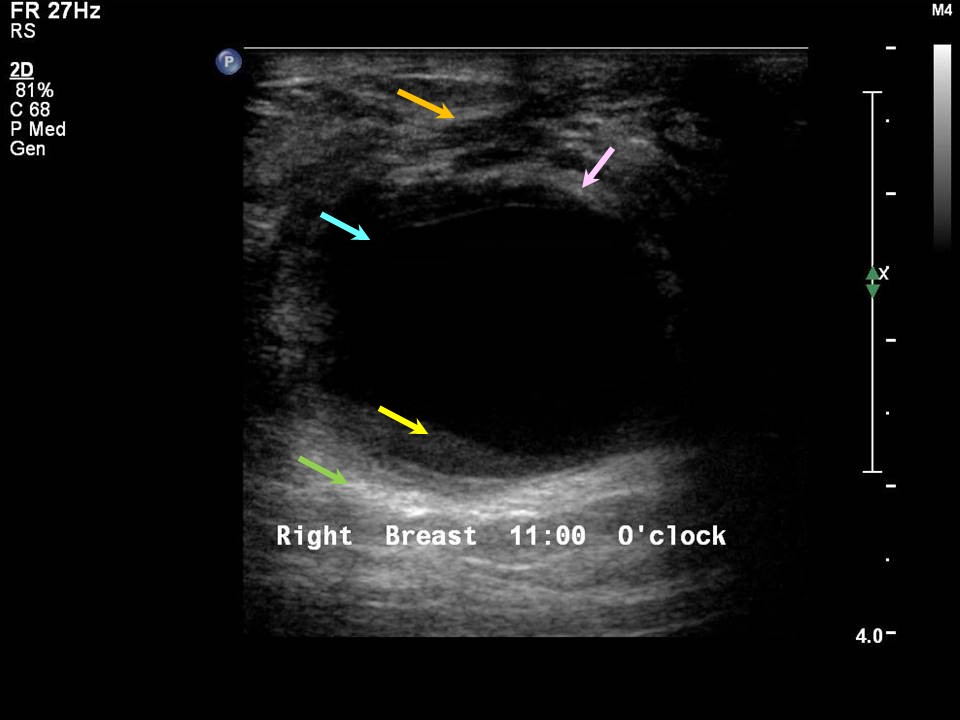
Simple cysts
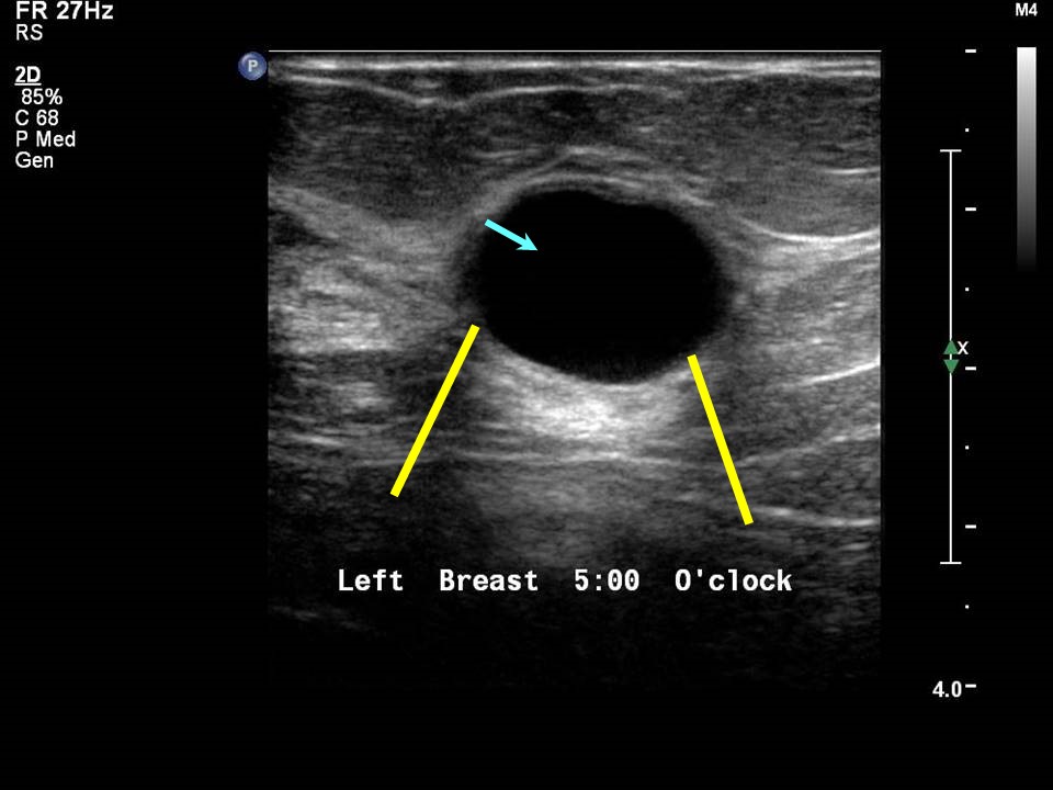
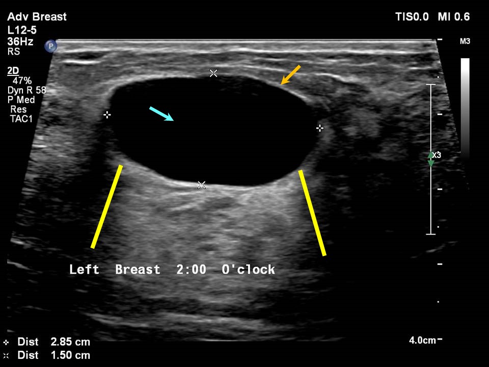
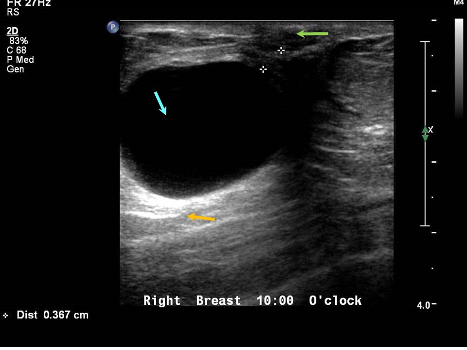
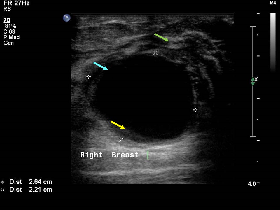
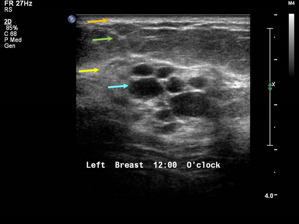
On ultrasound, a simple cyst appears as a well-circumscribed anechoic lesion (with no internal echoes), round or oval in shape, with sharp anterior and posterior borders and smooth walls. The cyst shows increased through transmission known as posterior acoustic enhancement. Lateral edge reverberations may be seen as sharply defined edges. On colour Doppler, no internal or peripheral vascularity is present. Cysts are a component of fibrocystic change and usually occur in women aged between 30 and 50 years. Simple cysts can be solitary but are often multiple and seen in groups. Multiple breast cysts may be seen in both breasts. The clinical presentation of a cyst is a lump, which may be associated with pain and discomfort. They are categorized as BI-RADS 2 (benign) .Complicated cysts Complicated cysts are associated with extensive fibrocystic changes. They show all the criteria of simple cysts except that they also contain low-level internal echoes, which may change with the patients position. This would indicate a benign cyst with secondary changes such as debris, haemorrhage, or infection. If the echoes are not mobile, it is important to rule out possibility of an intracystic mass, such as papilloma or intracystic carcinoma .Clustered microcysts Clustered microcysts are small simple cysts, < 3 mm and seen in a cluster. On ultrasound, they are visible as tiny anechoic lesions. On Doppler ultrasound, clustered microcysts with no colour flow suggest benign cysts. If there is increased central or peripheral vascularity, histopathology is recommended to rule out the possibility of malignancy. Clustered microcysts tend to form calcifications in the debris within the cysts, which is seen as microcalcifications on mammography. However, the calcifications are typically benign and seen as round acinar calcifications or milk-of-calcium sediments. Annual follow-up to rule out changes to suspicious calcifications is suggested. |
Click on the pictures to magnify and display the legends
Click on this icon to display a case study
25 avenue Tony Garnier CS 90627 69366, LYON CEDEX 07 France - Tel: +33 (0)4 72 73 84 85
© IARC 2025 - Terms of use - Privacy Policy.
© IARC 2025 - Terms of use - Privacy Policy.