


Home / Training / Manuals / Atlas of breast cancer early detection / Cases
Atlas of breast cancer early detection
Filter by language: English / Русский
Go back to the list of case studies
.png) Click on the pictures to magnify and display the legends
Click on the pictures to magnify and display the legends
| Case number: | 005 |
| Age: | 50 |
| Clinical presentation: | Woman who had undergone a hysterectomy, with average risk of breast cancer presented with an acute-onset painful lump in the upper outer quadrant of the right breast. Examination revealed a tender firm swelling 2.5 cm in diameter. Mammography was not done because of severe tenderness. |
Ultrasound:
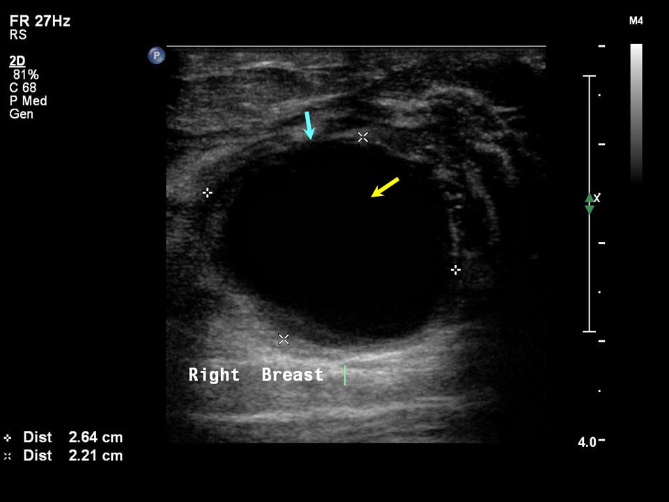 | 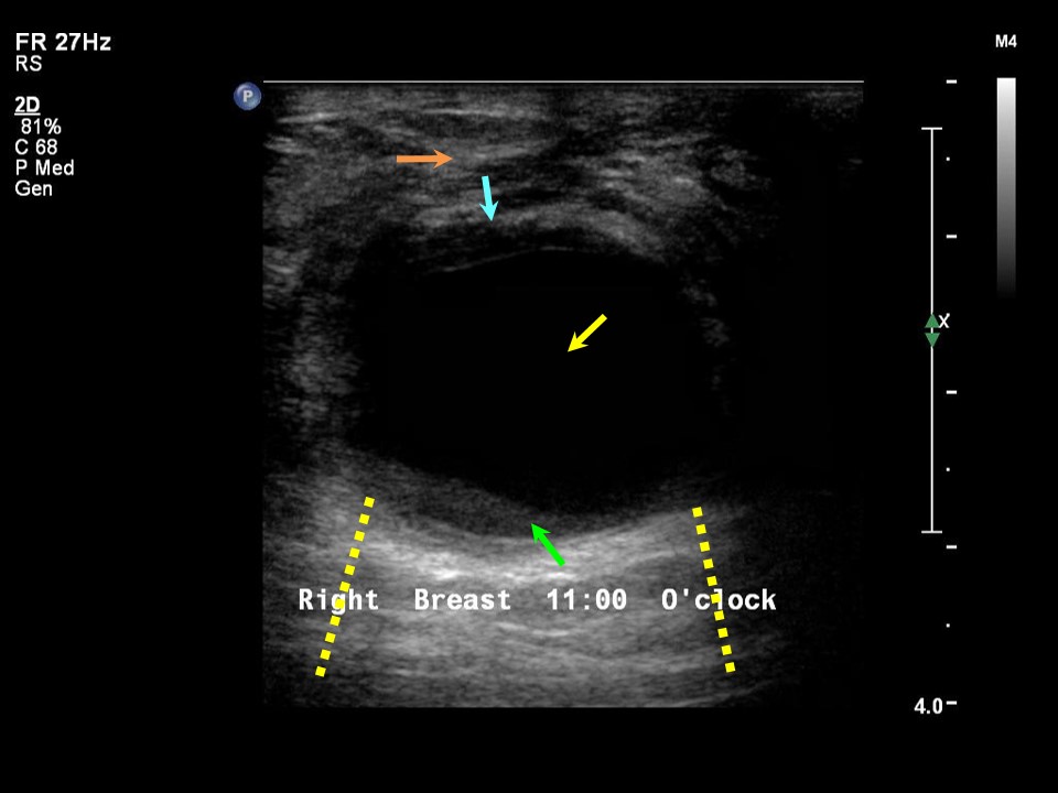 |
| Ultrasound features: Right breast, upper outer quadrant at 11 oclock | |
| ‣ Mass | |
| • Location: | Right breast, upper outer quadrant at 11 oclock |
| • Number: | 1 |
| • Size: | 2.6 × 2.2 cm |
| • Shape: | Oval |
| • Orientation: | Parallel |
| • Margins: | circumscribed |
| • Echo pattern: | Anechoic with internal echoes |
| • Posterior features: | Posterior Enhancement |
| ‣ Calcifications: | None |
| ‣ Associated features: | Oedema, internal vascularity, and vessels in rim |
| ‣ Special cases: | Complicated cyst |
BI-RADS:
BI-RADS Category: 2 (benign)Further assessment:
Further assessment advised: Referral for cytologyCytology:
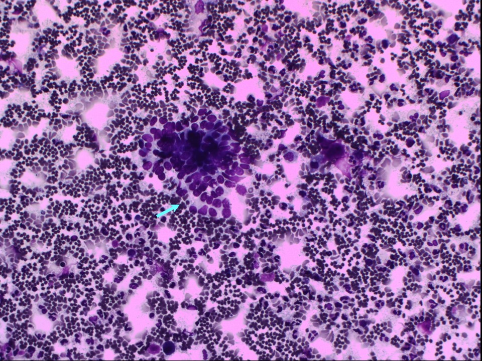 | 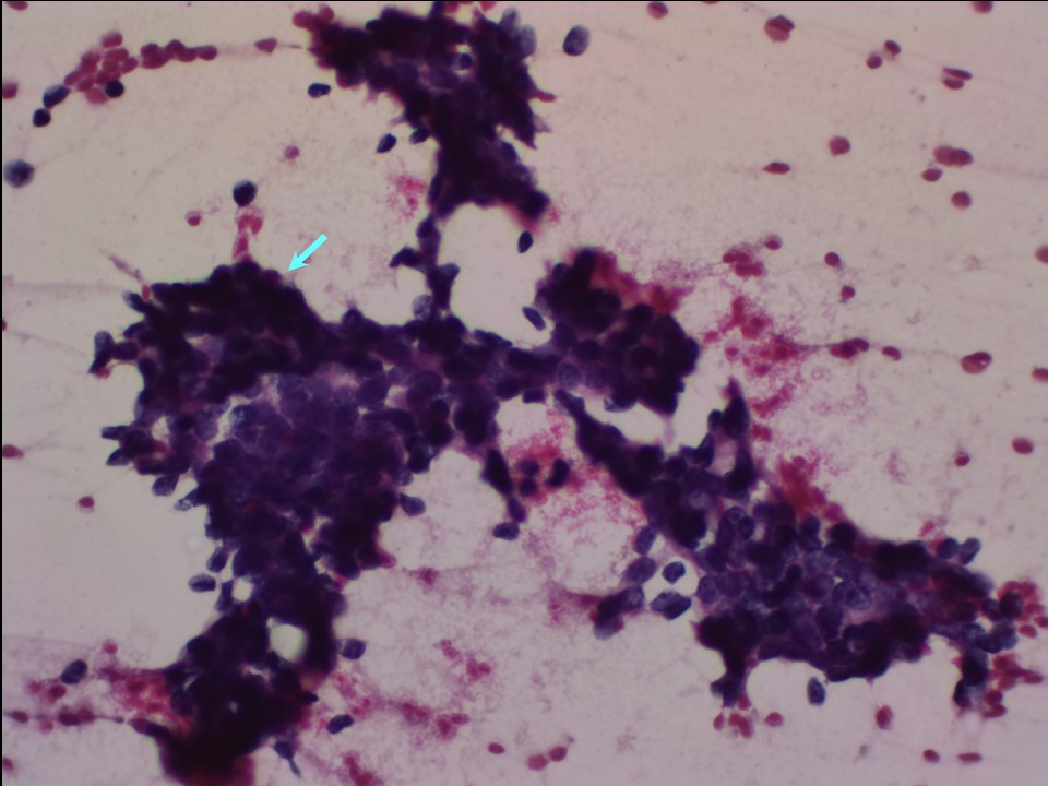 |
| Cytology features: | |
| ‣ Type of sample: | FNAC |
| ‣ Site of biopsy: | |
| • Laterality: | Right |
| • Quadrant: | Upper outer |
| • Localization technique: | Palpation |
| • Nature of aspirate: | 0.5 mL of brownish yellow fluid |
| ‣ Cytological description: | Cytospin preparation of the fluid aspirated shows a few benign epithelial cell clusters and many neutrophils, lymphocytes, and macrophages. Direct smears show many tightly cohesive, clustered benign ductal epithelial cells in flat sheets and as complex folded sheets with papillaroid edges. A few strands of fibrous stroma seen. Background shows proteinaceous material with many foamy histiocytes and neutrophils |
| ‣ Reporting category: | Benign |
| ‣ Diagnosis: | Benign proliferative breast lesion with fibrocystic change |
| ‣ Comments: | None |
Histopathology:
Lump excision
 |  |
 |  |
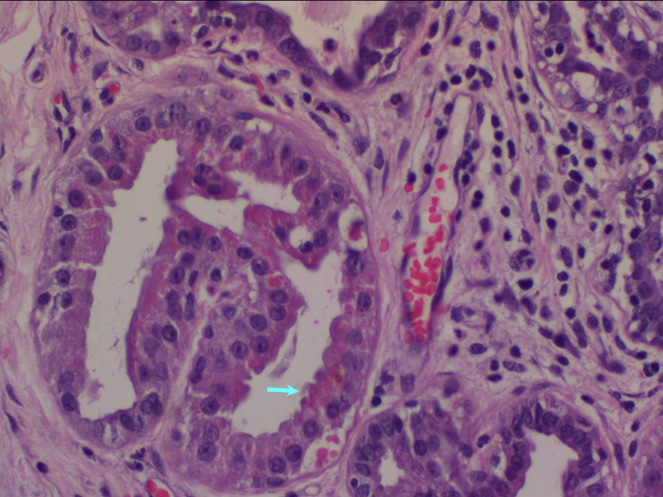 |
| Histopathology features: | |
| ‣ Specimen type: | Lump excision |
| ‣ Laterality: | Right |
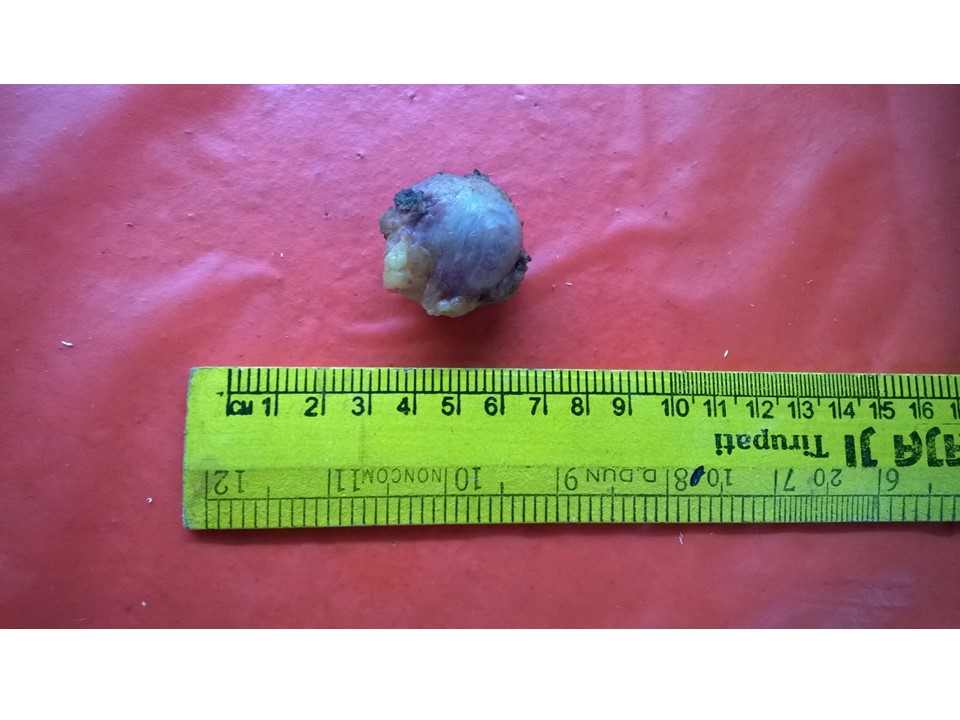
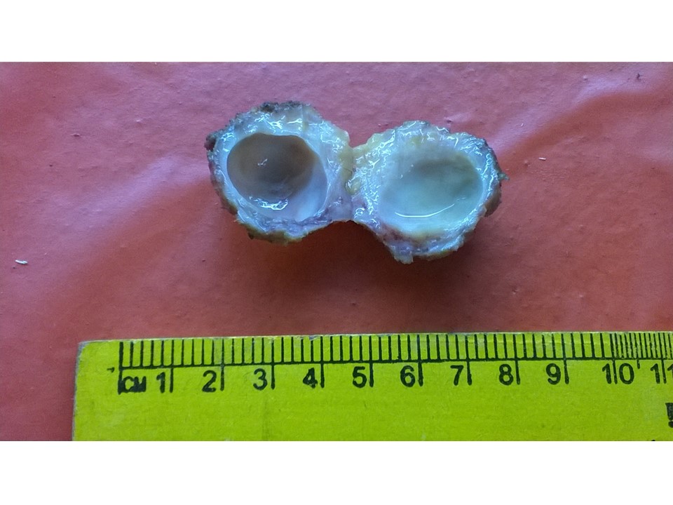
| ‣ Macroscopy: | Single cystic nodule (3.5 × 3.5 × 2.5 cm). Inner surface of cyst is very smooth, with no septations or solid areas seen. The cyst contained 2 mL of whitish turbid fluid. The outer surface of the cyst was brownish. The maximum thickness of the cyst wall was 10 mm |
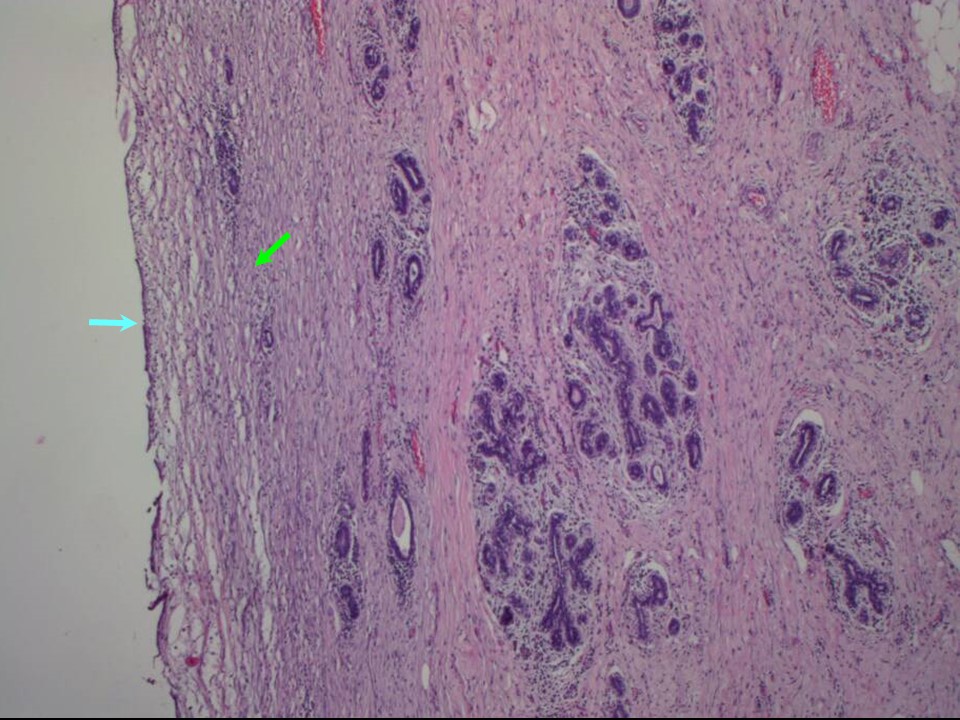
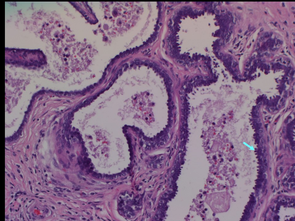
| ‣ Histological type: | The lining epithelium of the cyst wall is flattened epithelium and the wall is infiltrated with lymphocytes and histiocytes. Sections from the thickened part of the cyst wall shows adenosis, a few dilated ducts lined by metaplastic columnar epithelium and apocrine epithelium. Impression: Breast cyst showing changes of benign proliferative breast disease without atypia in the cyst wall |
| ‣ Histological grade: | |
| ‣ Mitosis: | |
| ‣ Maximum invasive tumour size: | |
| ‣ Lymph node status: | |
| ‣ Peritumoural lymphovascular invasion: | |
| ‣ DCIS/EIC: | |
| ‣ Margins: | |
| ‣ Pathological stage: | |
| ‣ Biomarkers: | |
| ‣ Comments: | Culture of the cystic fluid did not show any bacterial growth |
Case summary:
| Perimenopausal woman presented with acute-onset painful right breast lump. Diagnosed as complicated cyst, BI-RADS 2 on imaging and as benign proliferative breast disease on cytology and histopathology. |
Learning points:
|