Home / Training / Manuals / Atlas of breast cancer early detection / Learning

Atlas of breast cancer early detection

Breast imaging – Mammography technique – Mammography procedure – Additional mammography views – 90 degrees lateral views


A 90 degrees lateral view is the third most useful projection in mammography. It is used to visualize palpable breast lesions in the far medial, parasternal, or inner quadrant of the breast or in the medial quadrant in a patient with a pacemaker or port located in the upper quadrant of the breast, which limits a standard MLO view.

Two projections are used to obtain optimal geometric sharpness:

  • Mediolateral (ML) for a lesion in the lateral quadrant of the breast and
  • Lateromedial (LM) for a lesion in the medial quadrant of the breast.


Advantages
  • Localization and triangulation of the lesion.
  • Evaluation of calcifications. On 90 degrees lateral view, the milk-of-calcium calcifications can be seen precipitated and collected in a dependent portion of the cyst.
The medial quadrant lesion rises and the lateral quadrant lesion falls. In a 90 degree projection, a medial lesion will be seen in the upper quadrant and a lateral lesion in the lower quadrant. A lesion higher in ML view than in MLO view is in the medial quadrant in CC view, whereas a lesion lower in ML view is in the lateral quadrant in CC view. This can be remembered with the mnemonic “Muffins rise and lead falls”.

Steps to obtain the ML view
  1. Proceed to ML view after the bilateral breast MLO and CC projections have been obtained.
  2. Change the machine gantry angulation and patient positioning for the ML projection.
  3. Rotate the mammography unit tube gantry to 90 degrees so that breast compression is medial and at right angles to the CC view and the X-ray beam is parallel to the floor.
  4. Raise the patient’s arm and place it above the detector. Pull the right breast inward on the breast support to include as much breast tissue as possible with no angulation and patient body tilt. Apply gentle compression, keeping the right breast in position. Move the left breast aside with the left arm if necessary.
  5. When adequate compression (8–12 kg) has been achieved, step behind the protective lead shield and take the X-ray.


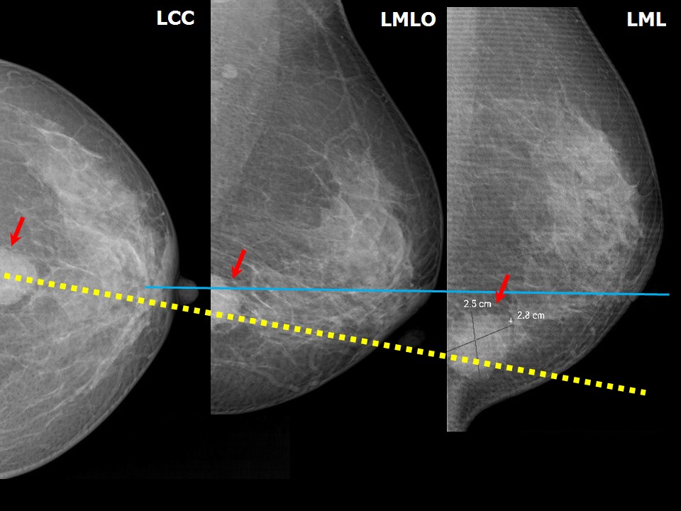
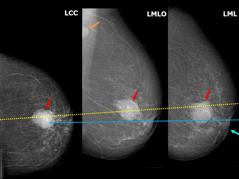
Quadrant localization


Place the CC view, the MLO view, and the 90 degrees lateral view in a row to be viewed with the nipple shadow at the same level along a straight line in all three images. If the lesion is lower on ML than on MLO, it lies in the lateral quadrant on CC. If the lesion is higher on ML than on MLO, it is in the medial quadrant on CC.


Click on the pictures to magnify and display the legends


Click on this icon to display a case study

IARC, 150 Cours Albert Thomas, 69372 Lyon CEDEX 08, France - Tel: +33 (0)4 72 73 84 85 - Fax: +33 (0)4 72 73 85 75
© IARC 2024 - All Rights Reserved.