Home / Training / Manuals / Atlas of breast cancer early detection / Learning

Atlas of breast cancer early detection

Breast pathology – Introduction


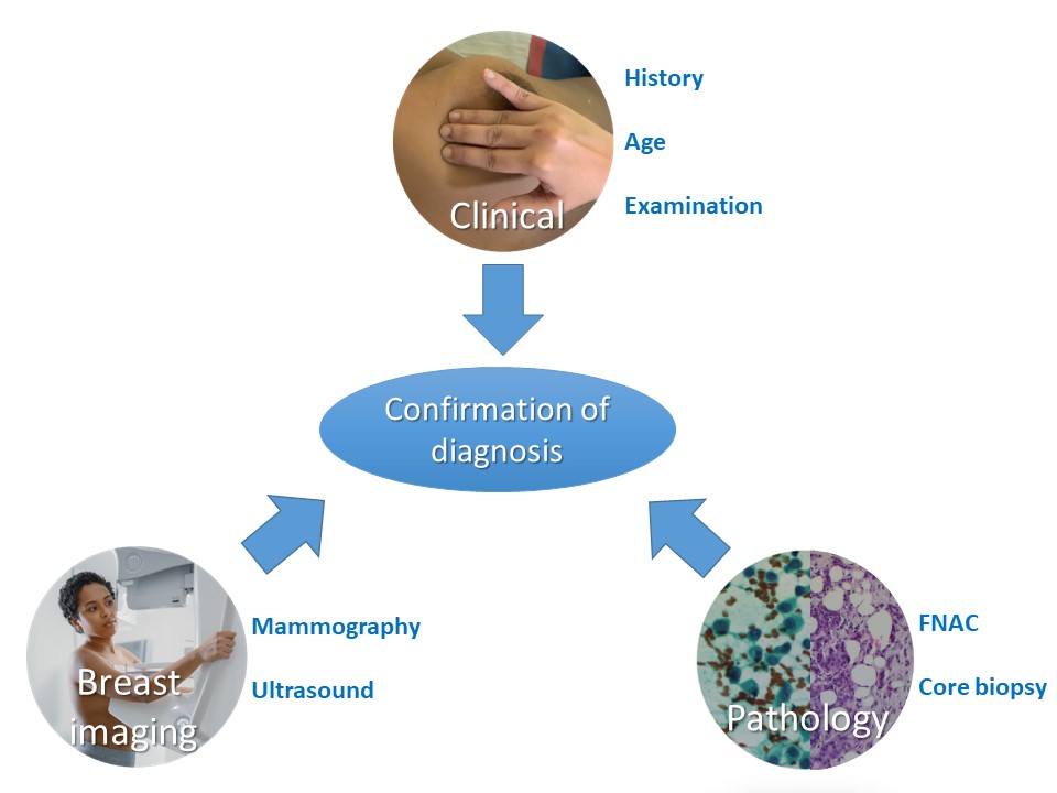
An accurate pathological diagnosis is crucial for determining the course of breast cancer treatment and estimating the prognosis with reasonable accuracy. Pathology is one of the key elements of “triple assessment” for the evaluation of a woman with an abnormal screening test or a woman with clinical and/or radiological suspicion of breast lesion. Ensuring quality at every step of breast pathology – from sample collection to final reporting – is absolutely essential to provide standardized evidence-based care to breast cancer patients.


Pathological evaluation of a suspected breast abnormality requires:
  • cells aspirated from a lesion (FNAC);
  • a core of abnormal breast tissue collected with a large-bore needle (core needle biopsy);
  • excision of the entire suspected malignant lesion of the breast (excisional biopsy); or
  • surgical removal of the entire breast (mastectomy specimen).


Core biopsy obtained with a needle (core needle biopsy) is the current standard of care for initial confirmation of diagnosis. It has the following advantages over FNAC:
  • Atypical hyperplasias and in situ carcinomas can be detected more efficiently.
  • The biological nature (type and differentiation of cancer) can be assessed.
  • Immunohistochemistry (IHC) can be used to study different molecular markers.


However, FNAC is still highly relevant in settings with limited resources. Most lesions detected in such settings are clinically palpable and studies have shown that the sensitivity and specificity of FNAC from such lesions is marginally lower than that of core biopsy. Moreover, FNAC is rapid, logistically simpler, and can be organized in secondary care facilities. The patient does not have to travel to a tertiary care facility for the confirmation of diagnosis. This avoids any delay in diagnosis. The advantages and disadvantages of the two techniques are compared in detail in the following table.


Comparison of FNAC and core needle biopsy as a diagnostic modality:

FNAC

Core needle biopsy

Sampling procedure

Time taken Quicker procedure to perform without need for local anaesthesia Longer, local anaesthesia required
Training and expertise of staff Training is essential in aspiration and preparation of quality smears Training is essential and more intensive
Complications of procedure Less traumatic because needle bore is smaller (23/24G), so may be appropriate in women taking anticoagulants More traumatic because needle bore is larger (14/16G). Associated with potential complications such as haematoma, haemorrhage, and, very rarely, implantation of tumour cells along the needle track
Yield of material Repeated sampling and moving the needle in different directions yields more material for assessment Sampling limited to the tissue removed in the biopsy cores

Diagnostic value in various breast lesions

Cystic lesions Indicated in the evaluation of cystic lesions of the breast, especially in ones with atypical imaging features Not useful in cystic lesions
Microcalcifications Inappropriate in assessment of microcalcifications detected on imaging because inadequate material may be aspirated from calcified areas Investigation of choice in evaluation of microcalcifications
DCIS Distinguishing between DCIS and invasive carcinoma is not possible because the cell morphology is similar in both Core biopsy, unlike FNAC, yields tissue fragments including architectural features, so it is possible to determine whether DCIS or invasive carcinoma is present
Papillary lesions Inaccurate for papillary lesions Useful to evaluate low-grade malignancies such as tubular carcinoma
Low-grade malignancies In the absence of architectural features, tumour cells with low-grade malignant features such as tubular carcinoma may be categorized as not malignant Useful to evaluate low-grade malignancies such as tubular carcinoma
Less-cellular breast lesions Not the sampling technique of choice for lesions that are relatively hypocellular and yield scanty epithelial material, such as sclerotic fibroadenomas, invasive breast carcinoma with extensive sclerosis, and infiltrating lobular carcinoma The diagnostic method of choice because the tissue section shows the sclerotic stromal tissue and intervening cell morphology

Turnaround time

Results are often available on the same day, possibly in a few hours Results are available after a few days. Tissue requires adequate fixation, then processing to make paraffin blocks and cutting before staining

Cost of procedure

Relatively inexpensive to perform More expensive than FNAC

Effect of procedure on interpretation of subsequent histopathology of the lesion

Does not significantly affect the subsequent histopathology of the sampled areas because of the small needle bore used For small lesions, the lesion seen on mammography may not be identified in subsequent open biopsy because it has been completely removed. The inflammation and fibrosis caused by the biopsy may interfere with the interpretation of subsequent excision biopsy, particularly with regard to grading and the estimation of the size of the lesion, especially in small lesions

Biomarker status assessment

Assessment of estrogen receptor (ER), progesterone receptor (PR), and human epidermal growth factor receptor 2 (HER2) status is not very reliable on cytology smears, although it is useful in cases of distant metastases and cases where tissue material is not available. However, on cell blocks with adequate cellularity, IHC is equivalent to a biopsy Assessment of ER, PR, and HER2 status is very reliable and is the standard practice


  • Most benign and malignant breast lesions can be reliably diagnosed by FNAC.
  • A positive diagnosis on FNAC is usually confirming for malignancy.
  • A negative cytological result does not guarantee that the lesion is benign. A mammographically or clinically suspicious lesion necessitates biopsy even when the cytology report is negative.
  • Image-guided FNAC significantly increases the accuracy of malignancy detection in non-palpable or small breast lesions.


The next few sections of the atlas deal with both breast cytopathology and histopathology. We will discuss the techniques of sample collection and handling, fixation, processing, and reporting. The atlas does not intend to deal with the interpretation of abnormal findings on cytology and histopathology in detail. That is in the domain of a trained pathologist.


Click on the pictures to magnify and display the legends


Click on this icon to display a case study

IARC, 150 Cours Albert Thomas, 69372 Lyon CEDEX 08, France - Tel: +33 (0)4 72 73 84 85 - Fax: +33 (0)4 72 73 85 75
© IARC 2024 - All Rights Reserved.