


Home / Training / Manuals / Atlas of breast cancer early detection / Learning
.png)
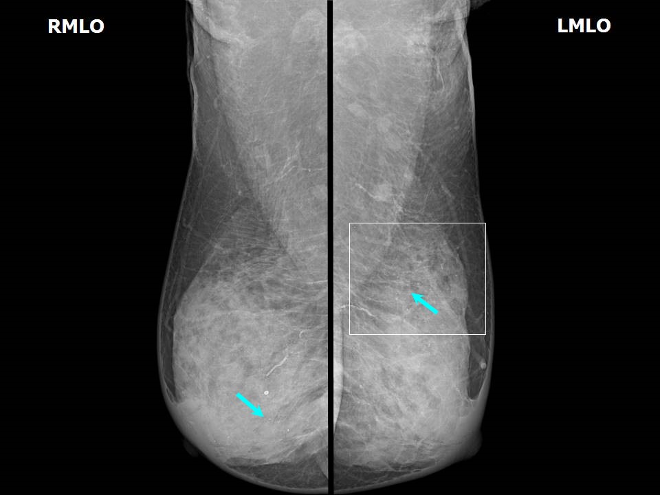
Click on the pictures to magnify and display the legends

Click on this icon to display a case study
Atlas of breast cancer early detection
Filter by language: English / РусскийBreast imaging Mammography interpretation Mammography lexicon Calcifications Suspicious morphology |
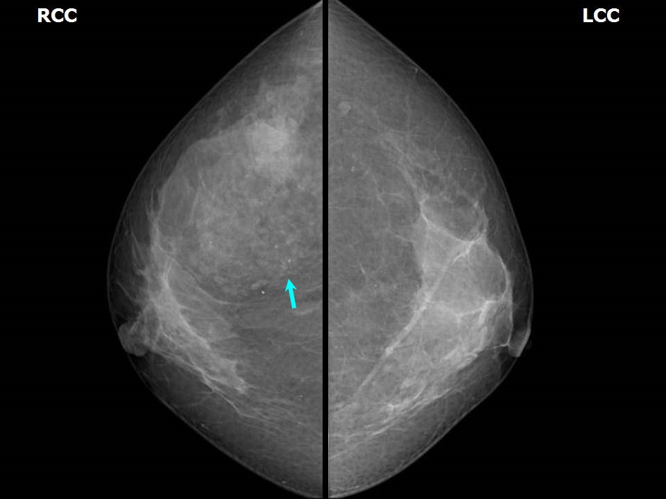
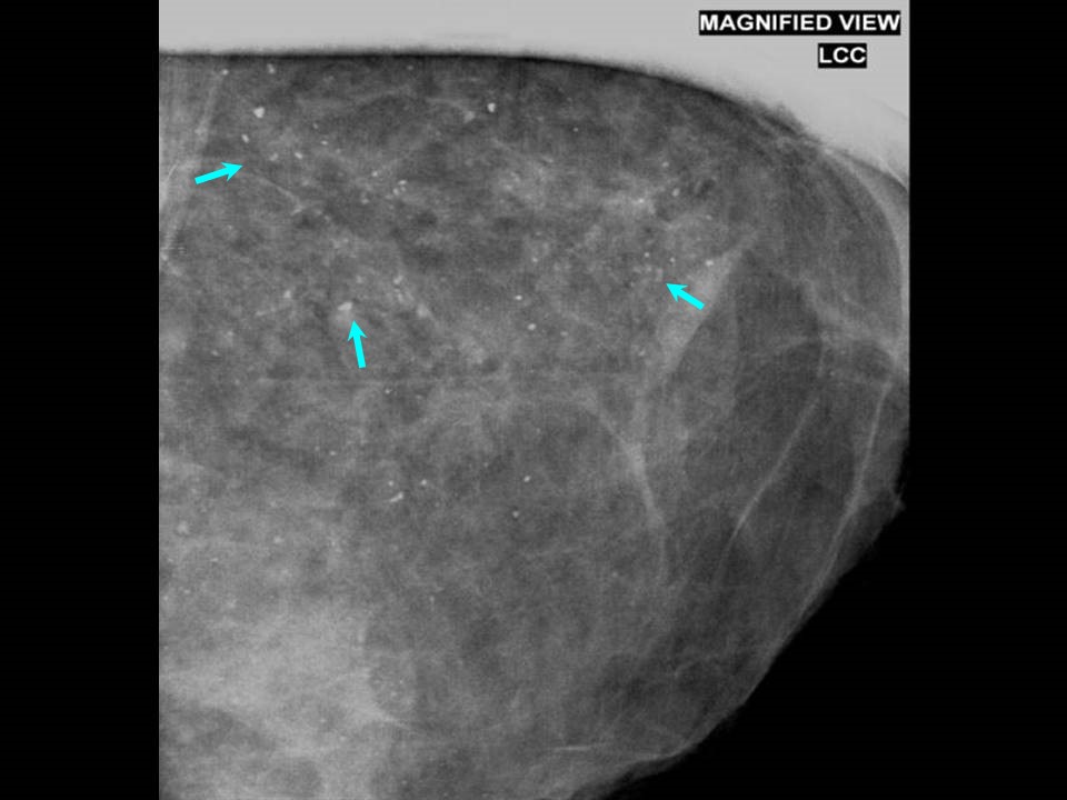
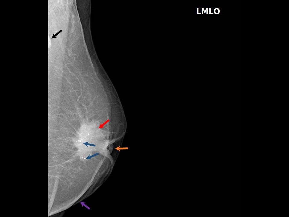
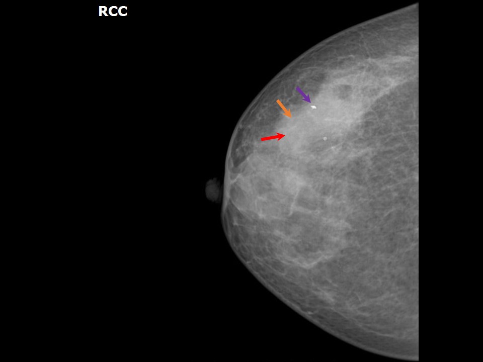
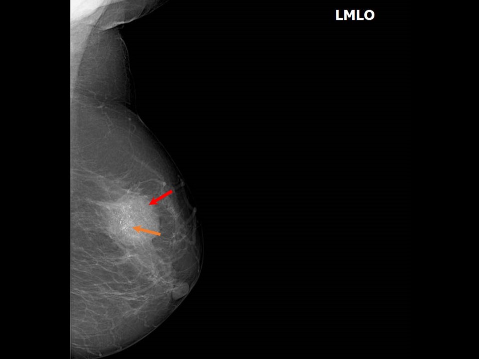
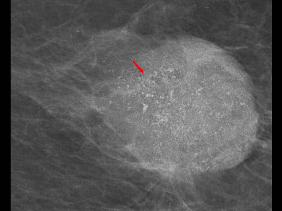
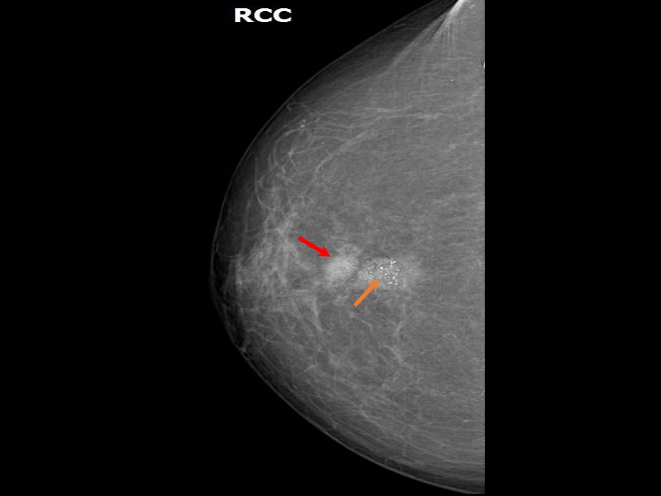
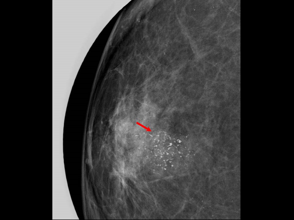
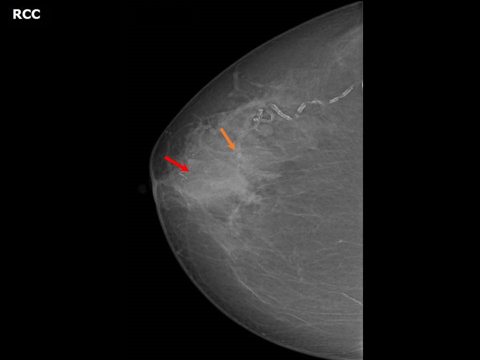
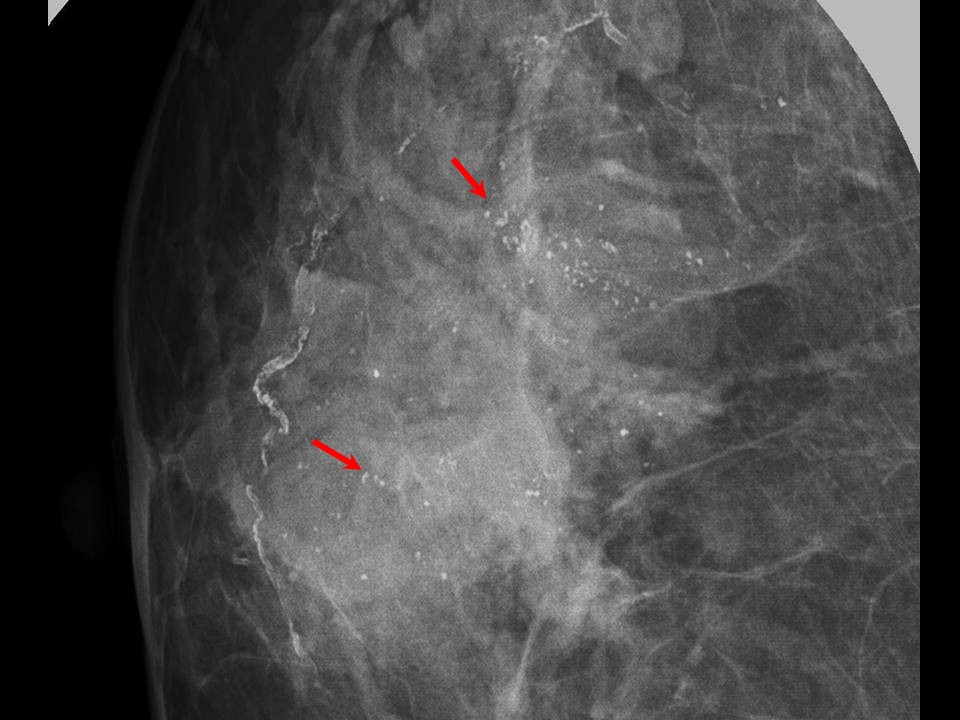
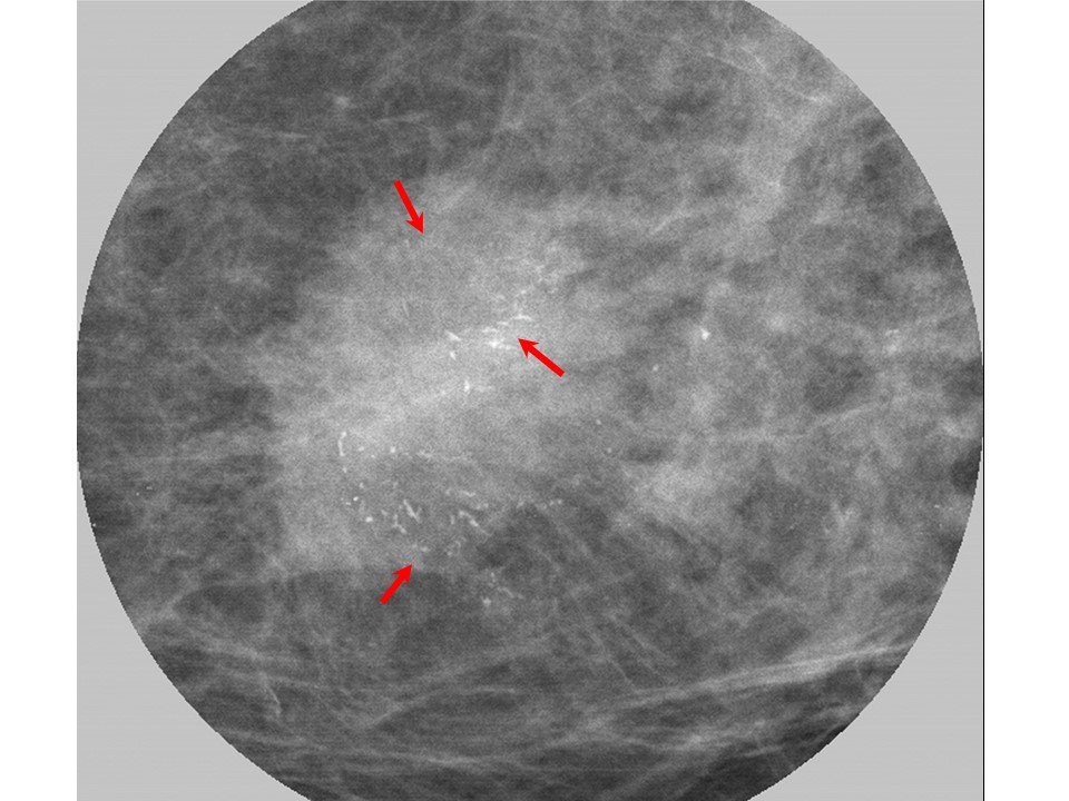
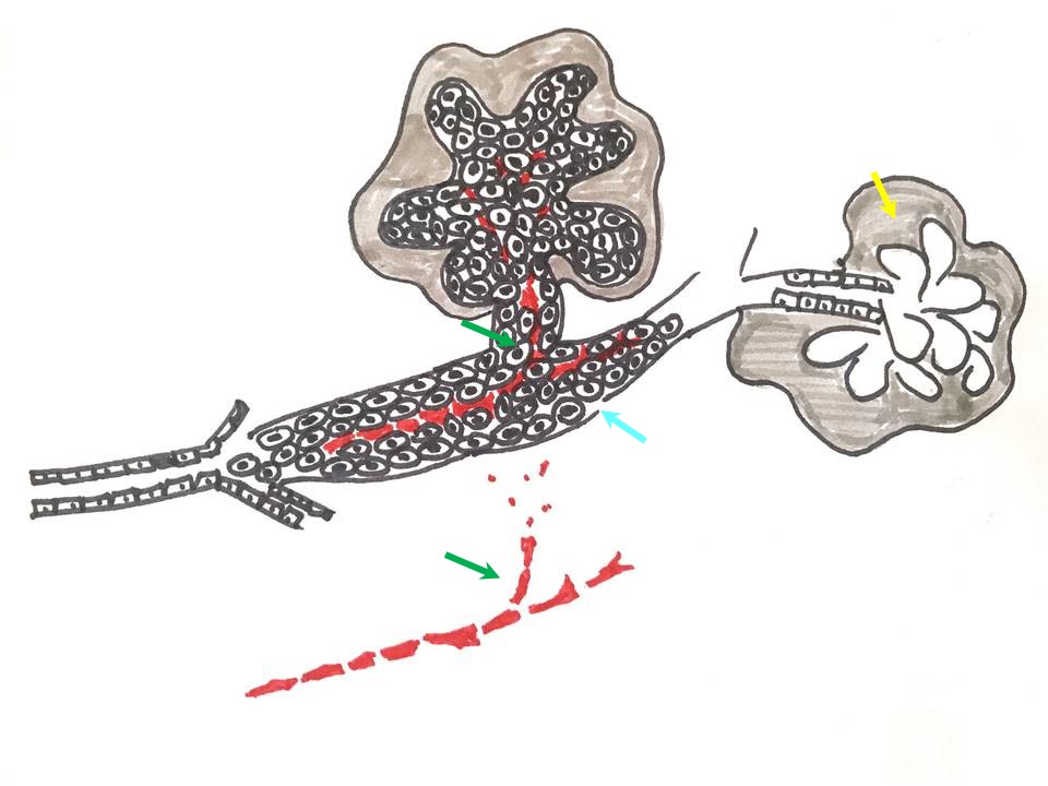
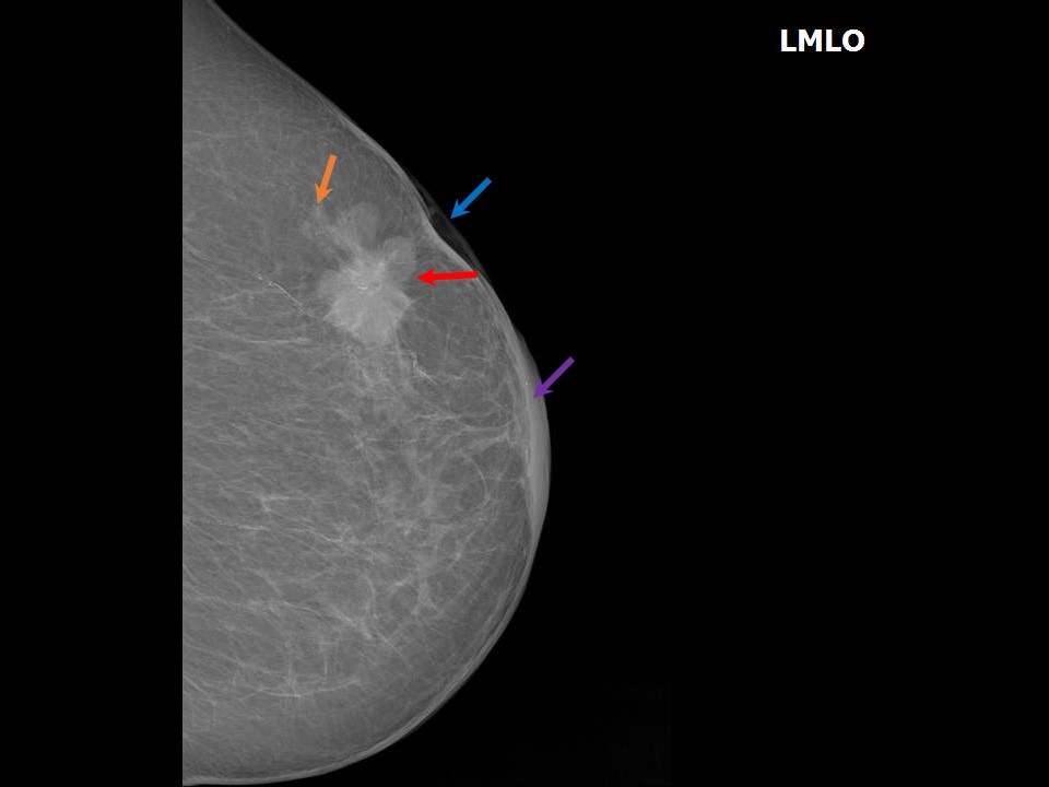
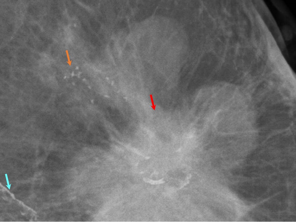
Suspicious morphology Several varieties may be seen in this category. 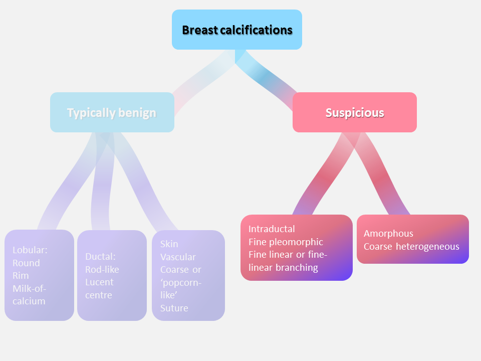 Amorphous calcifications These are small hazy calcifications of indistinct size, shape, and appearance. If seen in diffuse distribution, in one or both breasts, they represent benign amorphous calcifications. Multiple clustered amorphous calcifications are probably benign calcifications, whereas a solitary cluster of amorphous calcification or new calcifications are suspicious for malignancy .Coarse heterogeneous calcifications These are usually irregular calcifications > 0.5 mm in size but smaller than dystrophic calcifications .Fine pleomorphic calcifications These are < 0.5 mm across and are of varying shapes and sizes. They may be seen in a group or in regional distribution when entire duct distribution is involved. They may be within a mass. Fine pleomorphic calcifications usually represent likely malignant calcifications .Fine linear or fine-linear branching calcifications These are thin, linear, irregular, and usually branching discontinuous calcifications < 0.5 mm across. They form in the duct lumen, which is involved by the necrotic irregular cancer. Such calcifications may be seen with or without a mass . |
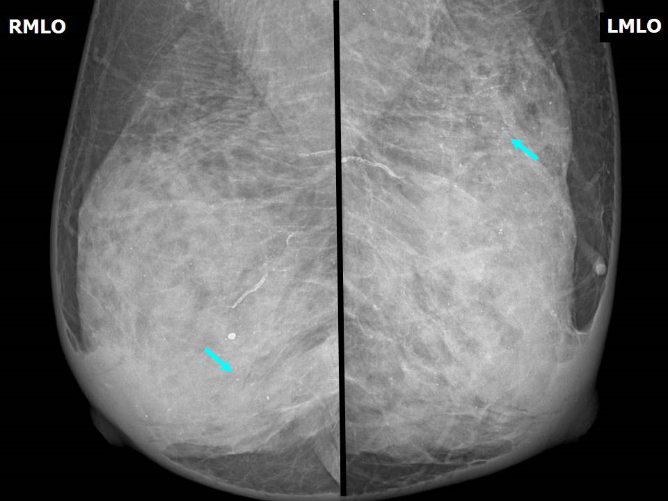

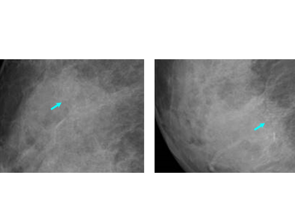


Click on the pictures to magnify and display the legends
Click on this icon to display a case study
25 avenue Tony Garnier CS 90627 69366, LYON CEDEX 07 France - Tel: +33 (0)4 72 73 84 85
© IARC 2025 - Terms of use - Privacy Policy.
© IARC 2025 - Terms of use - Privacy Policy.