


Home / Training / Manuals / Atlas of breast cancer early detection / Learning
.png)
Click on the pictures to magnify and display the legends

Click on this icon to display a case study
Atlas of breast cancer early detection
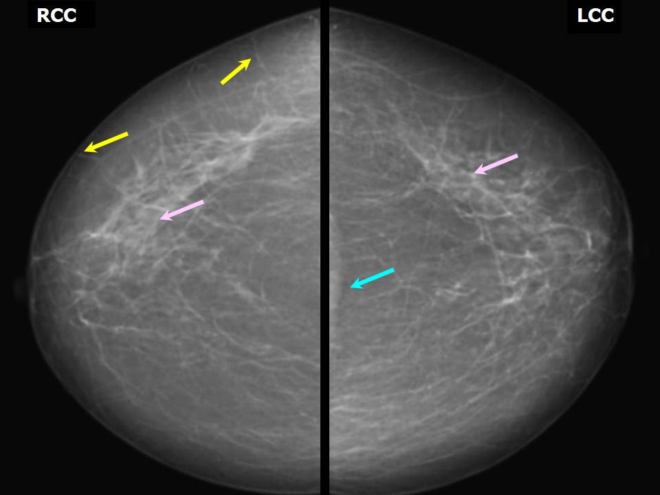
Filter by language: English / РусскийBreast imaging Mammography technique Mammography procedure Technical adequacy of acquired mammographic views Craniocaudal (CC) view |
The CC view complements the MLO view. The following criteria should be fulfilled to ensure that the CC image obtained is accurate and technically adequate to enable a diagnosis to be made:
Retromammary bursa Pectoralis shadow Bilateral symmetrical CC view Mirror image |


Click on the pictures to magnify and display the legends
Click on this icon to display a case study
25 avenue Tony Garnier CS 90627 69366, LYON CEDEX 07 France - Tel: +33 (0)4 72 73 84 85
© IARC 2025 - Terms of use - Privacy Policy.
© IARC 2025 - Terms of use - Privacy Policy.