


Home / Training / Manuals / Atlas of breast cancer early detection / Learning
.png)
Click on the pictures to magnify and display the legends

Click on this icon to display a case study
Atlas of breast cancer early detection
Filter by language: English / РусскийBreast imaging Breast ultrasound Ultrasound lexicon Calcifications |
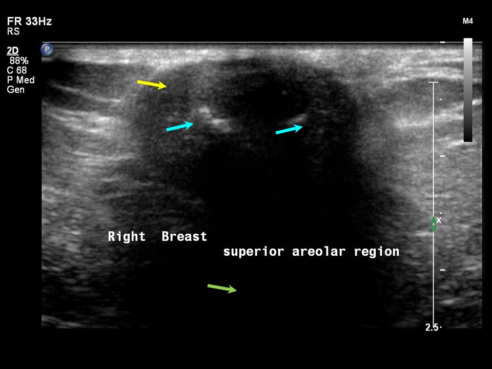
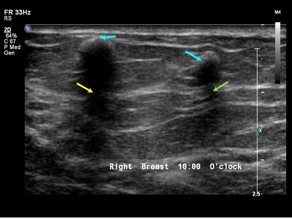
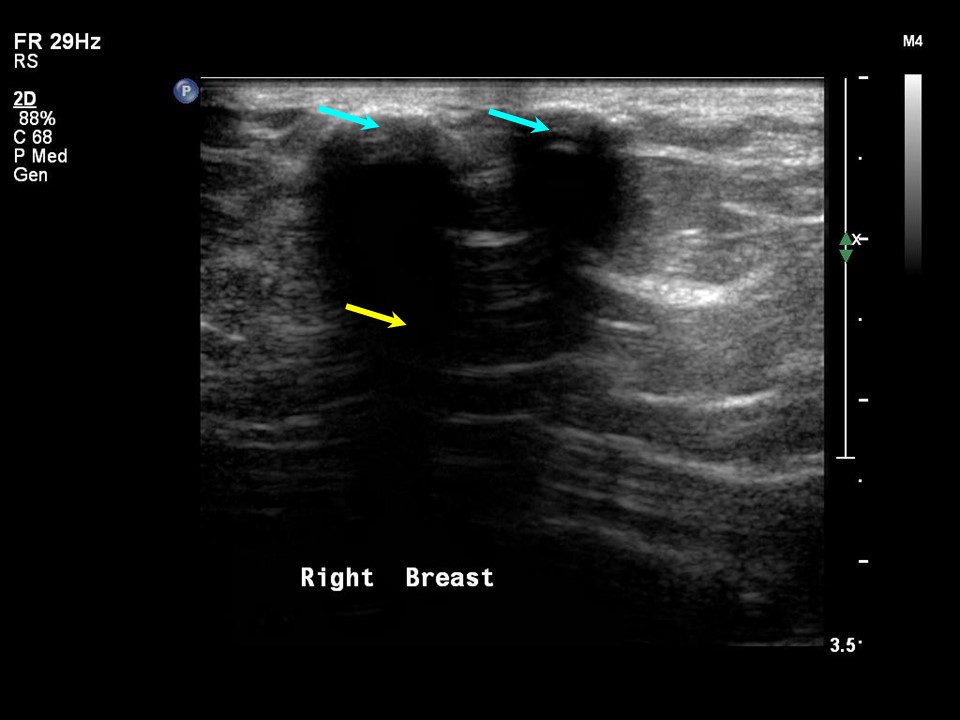
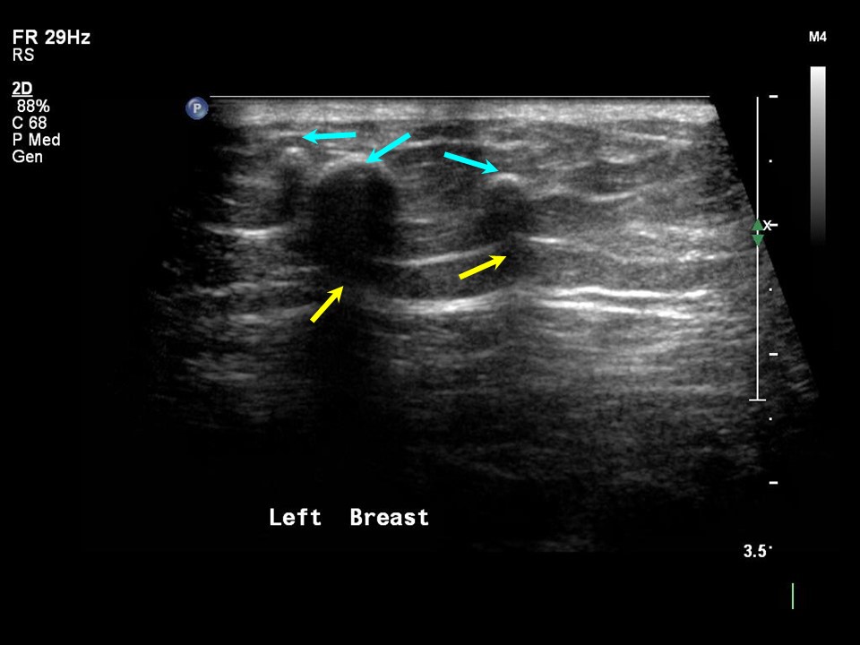
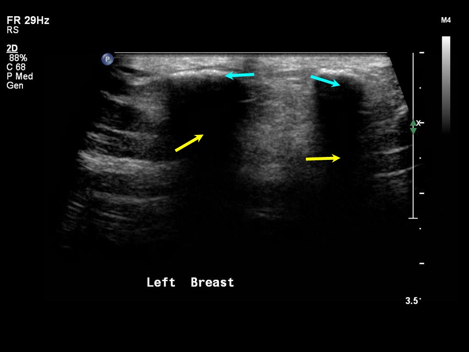
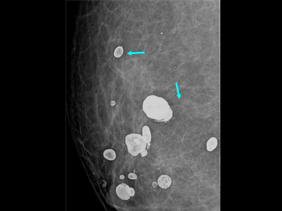
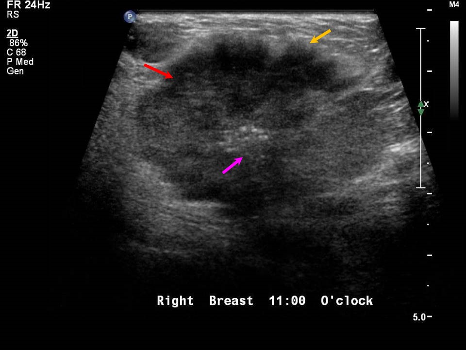
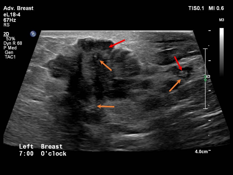
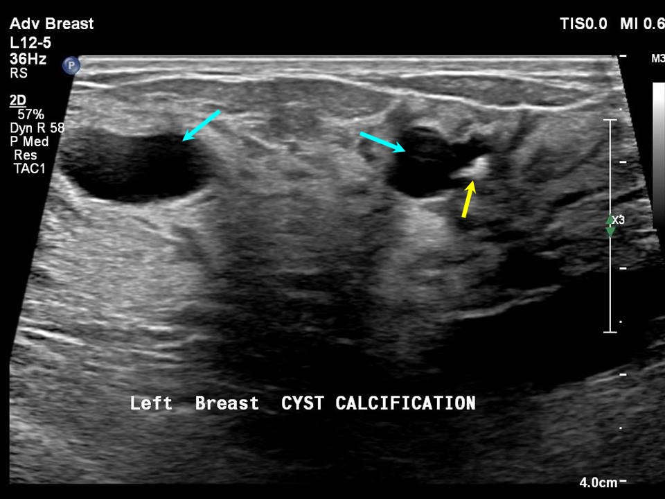
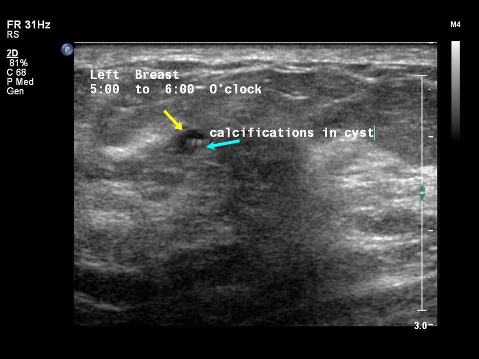
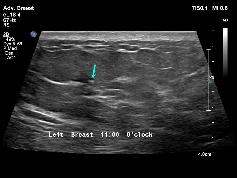
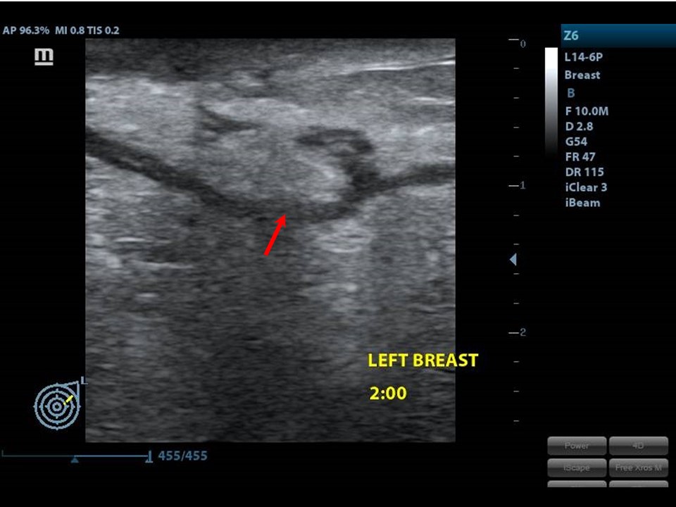
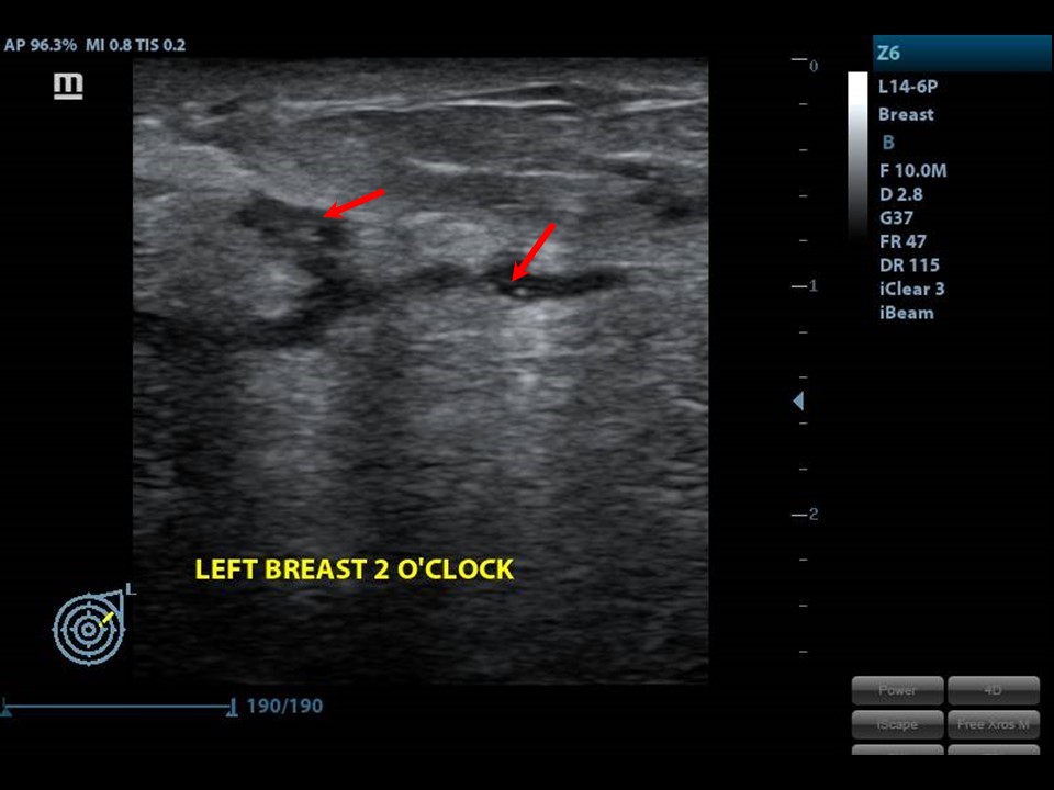
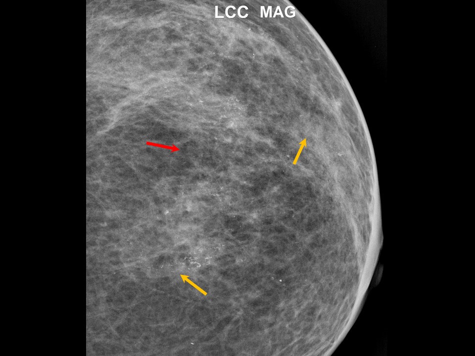
High-resolution ultrasound equipment used for diagnostic radiology can also show breast calcifications. Macrocalcifications are sonographically easy to locate in the breast parenchyma and also in the mass. Microcalcifications can be detected only if they are numerous and grouped together. Scattered microcalcifications are difficult to detect on ultrasound.
Calcifications on ultrasound are described as calcifications in a mass, outside a mass, or intraductal. The morphological appearance of ultrasound calcifications can be used to describe a mass as possibly benign or malignant. Macrocalcifications in benign lesions Macrocalcifications are commonly associated with benign lesions, such as involuting fibroadenoma. Scars from breast-conserving surgery may develop into dystrophic calcifications, which are also benign macrocalcifications .Macrocalcifications in breast parenchyma Microcalcifications Microcalcifications grouped in a segment or within a mass are suggestive of malignancy .Cysts Microcysts or large cysts can have eccentric wall calcifications, seen as rim calcifications on mammography. Fluid within the cysts may calcify over time. This is seen on mammography as classical milk-of-calcium calcifications or round acinar calcifications. Ectatic ducts with debris that becomes calcified over time are seen as intraductal calcifications. Intraductal calcifications Intraductal calcifications are seen in DCIS. |


Click on the pictures to magnify and display the legends
Click on this icon to display a case study
25 avenue Tony Garnier CS 90627 69366, LYON CEDEX 07 France - Tel: +33 (0)4 72 73 84 85
© IARC 2025 - Terms of use - Privacy Policy.
© IARC 2025 - Terms of use - Privacy Policy.