


Home / Training / Manuals / Atlas of breast cancer early detection / Learning
.png)
Click on the pictures to magnify and display the legends

Click on this icon to display a case study
Atlas of breast cancer early detection
Filter by language: English / РусскийBreast imaging Breast ultrasound Ultrasound lexicon Special cases Lymph nodes Intramammary |
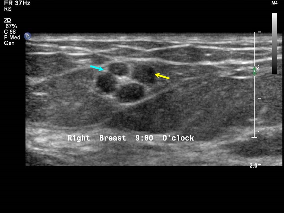
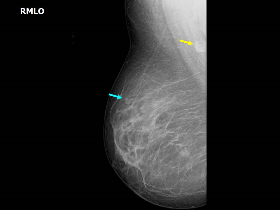
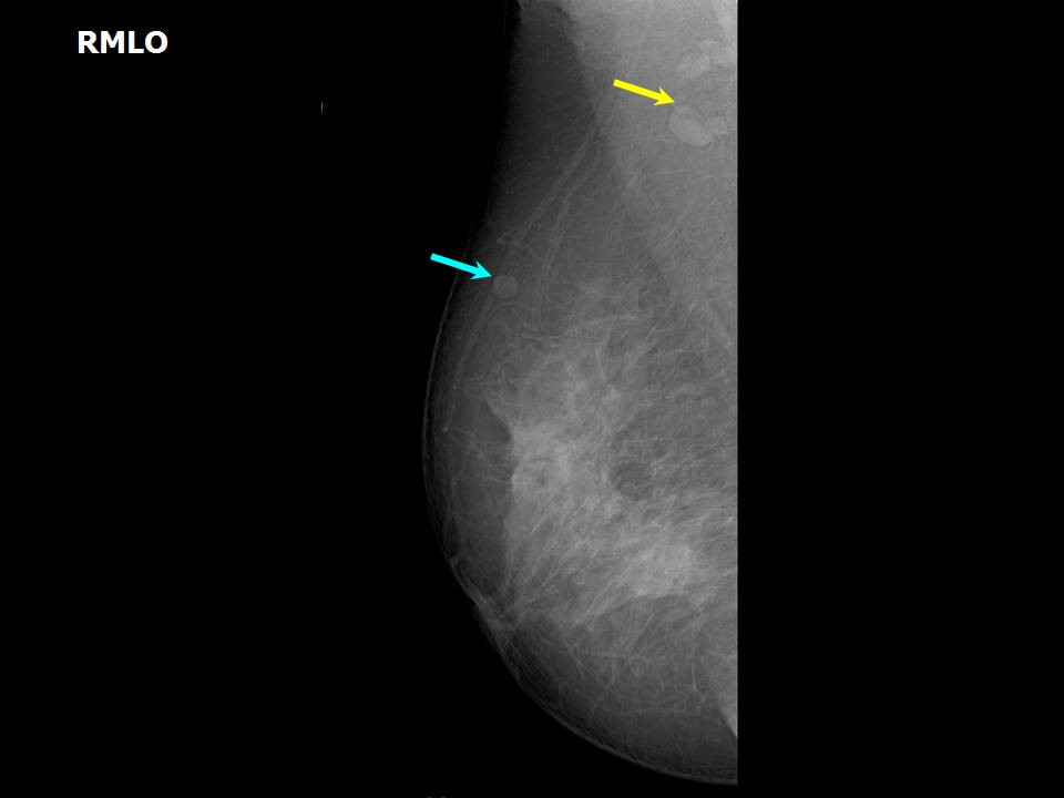
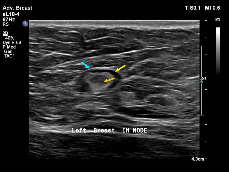
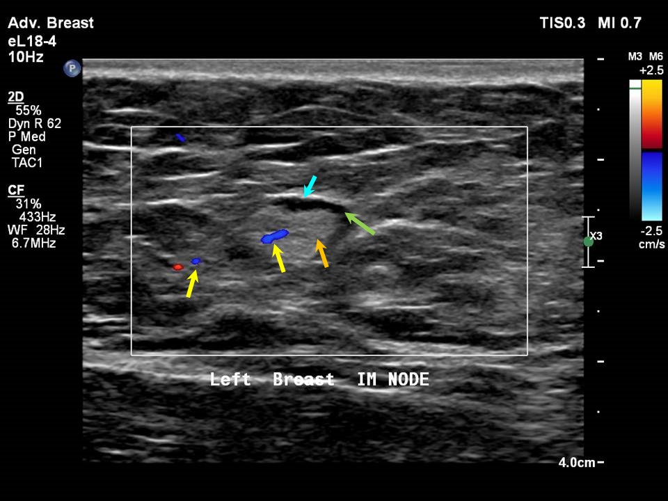
Lymph nodes within the breast tissue are called intramammary nodes. The nodes are commonly seen in the upper outer quadrant of the breast. They are oval, round, or uniform in shape, < 1 cm in diameter, with circumscribed margins and a central fatty hilum. They remain stable on follow-up mammogram. At times, a feeding vessel to the intramammary node may be seen .Case 1: Case 2: Case 3: Metastatic intramammary nodes
|

Click on the pictures to magnify and display the legends
Click on this icon to display a case study
25 avenue Tony Garnier CS 90627 69366, LYON CEDEX 07 France - Tel: +33 (0)4 72 73 84 85
© IARC 2025 - Terms of use - Privacy Policy.
© IARC 2025 - Terms of use - Privacy Policy.