


Home / Training / Manuals / Atlas of breast cancer early detection / Learning
.png)
Click on the pictures to magnify and display the legends

Click on this icon to display a case study
Atlas of breast cancer early detection
Filter by language: English / РусскийBreast imaging Breast ultrasound Ultrasound lexicon Special cases Fat necrosis |
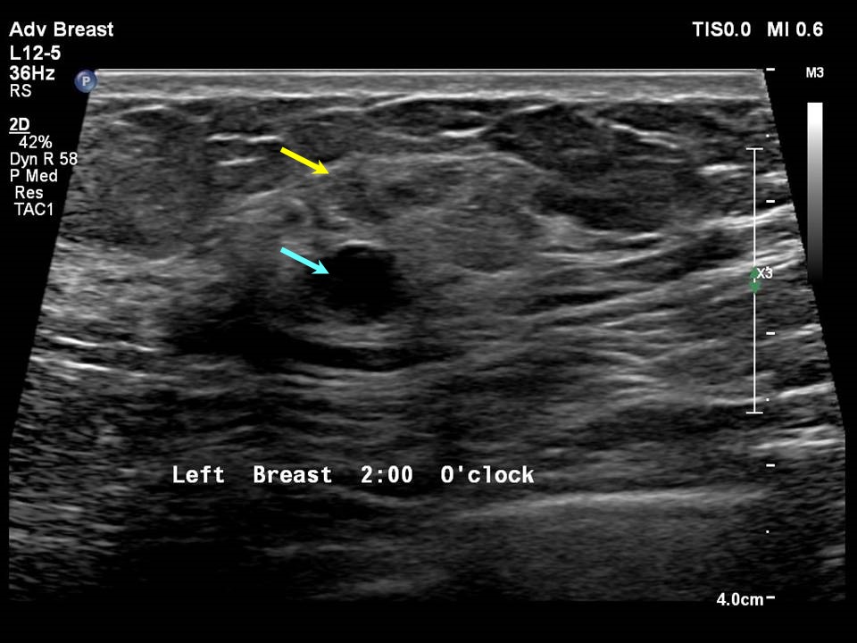
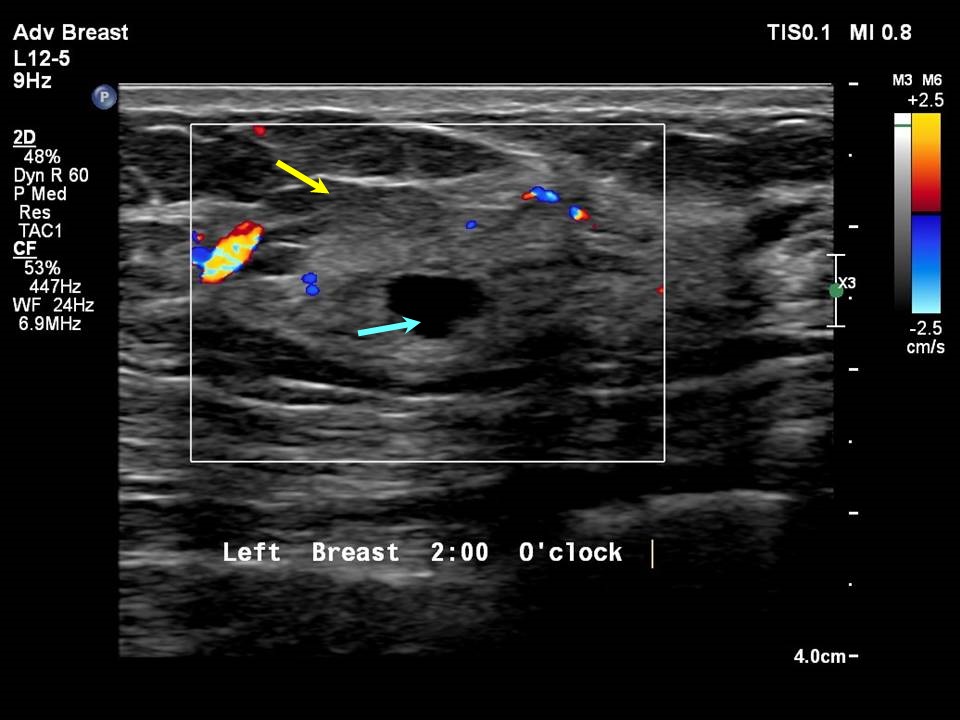
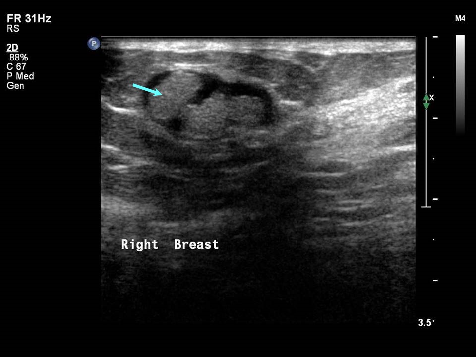
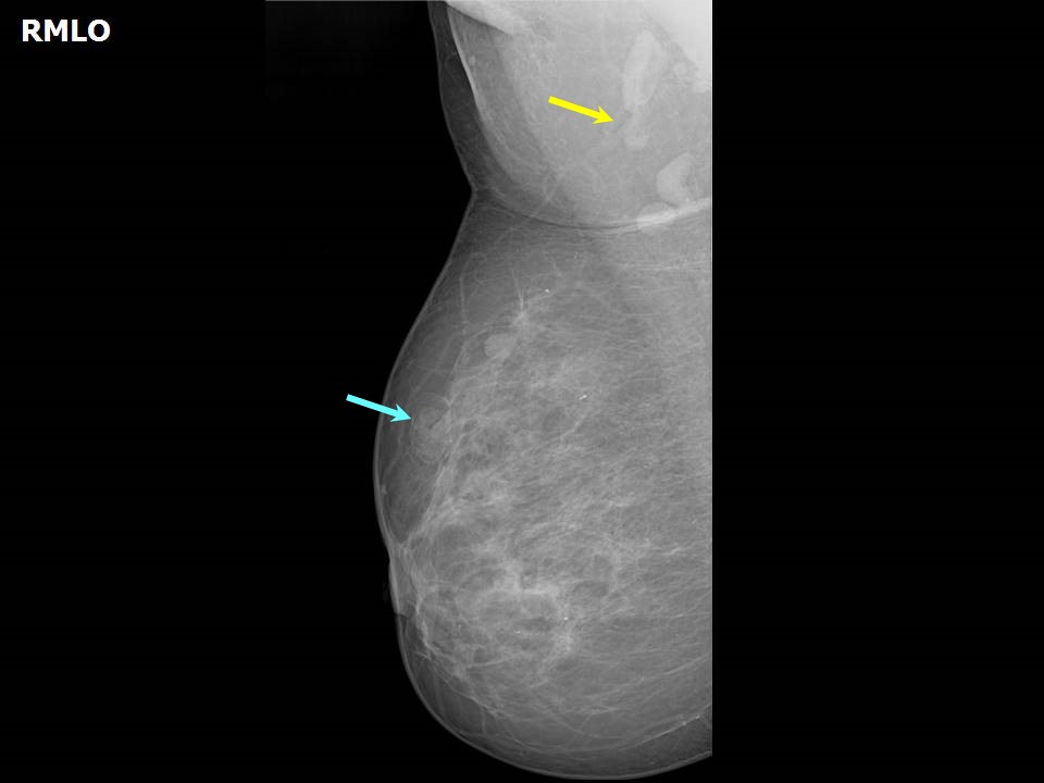
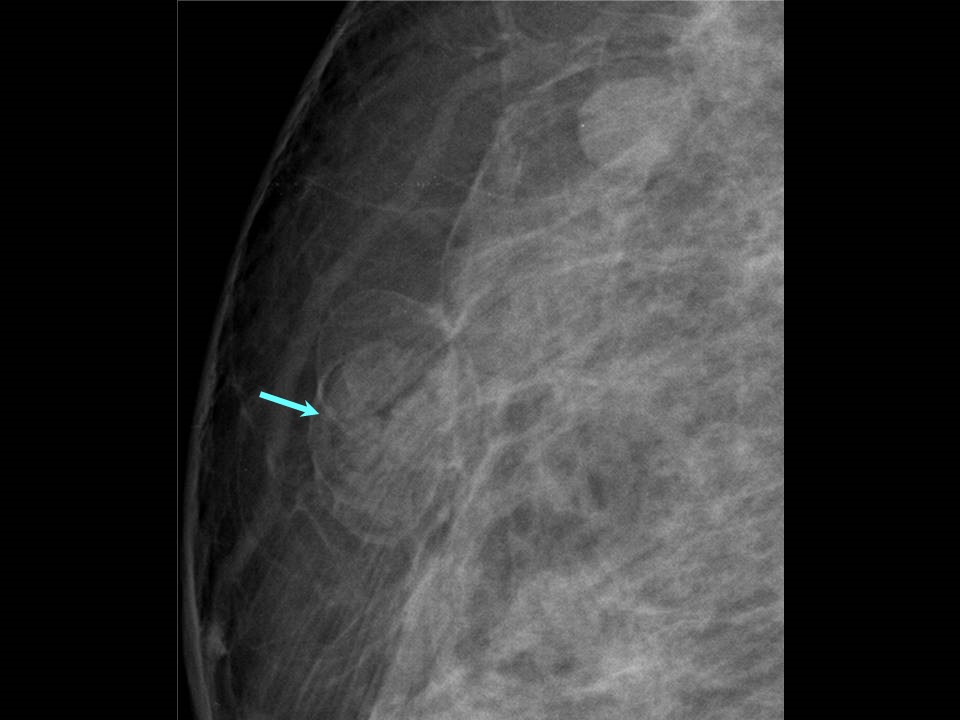
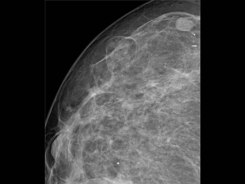
Fat necrosis develops spontaneously or as a consequence of breast trauma, surgery, biopsy, or radiotherapy. The acute stage shows disrupted fatty tissue with haemorrhage and inflammatory reaction. Over time, underlying inflammatory response changes occur; stromal fibrosis and calcification may become noticeable and a clinically palpable lump may develop at the site of fat necrosis. Pseudo mass formation may be visible on ultrasound as a circumscribed fat-containing lesion with thin walls. Calcifications with lucent centres are seen . |
Click on the pictures to magnify and display the legends
Click on this icon to display a case study
25 avenue Tony Garnier CS 90627 69366, LYON CEDEX 07 France - Tel: +33 (0)4 72 73 84 85
© IARC 2025 - Terms of use - Privacy Policy.
© IARC 2025 - Terms of use - Privacy Policy.