


Home / Training / Manuals / Atlas of breast cancer early detection / Cases
Atlas of breast cancer early detection
Filter by language: English / Русский
Go back to the list of case studies
.png) Click on the pictures to magnify and display the legends
Click on the pictures to magnify and display the legends
| Case number: | 165 |
| Age: | 56 |
| Clinical presentation: | Postmenopausal woman with average risk of breast cancer presented with a left breast lump and mastalgia noticed 1 week ago. |
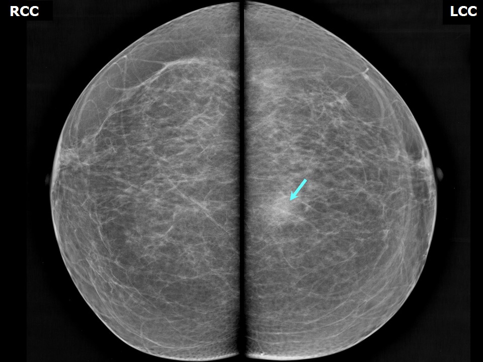
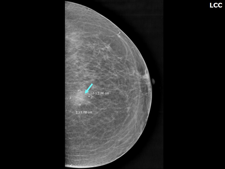
Mammography:
 |  |
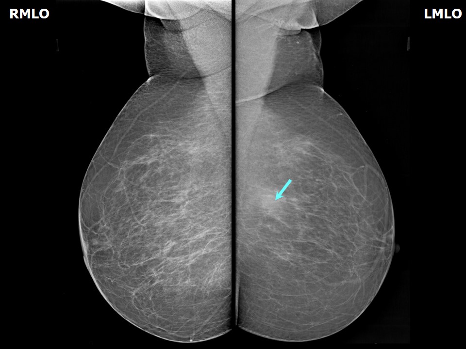 |
| Breast composition: | ACR category b (there are scattered areas of fibroglandular density) | Mammography features: |
| ‣ Location of the lesion: | Left breast, upper inner quadrant at 11 oclock, posterior third |
| ‣ Mass: | |
| • Number: | 1 |
| • Size: | 2.6 × 1.7 cm |
| • Shape: | Irregular |
| • Margins: | Indistinct |
| • Density: | Equal |
| ‣ Calcifications: | |
| • Typically benign: | None |
| • Suspicious: | None |
| • Distribution: | None |
| ‣ Architectural distortion: | None |
| ‣ Asymmetry: | None |
| ‣ Intramammary node: | None |
| ‣ Skin lesion: | None |
| ‣ Solitary dilated duct: | None |
| ‣ Associated features: | None |
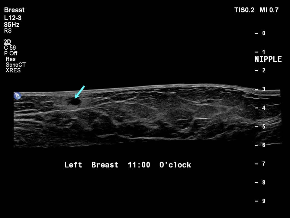
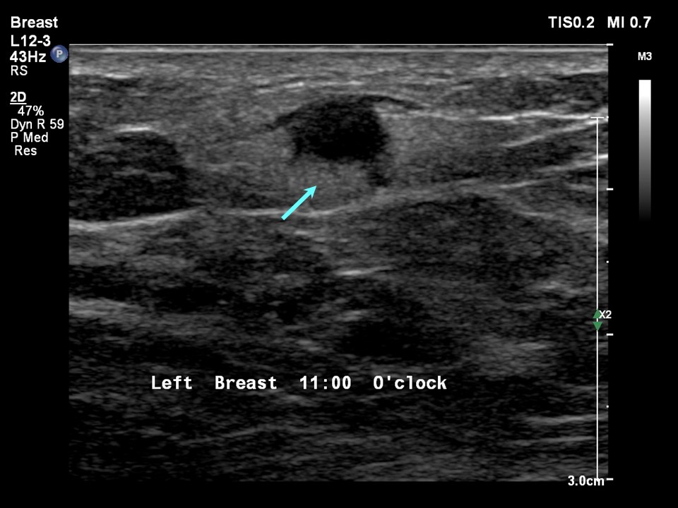
Ultrasound:
 |  |
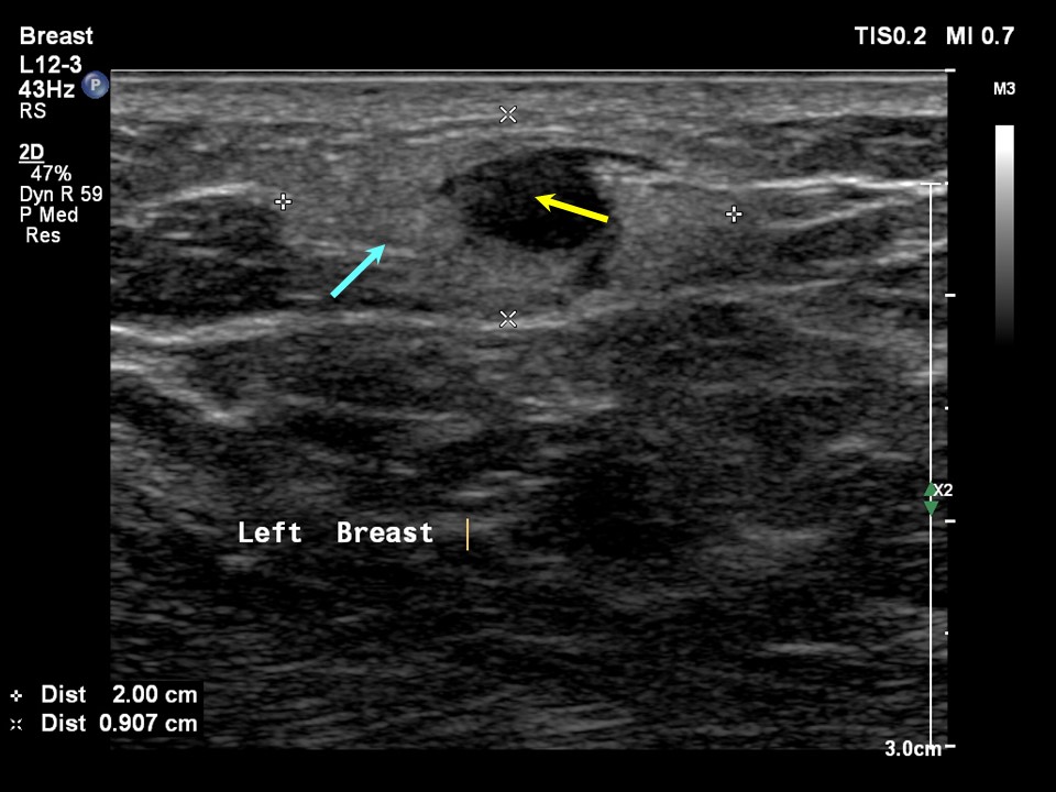 |
| Ultrasound features: Left breast, upper inner quadrant at 11 oclock, 10.2 cm from the nipple and at 1.0 cm skin depth | |
| ‣ Mass | |
| • Location: | Left breast, upper inner quadrant at 11 oclock, 10.2 cm from the nipple and at 1.0 cm skin depth |
| • Number: | 1 |
| • Size: | 2.0 × 1.0 cm |
| • Shape: | Oval |
| • Orientation: | Parallel |
| • Margins: | Circumscribed, 23 lobulations present |
| • Echo pattern: | Heteroechoic with central anechoic component |
| • Posterior features: | No posterior features |
| ‣ Calcifications: | None |
| ‣ Associated features: | None |
| ‣ Special cases: | None |
BI-RADS:
BI-RADS Category: 2 (benign)Further assessment:
Further assessment advised: Referral for cytologyCytology:
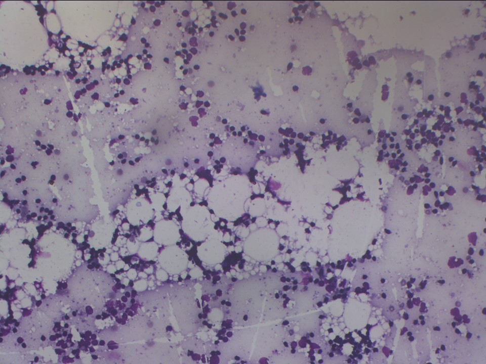 |
| Cytology features: | |
| ‣ Type of sample: | FNAC |
| ‣ Site of biopsy: | |
| • Laterality: | Left |
| • Quadrant: | Upper inner |
| • Localization technique: | Palpation |
| • Nature of aspirate: | 0.5 mL of thin greyish fluid |
| ‣ Cytological description: | Smears reveal foamy macrophages, a few multinucleated giant cells, numerous lymphocytes, and occasional ductal epithelial cells on a background of granular debris, fat droplets, and fragments of adipose tissue |
| ‣ Reporting category: | Benign |
| ‣ Diagnosis: | Consistent with fat necrosis |
| ‣ Comments: | None |
Case summary:
| Postmenopausal woman presented with a painful left breast lump. Diagnosed as fat necrosis, BI-RADS 2 on imaging and as benign fat necrosis on cytology. |
Learning points:
|