


Home / Training / Manuals / Atlas of breast cancer early detection / Learning
.png)
Click on the pictures to magnify and display the legends

Click on this icon to display a case study
Atlas of breast cancer early detection
Filter by language: English / РусскийBreast imaging Breast ultrasound Ultrasound lexicon Special cases Lymph nodes Axillary |
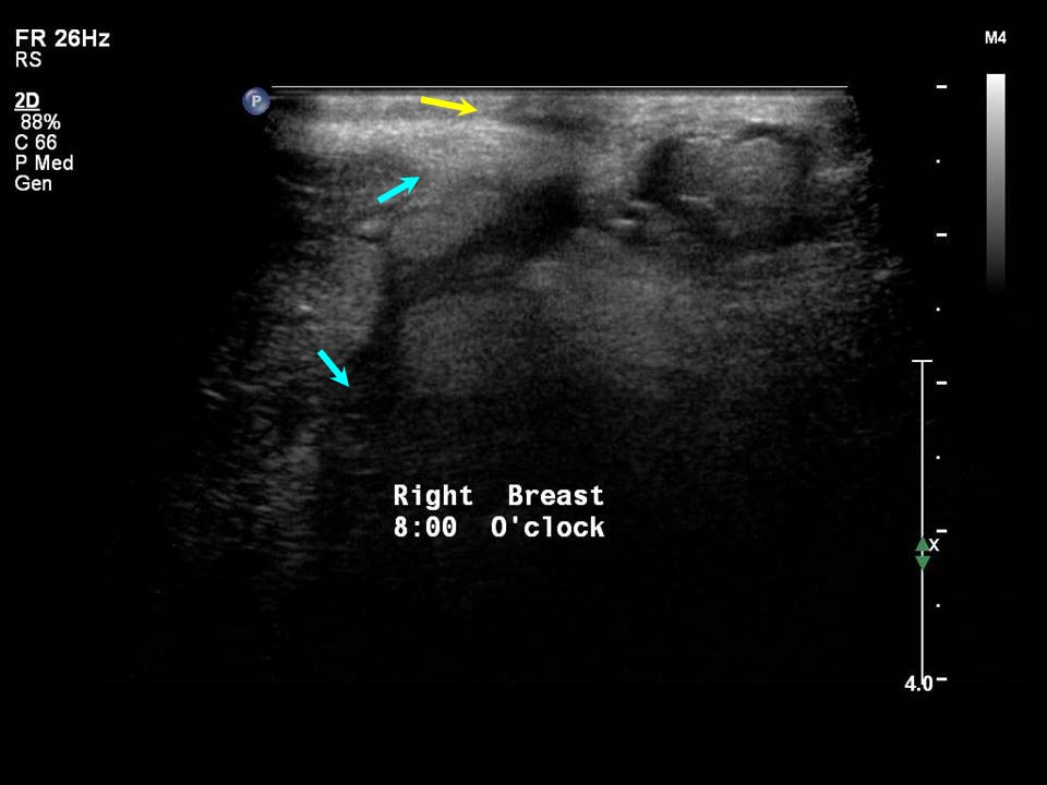
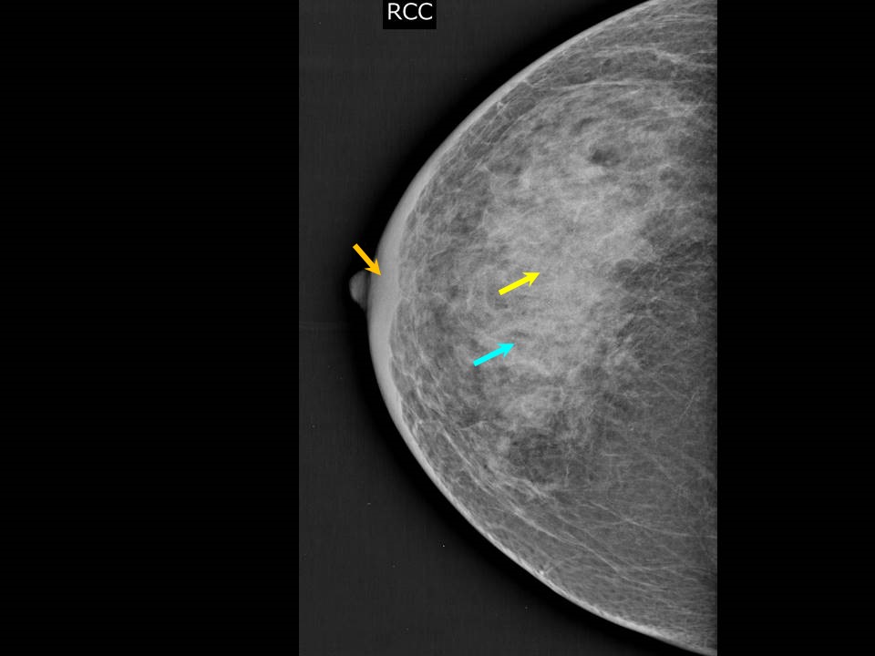
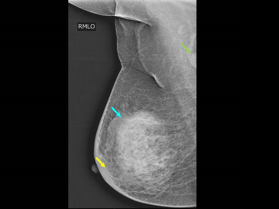
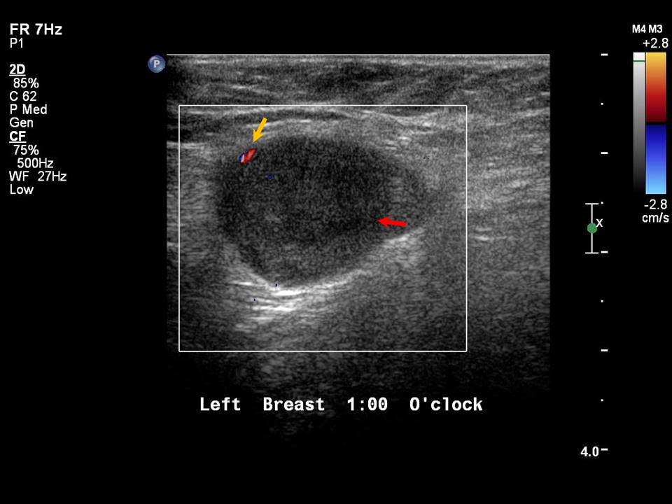
Normal axillary lymph nodes Axillary lymph nodes in both axillae facilitate the lymphatic drainage of the breast as discussed earlier (Breast anatomy). Level I axillary lymph nodes can be evaluated on breast ultrasound and mammography. Level II and III axillary lymph nodes are not commonly seen on ultrasound, though large reactive level II nodes may sometimes be visualized. Ultrasound is the first-line examination for imaging the axillae. High-frequency ultrasound examination evaluates level I nodes located in the lower part of the axilla, posterolateral to the lateral edge of the pectoralis major muscle. The evaluation of the node includes:
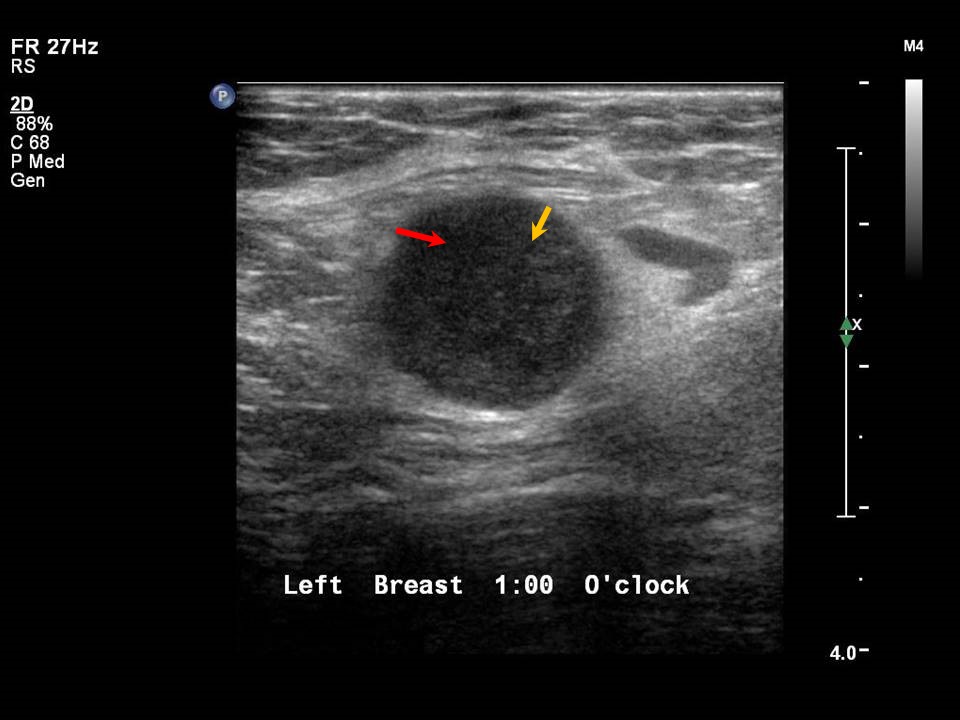
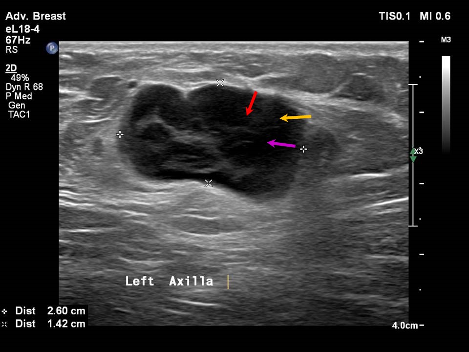
Case 1: Case 2: Case 3: Abnormal axillary lymph nodes Diseases of the lymph node begin from the periphery and an abnormal axillary lymph node will show altered morphological features such as:
Differential diagnoses of axillary lymphadenopathy include:
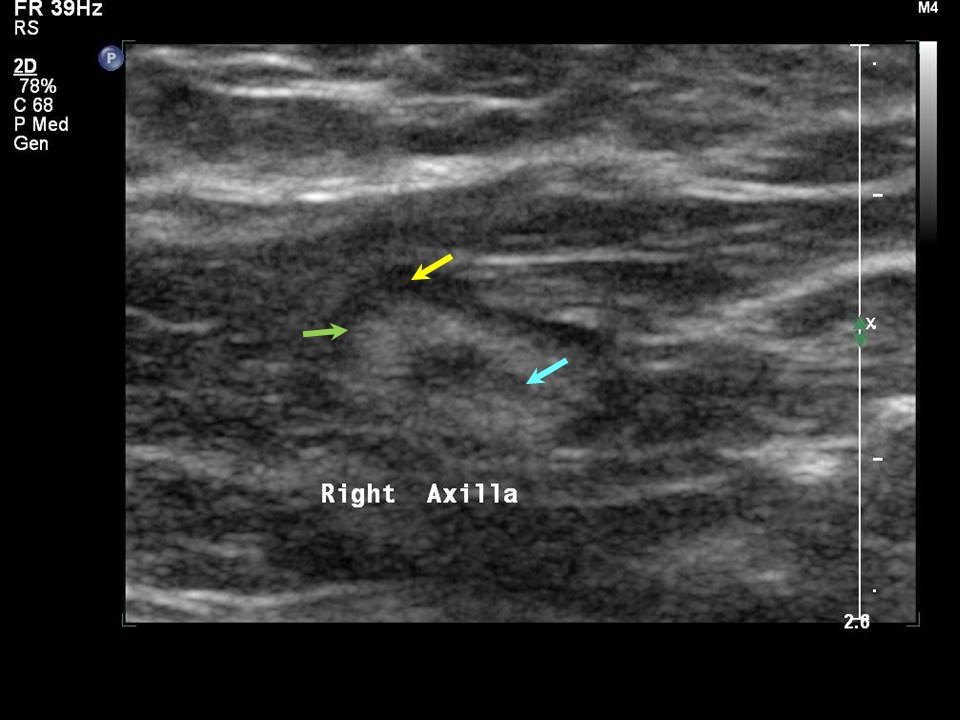
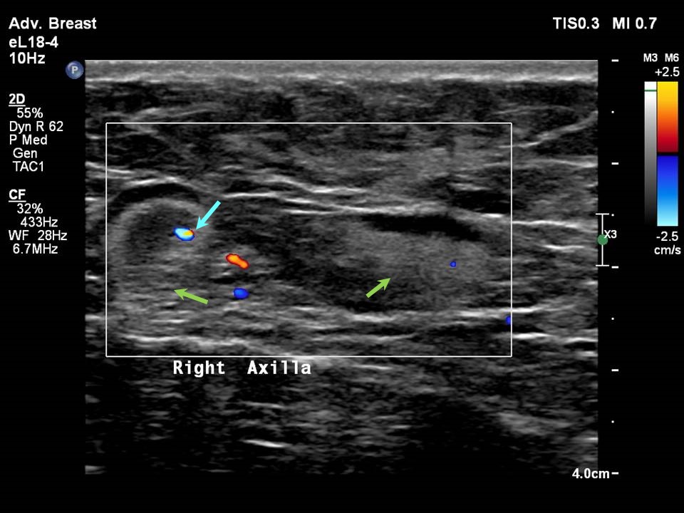
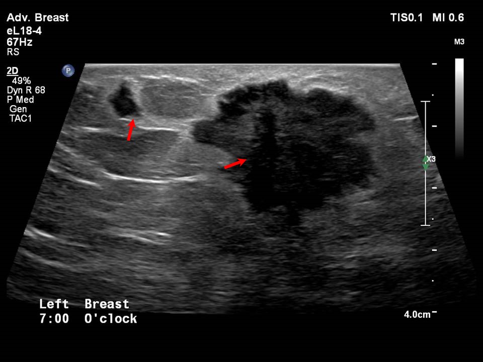
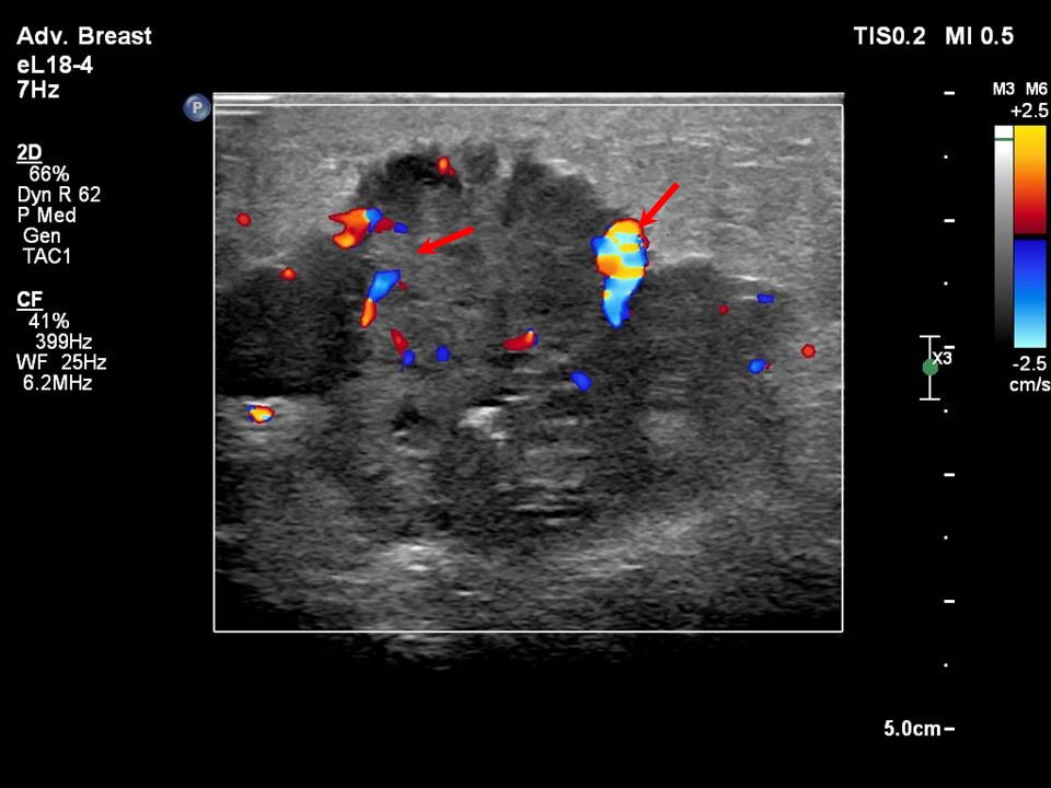
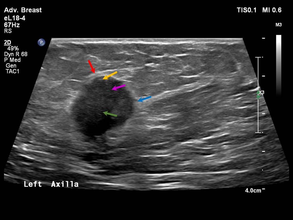
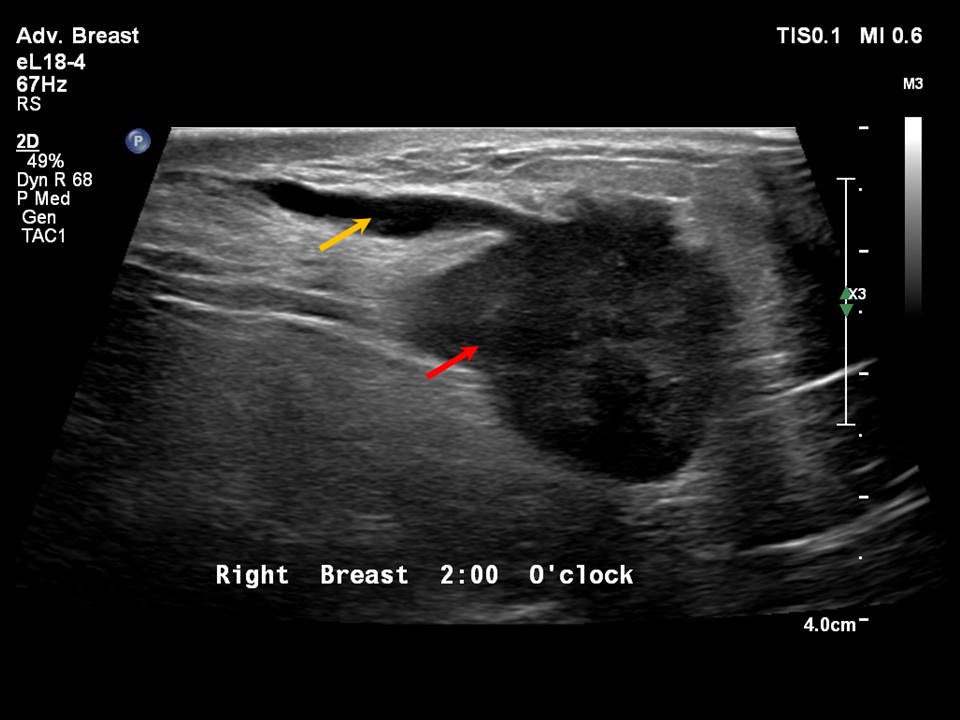
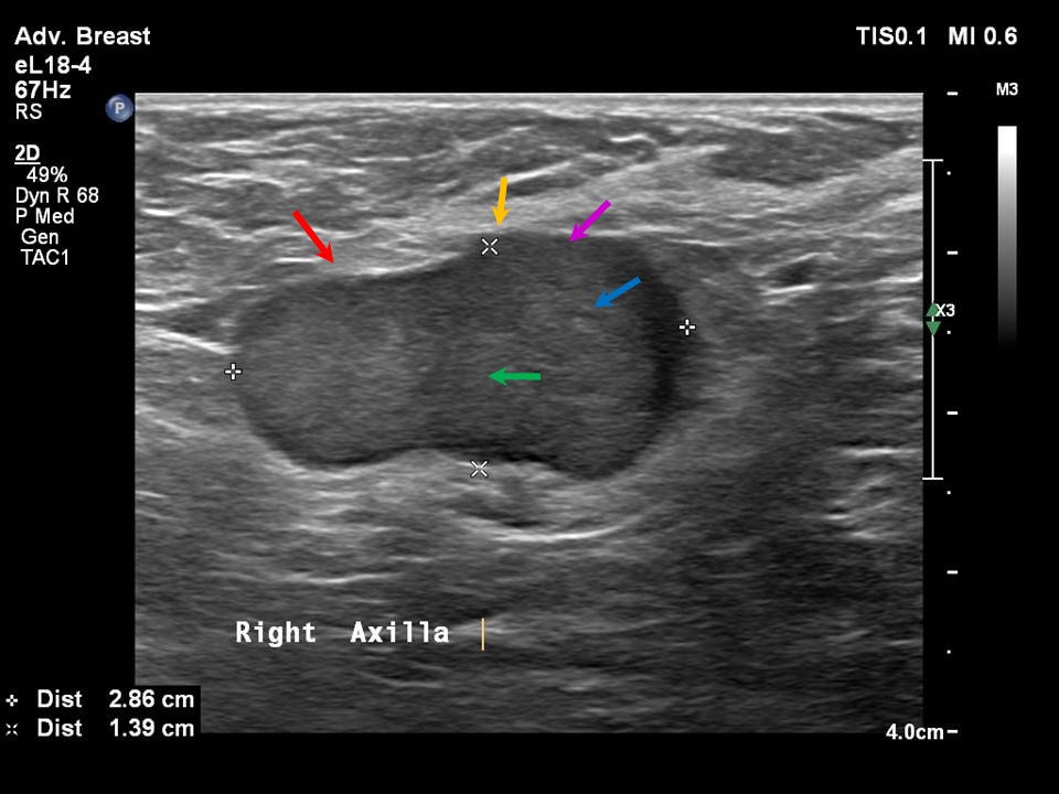
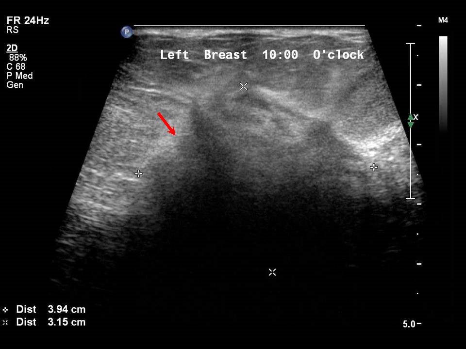
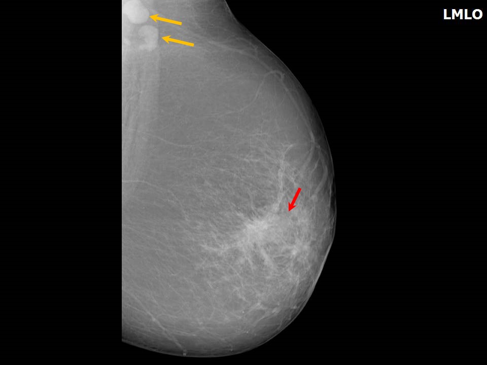
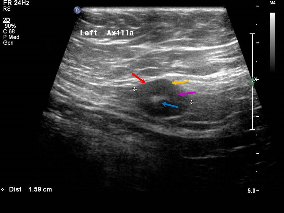
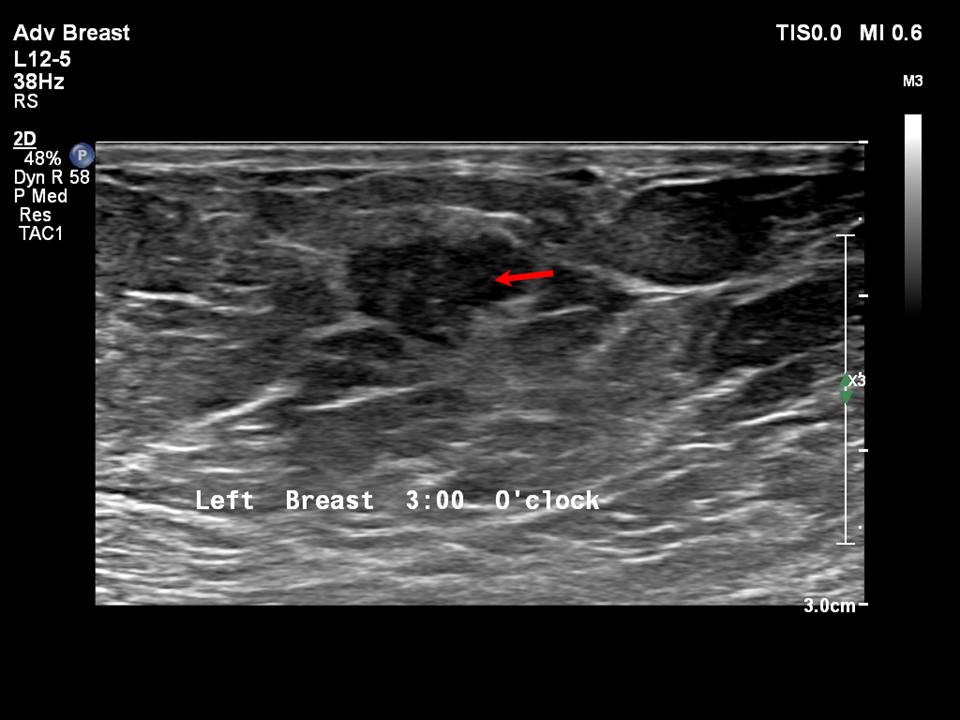
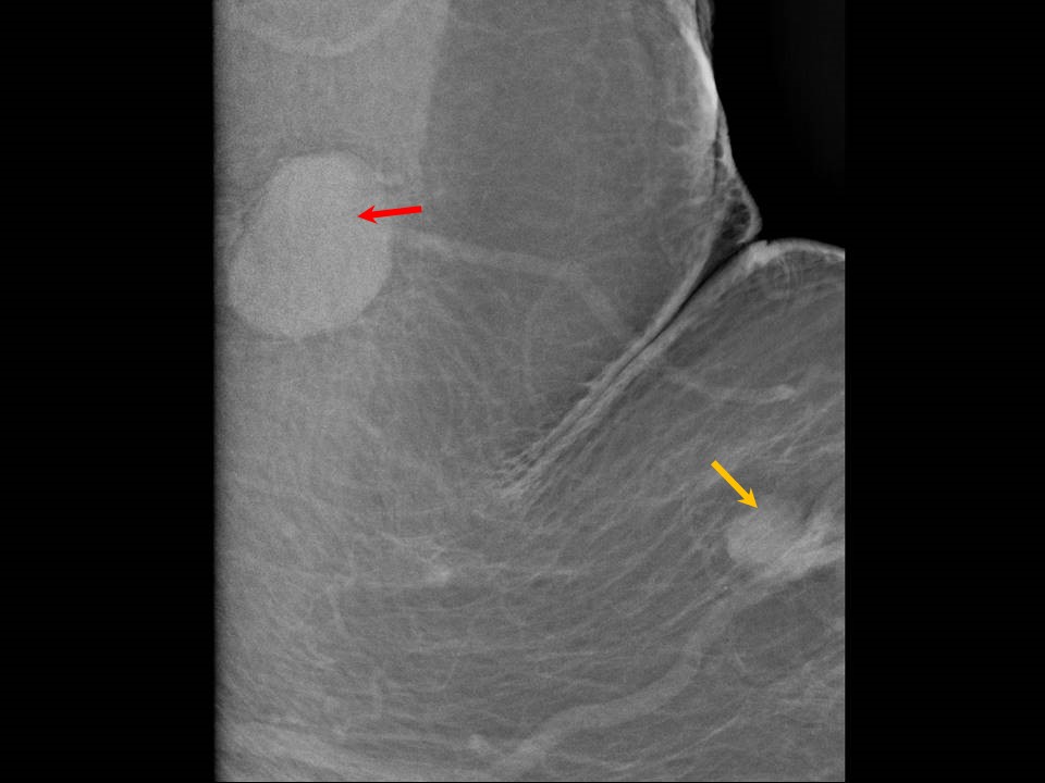
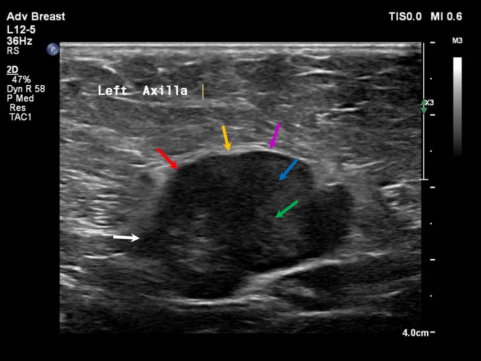
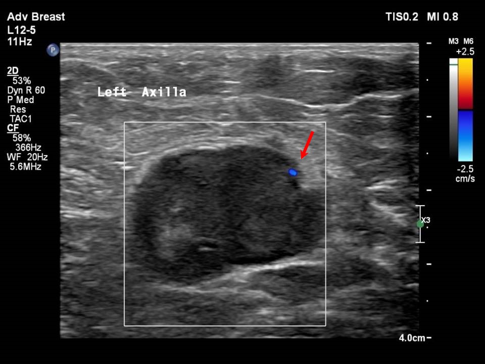
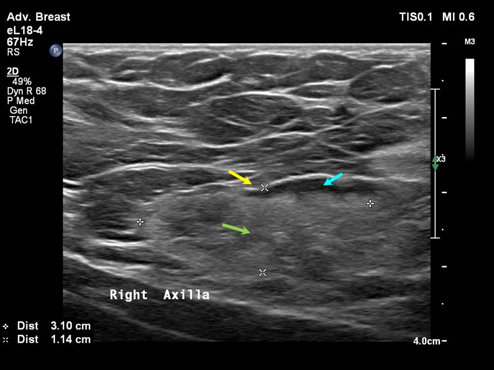
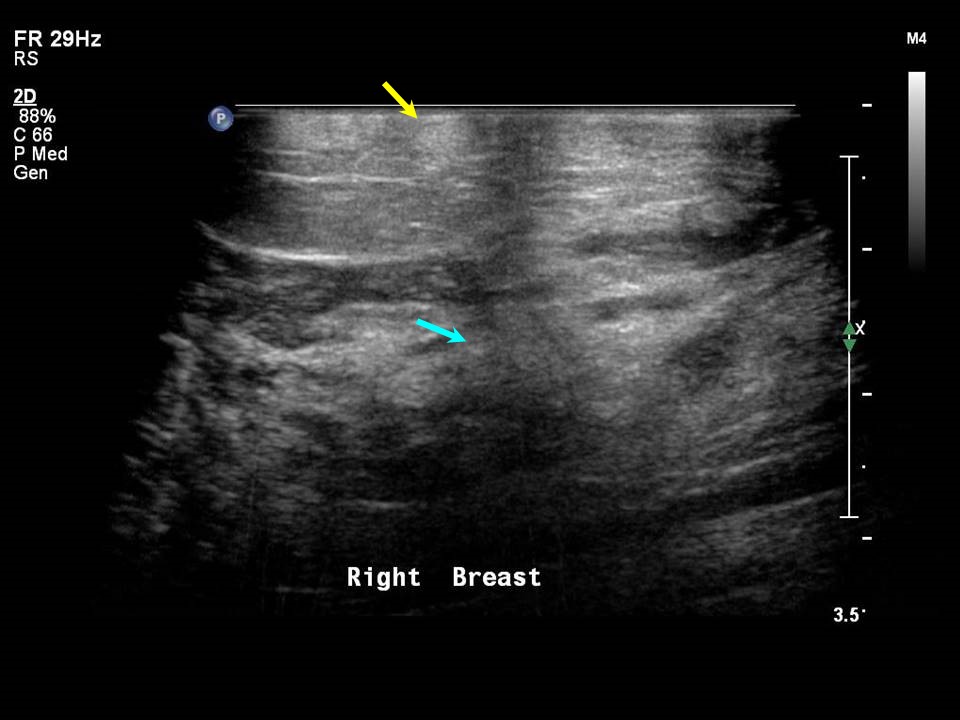
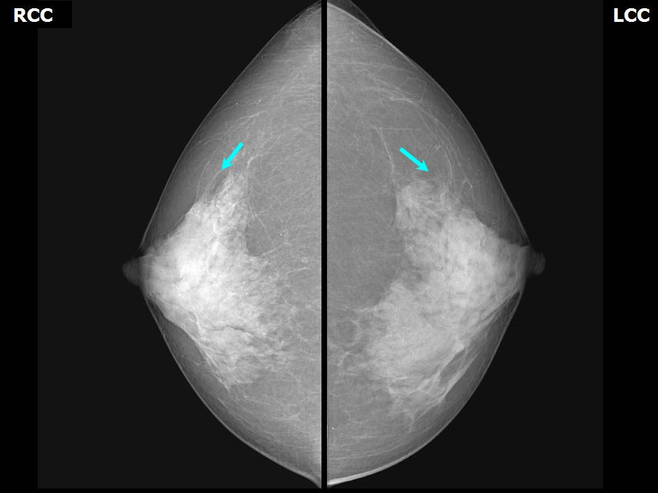
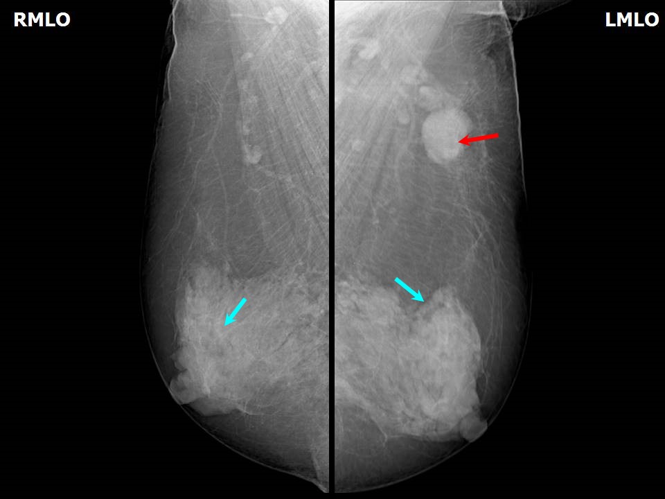
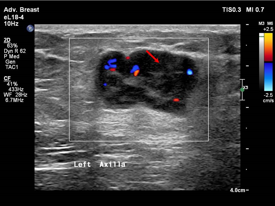
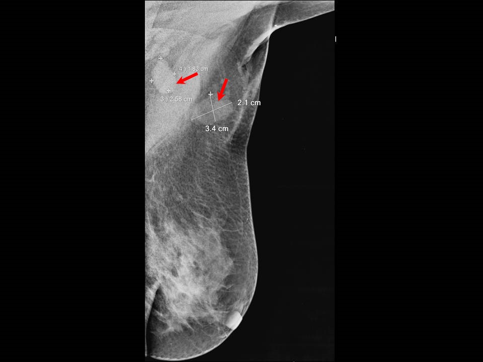
Breast cancer with axillary metastatic node The morphological characteristics of metastatic lymph nodes are enlarged size, round shape, absence of the fatty hilum, and concentric or focal cortical thickness greater than 3 mm. Case 1 Case 2 Case 3 Case 4 Infection Hodgkin lymphoma or non-Hodgkin lymphoma Inflammatory mastitis Metastatic nodes from distant malignancy Lymphoid hyperplasia |


Click on the pictures to magnify and display the legends
Click on this icon to display a case study
25 avenue Tony Garnier CS 90627 69366, LYON CEDEX 07 France - Tel: +33 (0)4 72 73 84 85
© IARC 2025 - Terms of use - Privacy Policy.
© IARC 2025 - Terms of use - Privacy Policy.