Go back to the list of case studies .png) Click on the pictures to magnify and display the legends
Click on the pictures to magnify and display the legends| Case number: | 080 |
| Age: | 64 |
| Clinical presentation: | Postmenopausal woman with average risk of developing breast cancer presented with redness of the skin over the right breast and lump in the same area. The duration of breast lump was 2 years whereas the duration of skin changes were for past 7 days. On clinical examination, a hard lump was palpable in the central quadrant of the right breast with overlying skin thickening. |
Mammography:
| Breast composition: | ACR category b (there are scattered areas of fibroglandular density) |
Mammography features: | |
|
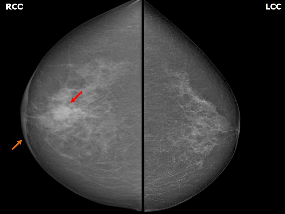
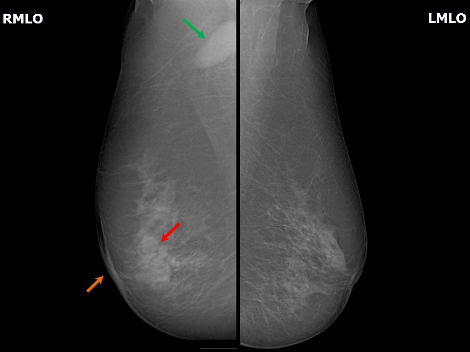
| ‣ Location of the lesion: | May 2012: Right breast, central portion of the breast, central zone, anterior and middle thirds |
| ‣ Mass: | |
| • Number: | 1 |
| • Size: | 4.0 × 2.5 cm |
| • Shape: | Irregular |
| • Margins: | Indistinct |
| • Density: | High |
| ‣ Calcifications: | |
| • Typically benign: | None |
| • Suspicious: | None |
| • Distribution: | None |
| ‣ Architectural distortion: | Present |
| ‣ Asymmetry: | Focal |
| ‣ Intramammary node: | None |
| ‣ Skin lesion: | None |
| ‣ Solitary dilated duct: | None |
| ‣ Associated features: | Skin thickening, trabecular thickening, axillary adenopathy, enlarged node (3.8 × 1.2 cm) with loss of fatty hilum, and architectural distortion. |
| Breast composition: | ACR category b (there are scattered areas of fibroglandular density) |
Mammography features: | |
|
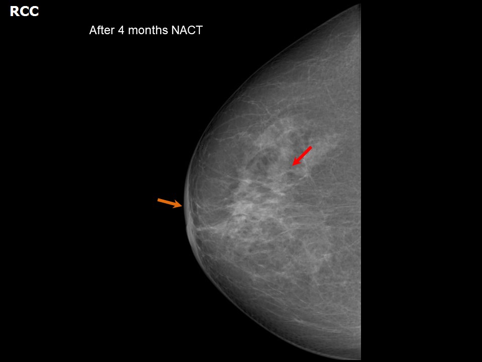
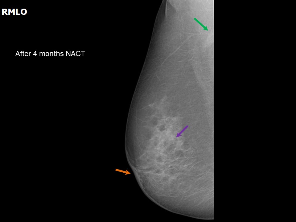
| ‣ Location of the lesion: | September 2012: Right breast, central portion of the breast, central zone, middle third |
| ‣ Mass: | |
| • Number: | 1 |
| • Size: | 2.3 × 1.0 cm |
| • Shape: | Irregular |
| • Margins: | Indistinct |
| • Density: | High |
| ‣ Calcifications: | |
| • Typically benign: | None |
| • Suspicious: | None |
| • Distribution: | None |
| ‣ Architectural distortion: | Present |
| ‣ Asymmetry: | Focal |
| ‣ Intramammary node: | None |
| ‣ Skin lesion: | None |
| ‣ Solitary dilated duct: | None |
| ‣ Associated features: | Skin thickening, trabecular thickening, axillary adenopathy, enlarged node (1.6 × 0.8 cm) with loss of fatty hilum, and architectural distortion |
| Breast composition: | ACR category b (there are scattered areas of fibroglandular density) |
Mammography features: | |
|
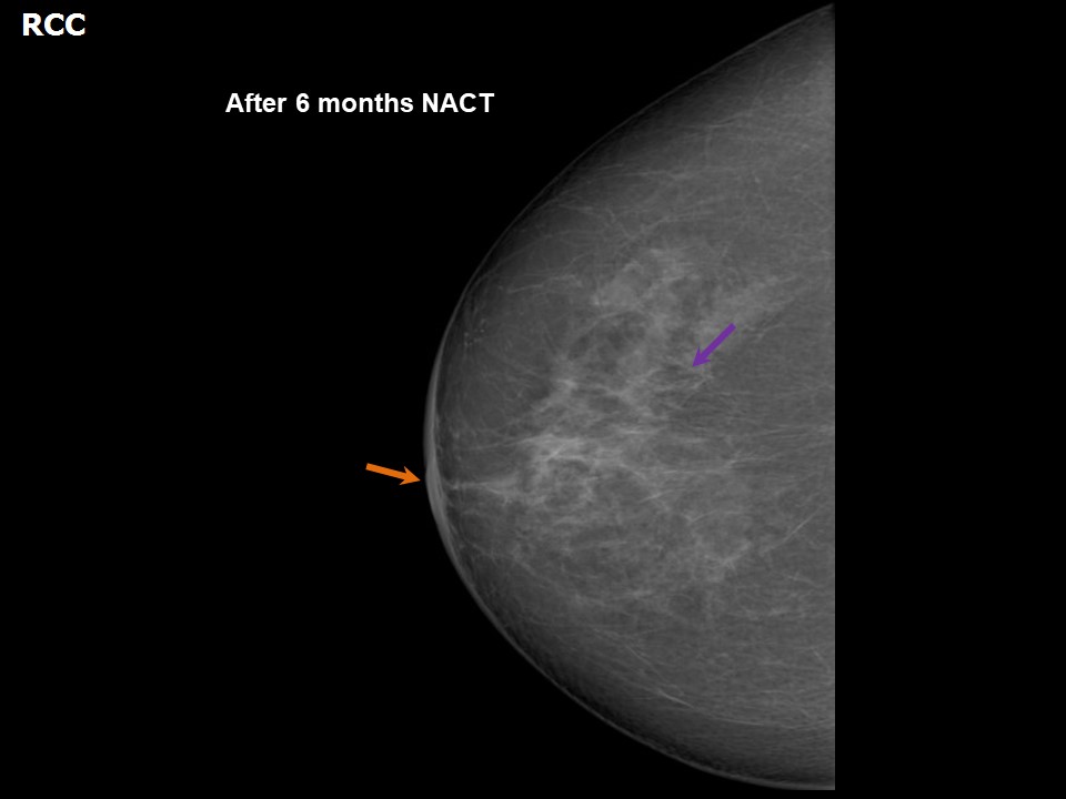
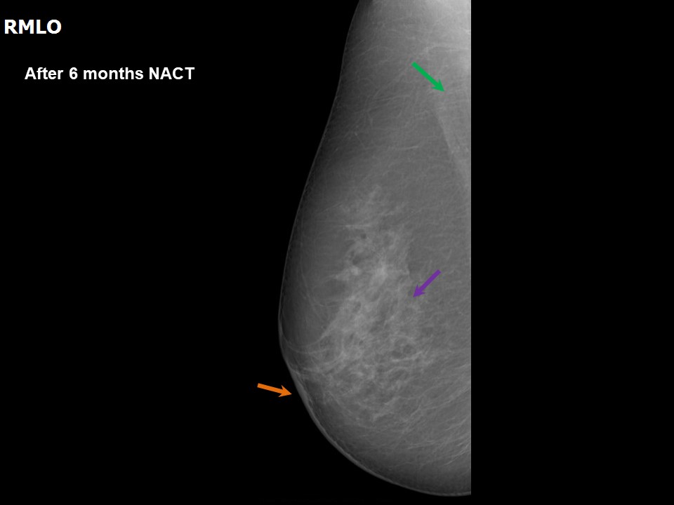
| ‣ Location of the lesion: | December 2012: Right breast, central portion of the breast, central zone, middle third |
| ‣ Mass: | |
| • Number: | 1 |
| • Size: | 1.2 × 0.7 cm |
| • Shape: | Irregular |
| • Margins: | Indistinct |
| • Density: | High |
| ‣ Calcifications: | |
| • Typically benign: | None |
| • Suspicious: | None |
| • Distribution: | None |
| ‣ Architectural distortion: | Present |
| ‣ Asymmetry: | Focal |
| ‣ Intramammary node: | None |
| ‣ Skin lesion: | None |
| ‣ Solitary dilated duct: | None |
| ‣ Associated features: | Skin thickening, trabecular thickening, axillary adenopathy, enlarged node (0.9 cm) with loss of fatty hilum, and architectural distortion |
Ultrasound:
| Ultrasound features: May 2012: Right breast, central portion of the breast, upper quadrant |
|
| ‣ Mass | |
| • Location: | May 2012: Right breast, central portion of the breast, upper quadrant |
| • Number: | 1 |
| • Size: | 3.8 × 2.5 cm |
| • Shape: | Irregular |
| • Orientation: | Not parallel |
| • Margins: | Angular and spiculated |
| • Echo pattern: | Hypoechoic |
| • Posterior features: | Posterior shadowing |
| ‣ Calcifications: | None |
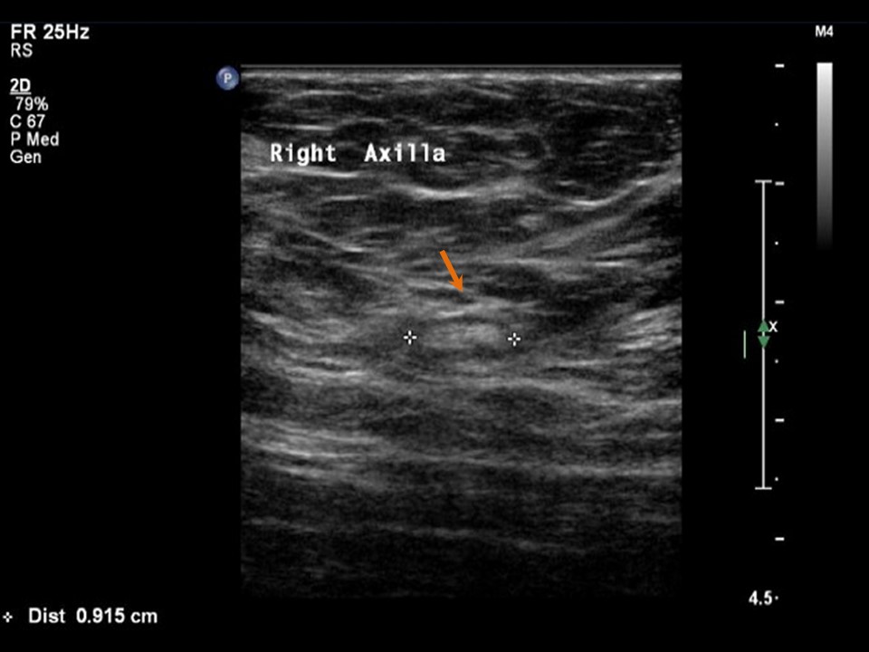
| ‣ Associated features: | Architectural distortion, skin thickening, internal vascularity, and axillary lymphadenopathy node (3.6 × 1.8 cm) |
| ‣ Special cases: | None |
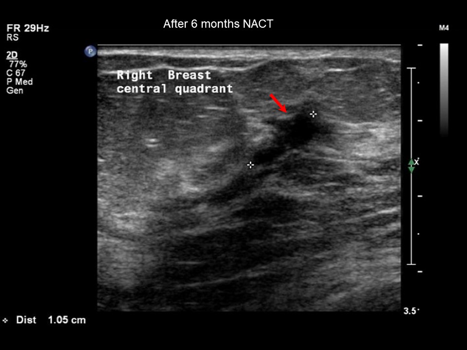
| Ultrasound features: September 2012, neo-adjuvant chemotherapy follow-up: Right breast, central portion of the breast |
|
| ‣ Mass | |
| • Location: | September 2012, neo-adjuvant chemotherapy follow-up: Right breast, central portion of the breast |
| • Number: | 1 |
| • Size: | 2.3 × 0.8 cm |
| • Shape: | Irregular |
| • Orientation: | Not parallel |
| • Margins: | Angular and spiculated |
| • Echo pattern: | Hypoechoic |
| • Posterior features: | Posterior shadowing |
| ‣ Calcifications: | None |
| ‣ Associated features: | Architectural distortion, skin thickening, internal vascularity, and axillary lymphadenopathy |
| ‣ Special cases: | None |
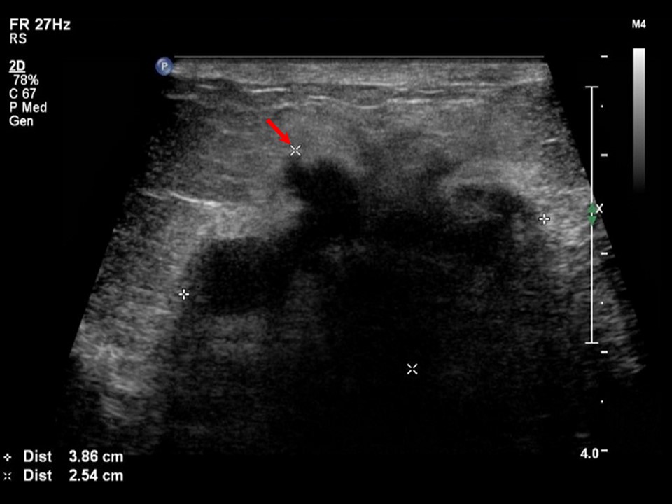
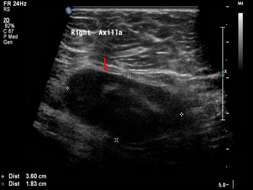
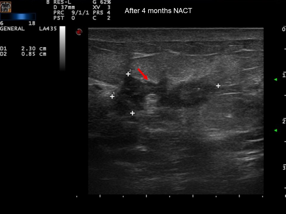
| Ultrasound features: December 2012, neo-adjuvant chemotherapy follow-up: Right breast, central portion of the breast |
|
| ‣ Mass | |
| • Location: | December 2012, neo-adjuvant chemotherapy follow-up: Right breast, central portion of the breast |
| • Number: | 1 |
| • Size: | 1.2 × 0.7 cm |
| • Shape: | Irregular |
| • Orientation: | Not parallel |
| • Margins: | Spiculated |
| • Echo pattern: | Hypoechoic |
| • Posterior features: | No posterior features |
| ‣ Calcifications: | None |
| ‣ Associated features: | Architectural distortion, skin thickening, internal vascularity, and axillary node (1.0 cm) |
| ‣ Special cases: | None |
BI-RADS:
BI-RADS Category (May 2012): 5 (highly suggestive of malignancy)
BI-RADS Category (September 2012): 6 (known biopsy-proven malignancy)
BI-RADS Category (December 2012): 6 (known biopsy-proven malignancy)
Further assessment:
Further assessment advised: Referral for cytology
Cytology:
| Cytology features: | |
|
| ‣ Type of sample: | FNAC (solid lesion) |
| ‣ Site of biopsy: | |
| • Laterality: | Right |
| • Quadrant: | Central zone retroareolar |
| • Localization technique: | palpation |
| • Nature of aspirate: | Whitish |
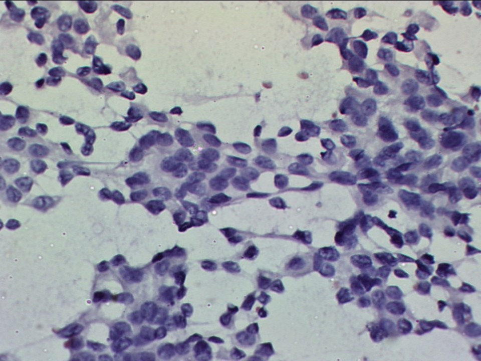
| ‣ Cytological description: | Smears show plenty of dyscohesive clusters of malignant ductal epithelial cells. Single isolated malignant cells are also seen. The cells show marked pleomorphism and prominent nucleoli |
| ‣ Reporting category: | Malignant |
| ‣ Diagnosis: | Carcinoma high grade |
| ‣ Comments: | None |
Histopathology:
Breast-conserving surgery
| Histopathology features: | |
|
| ‣ Specimen type: | Breast-conserving surgery |
| ‣ Laterality: | Right |
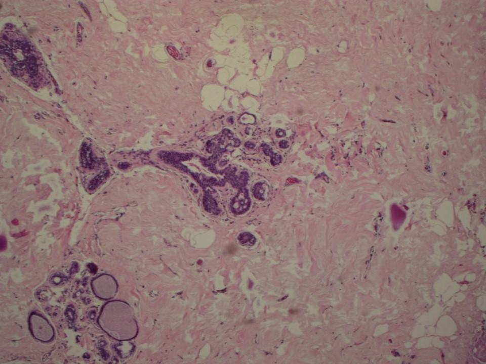
| ‣ Macroscopy: | Serial sectioning revealed on the cut surface shows a fibrous rubbery area (1.5 × 1.0 × 1.0 cm), which is firm to hard and greyish white. This area is 2.5 cm from the base, 2.8 cm from the skin, 2.0 cm from the superior margin, 5.0 cm from the inferior margin, 4.0 cm from the lateral margin, and 3.0 cm from the medial margins. Another firm area felt at the superior margin is 0.5 × 0.3 × 0.2 cm. Diffuse areas of fibrosis are seen in the surrounding breast |
| ‣ Histological type: | No residual tumour identified. Extensive dense sclerosed areas seen in the breast parenchyma. A few ducts are cystically dilated and lined by flattened benign epithelium |
| ‣ Histological grade: | |
| ‣ Mitosis: | |
| ‣ Maximum invasive tumour size: | |
| ‣ Lymph node status: | 0/4 |
| ‣ Peritumoural lymphovascular invasion: | Not identified |
| ‣ DCIS/EIC: | Not identified |
| ‣ Margins: | |
| ‣ Pathological stage: | |
| ‣ Biomarkers: | |
| ‣ Comments: | Pathological complete response |
Case summary:
| Postmenopausal woman presented with right breast lump and redness of overlying skin. Diagnosed as right breast carcinoma with areolar skin thickening and right axillary metastatic node, BI-RADS 5 on imaging and as right breast carcinoma, high grade on cytology. Follow-up after neo-adjuvant chemotherapy showed significant tumour regression, BI-RADS 6 on imaging. Operative specimen after neo-adjuvant chemotherapy revealed pathological complete response. No residual tumour identified. Extensive dense sclerosed areas seen in the breast parenchyma on histopathology. |
Learning points:
- BI-RADS 6 category is used in patients on follow-up to assess response to neo-adjuvant chemotherapy. Mammography and ultrasound follow-up are used to assess the size reduction and nodal response in locally advanced breast carcinoma and to plan further definitive surgical management.
- Pathological examination of the excised tumour bed is the reference standard and is essential for identifying patients with a pathological complete response to treatment or partial response to treatment.
|