


Home / Training / Manuals / Atlas of breast cancer early detection / Learning
.png)
Click on the pictures to magnify and display the legends

Click on this icon to display a case study
Atlas of breast cancer early detection
Filter by language: English / РусскийBreast imaging Mammography interpretation Understanding the normal mammogram Normal mammogram |
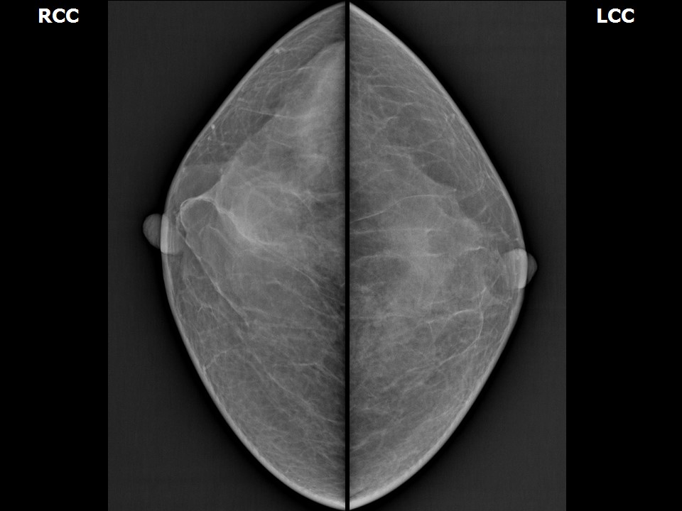
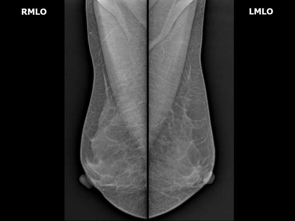
A clear understanding of the normal appearance of breast tissue patterns is important to detect an abnormal finding on mammography.
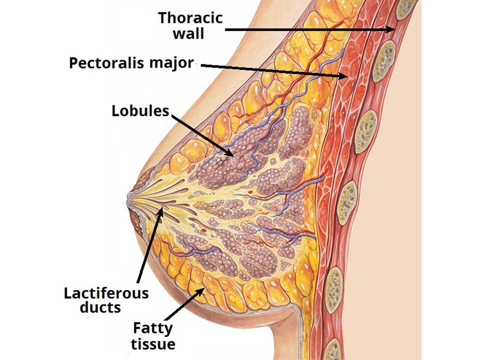
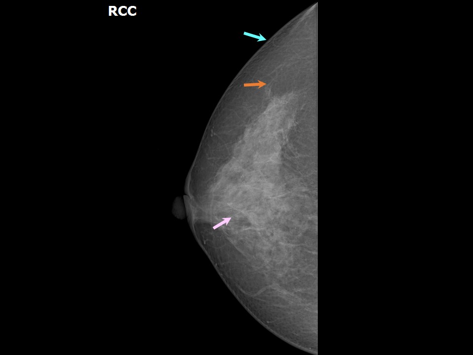
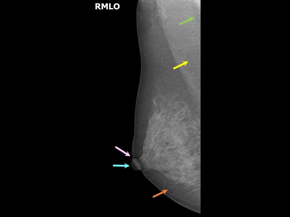
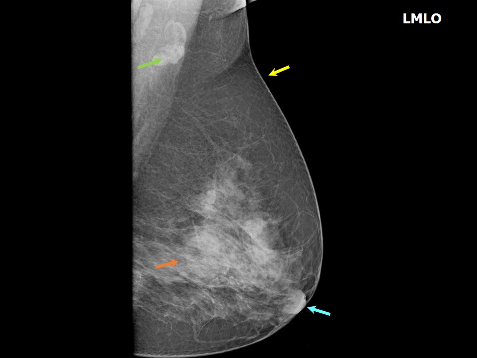
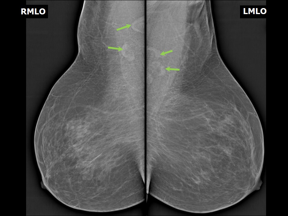
Normal mammogram Normal breast anatomy on mammography In a mammographic image the following features should be examined carefully before reporting:
Mammograms of the normal breast showing the anatomy |
Click on the pictures to magnify and display the legends
Click on this icon to display a case study
25 avenue Tony Garnier CS 90627 69366, LYON CEDEX 07 France - Tel: +33 (0)4 72 73 84 85
© IARC 2025 - Terms of use - Privacy Policy.
© IARC 2025 - Terms of use - Privacy Policy.