


Home / Training / Manuals / Atlas of breast cancer early detection / Learning
.png)
Click on the pictures to magnify and display the legends

Click on this icon to display a case study
Atlas of breast cancer early detection
Filter by language: English / РусскийBreast imaging Mammography technique Mammography procedure Additional mammographic views Axillary tail view |
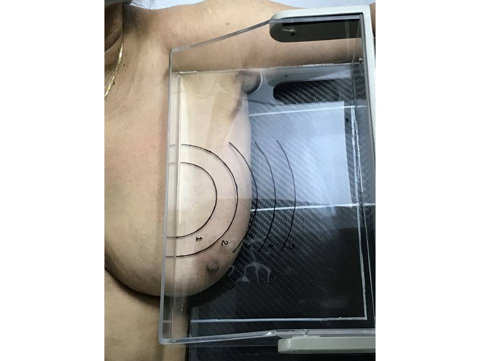
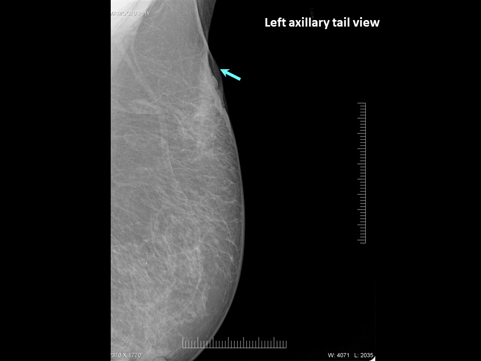
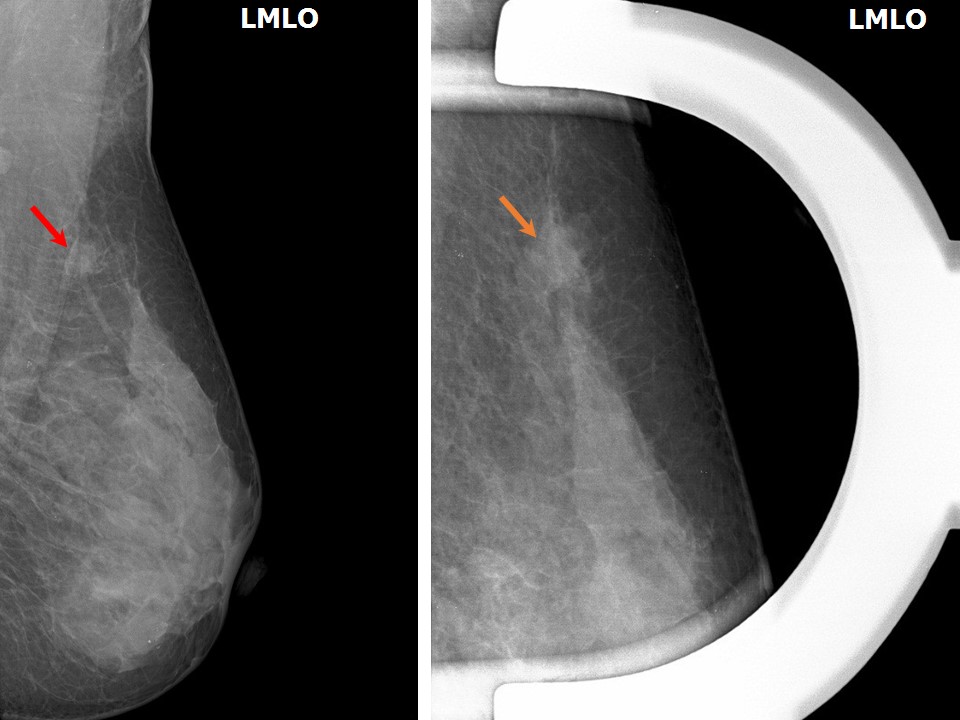
Indication The axillary tail view (previously known as the Cleopatra view) is used to isolate a lesion in the tail of the breast. The view is taken primarily to confirm the location of the lesion in the tail of the breast. Steps to obtain the axillary tail view
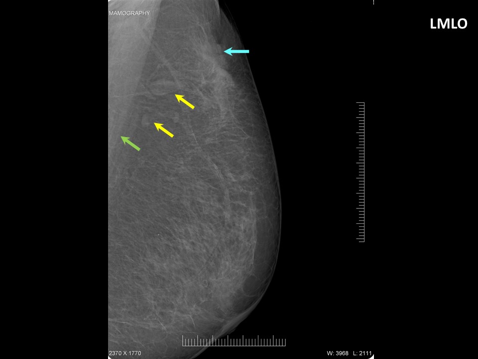
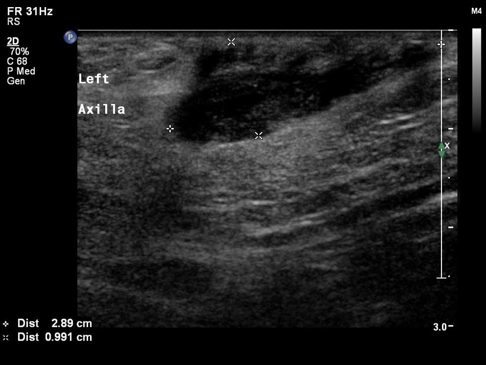
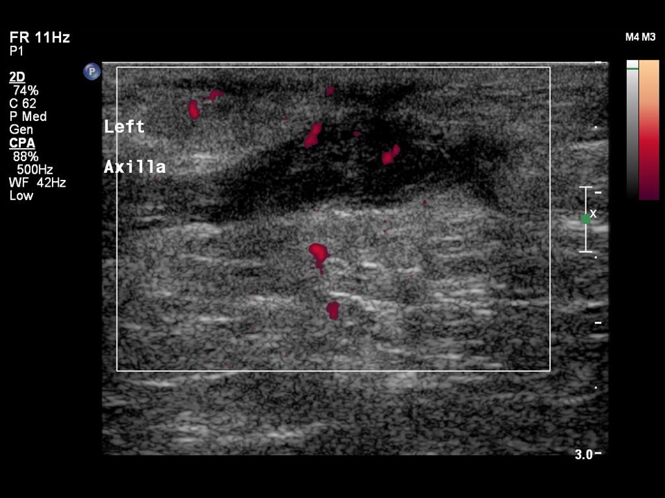
Patient position Mammogram axillary tail view Left breast axillary tail inflammation Ultrasound for left breast axillary tail inflammation Left breast axillary tail carcinoma |

Click on the pictures to magnify and display the legends
Click on this icon to display a case study
25 avenue Tony Garnier CS 90627 69366, LYON CEDEX 07 France - Tel: +33 (0)4 72 73 84 85
© IARC 2025 - Terms of use - Privacy Policy.
© IARC 2025 - Terms of use - Privacy Policy.