Home / Training / Manuals / Atlas of breast cancer early detection / Learning

Atlas of breast cancer early detection

Breast imaging – Mammography interpretation – Getting organized


Prerequisites for image interpretation
  • Mammographic images, standard views, and additional views, if necessary, are acquired as described earlier. The mammography films are then viewed on a view box or a medical monitor in a standard and consistent sequence.
  • Left and right CC images should be viewed base-to-base as mirror images with the lateral (axillary) portion of both at the top.
  • Left and right MLO images should be viewed back-to-back as mirror images.
  • Check the breast positioning and symmetry and that enough breast tissue has been included in the acquired image. The adequacy criteria of CC and MLO views are described in an earlier section.
  • A view box with two rows is useful to compare older mammography films with the new images. New findings become more apparent if previous images are toggled rapidly in the same field of view.
  • Digital mammography viewed on medical monitors enables the use of in-built applications such as zoom, cropped magnification views, contrast adjustment, and measurement tools.
  • Mount the older mammogram and the recent mammogram on the view box.
  • If possible, sit back and view images at a distance of 15–45 cm to evaluate tissue details.
  • Closer evaluation to view subtle details is then done with the help of the zoom feature.
  • Avoid overlap of screen and body artefacts. They may interfere with image interpretation.
  • Detailed viewing is carried out with the normal variants in mind.
  • Localize the lesion by o’clock positioning.
  • Special attention is given to the retroareolar region, deeper tissues, the axilla, and the inframammary fold to avoid overlooking a lesion.

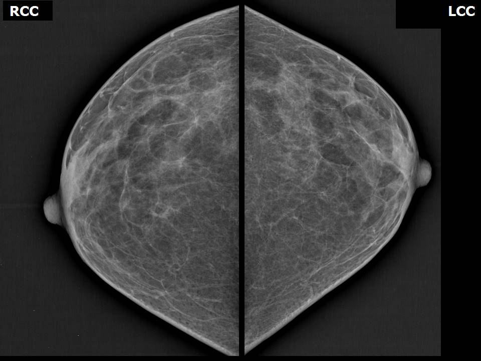
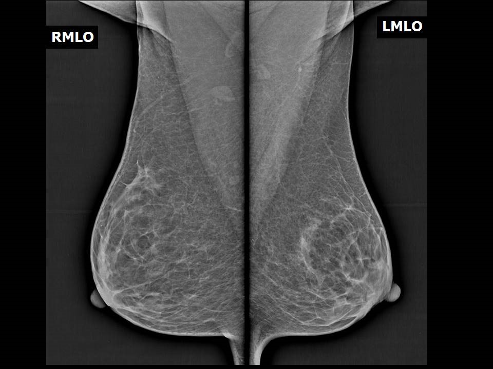
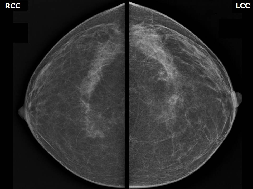
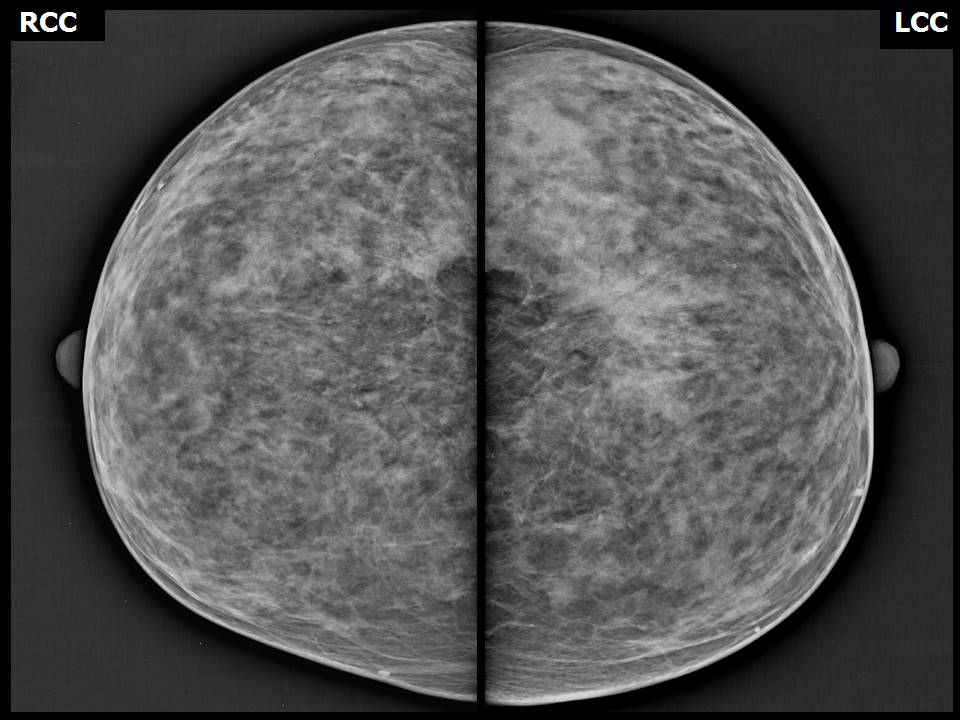
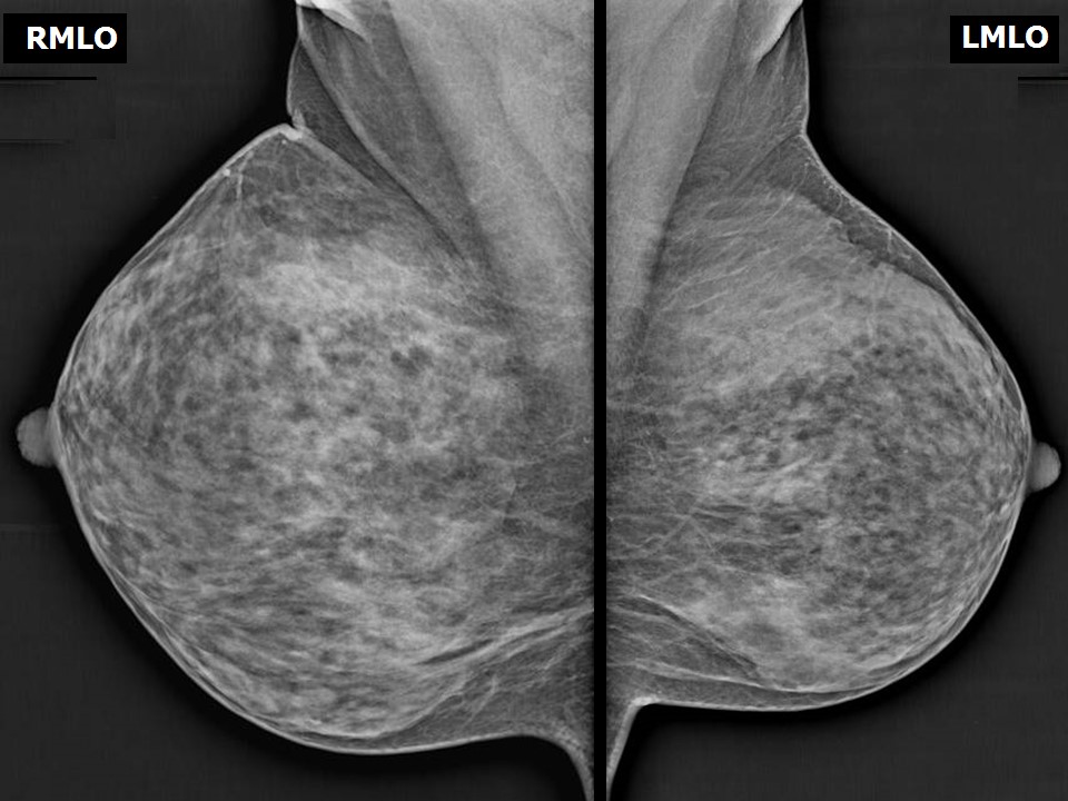
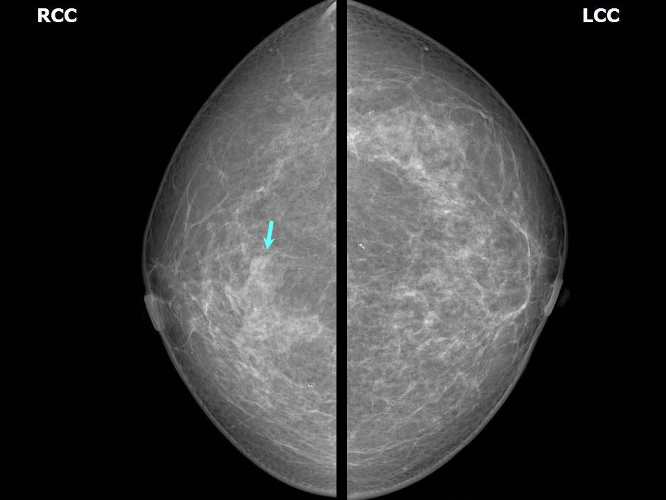
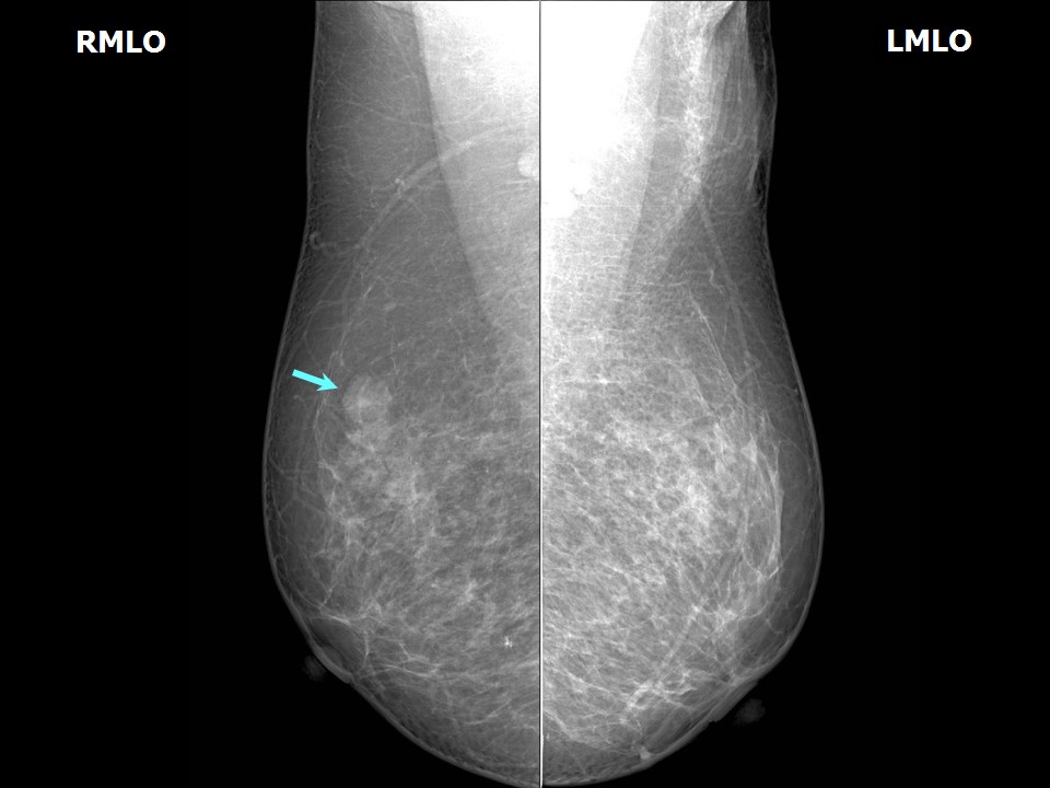
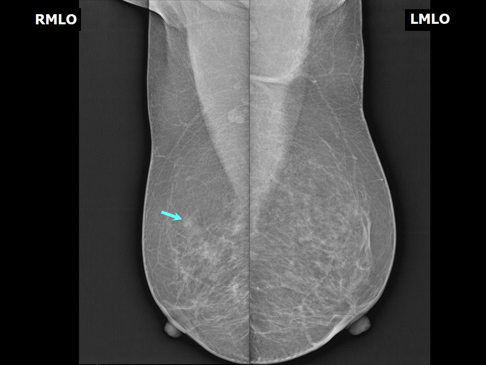
Normal mammogram


Bilateral breast mirror images


Bilateral symmetrical breast tissue


Asymmetrical breast tissue
Developing asymmetry
For women with a high risk of developing malignancy who attend for regular screening mammography, it is important to retain earlier mammographic images for accurate interpretation. The images are viewed sequentially, the older CC with the recent CC views and the same for the MLO views. Clinically occult small early cancer may be detected as a developing asymmetry.


Focal asymmetry
Screening mammograms in women with high risk of developing breast cancer may reveal focal asymmetrical breast tissue. Regular follow-ups at short intervals are recommended to monitor the stability or regression of these mammography findings. The likelihood of breast cancer can thus be ruled out.


Click on the pictures to magnify and display the legends


Click on this icon to display a case study

IARC, 150 Cours Albert Thomas, 69372 Lyon CEDEX 08, France - Tel: +33 (0)4 72 73 84 85 - Fax: +33 (0)4 72 73 85 75
© IARC 2024 - All Rights Reserved.