


Home / Training / Manuals / Atlas of breast cancer early detection / Learning
.png)
Click on the pictures to magnify and display the legends

Click on this icon to display a case study
Atlas of breast cancer early detection
Filter by language: English / РусскийBreast imaging Mammography technique Mammography procedure Additional mammographic views Cleavage view |
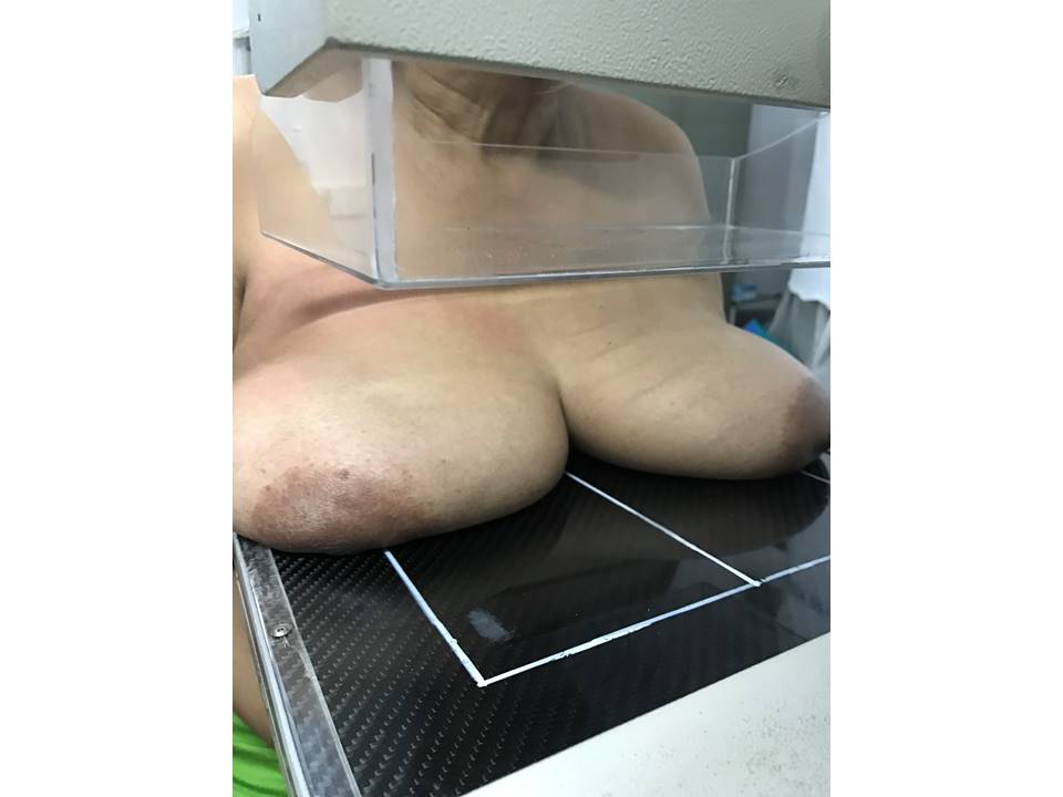
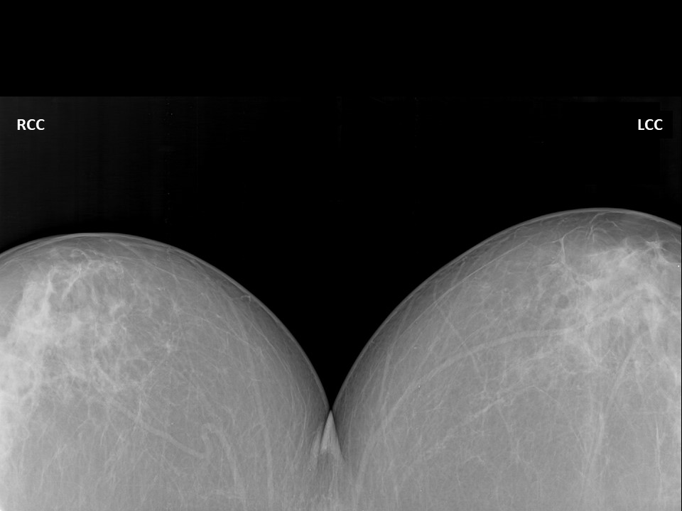
The cleavage view, also called the valley view, shows the medial breast tissues adjacent to the sternum. The medial breast tissue is almost always completely included in the CC view. However, if a medial lesion is suspected, the cleavage view is a better option than the XCCM view for maximum medial breast tissue visualization and is easily positioned for lesions at the sternal end of the breast, which are sometimes missed in CC views. Steps to obtain the cleavage view
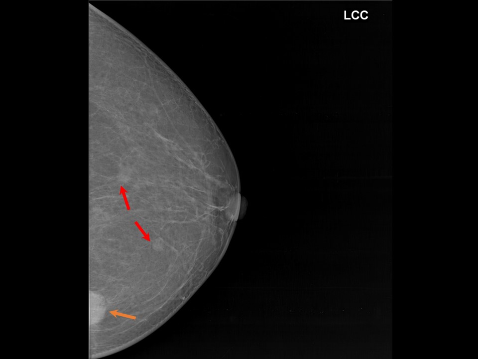
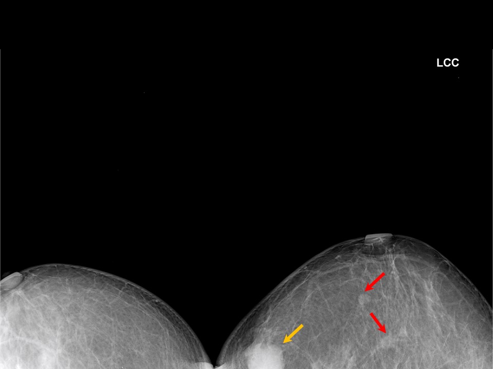
Mammogram cleavage view Cleavage view of the left breast showing a medial quadrant lesion Cleavage view of the left breast showing a medial quadrant lesion |

Click on the pictures to magnify and display the legends
Click on this icon to display a case study
25 avenue Tony Garnier CS 90627 69366, LYON CEDEX 07 France - Tel: +33 (0)4 72 73 84 85
© IARC 2024 - Terms of use - Privacy Policy.
© IARC 2024 - Terms of use - Privacy Policy.