|
Breast imaging – Mammography technique – Description of a mammography unit |   |
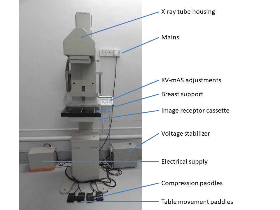
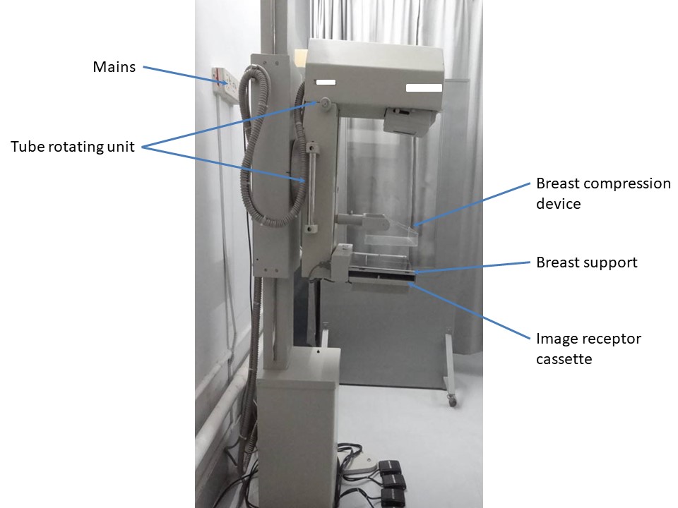
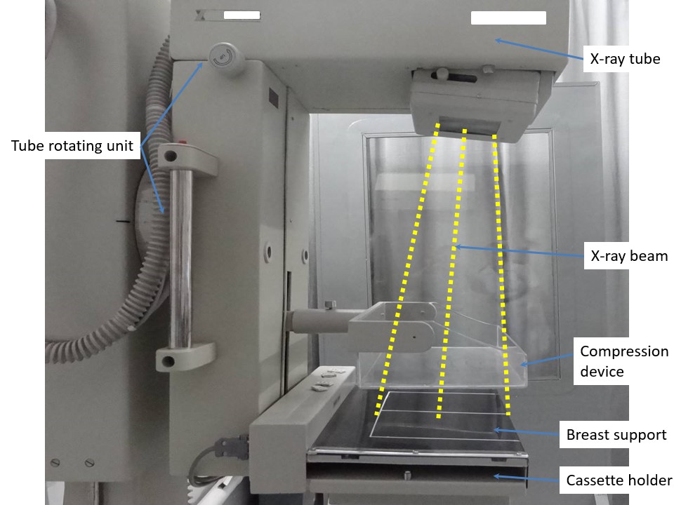
A mammography radiographic unit includes the following:
- X-ray tube with dual microfocus (0.1 mm and 0.3 mm) and a rotating molybdenum anode;
- X-ray generator that transforms low voltage (200–240 V) into high voltage to feed the X-ray tube;
- voltage stabilizer;
- molybdenum filter that absorbs high-energy photons of energy exceeding 20 keV to produce softer radiation from low-energy photons (17.9 keV and 19.5 keV) for better subject contrast;
- beryllium window to control the photons reaching the breast tissue;
- gantry;
- breast support for placement of the breast and a compression device;
- high-resolution phosphorescent screen with phosphor crystals that emit light when exposed to X-rays in screen-film mammography;
- digital cassette and digital cassette reader (replacing screen-film) in computed radiography digital mammography;
- automatic exposure control (AEC) sensor at the base of the image receptor cassette to measure the radiation passing through the breast and cassette. The AEC sensor is calibrated so that when a sufficient number of X-ray photons have reached the sensor, exposure is automatically terminated. There is no need for manual adjustment and judgement of exposure factors, such as kilovoltage and milliampere-seconds, for variations in breast size and the compressed thickness of the breast. The radiation dose to the breast is automatically controlled to ensure an image of optimal contrast.
|   |
|
.png)
Click on the pictures to magnify and display the legends

Click on this icon to display a case study
25 avenue Tony Garnier CS 90627 69366, LYON CEDEX 07 France - Tel: +33 (0)4 72 73 84 85 © IARC 2025 - Terms of use - Privacy Policy.
| .png)


