Home / Training / Manuals / Atlas of breast cancer early detection / Learning

Atlas of breast cancer early detection

Breast imaging – Breast ultrasound – Technique – Procedural steps


  1. A linear-array high-frequency 18–5 MHz transducer is ideal for breast ultrasound. Apply ultrasound gel directly on the skin of the breast. High-frequency sound waves are transmitted from the probe through the gel into the body. The transducer collects the reflected sound signals and a computer is used to convert them into a diagnostic image.
  2. Perform two-dimensional grey-scale ultrasound with uniform pressure of the hand and probe. Place the transducer on the breast and move cranial to caudal in a longitudinal and/or transverse movement over the entire breast for both breasts until the desired images are captured.
  3. A radial movement of the transducer in the pattern of dial of a clock can also be performed, although it is used less often.
  4. Evaluate the echogenicity of the breast tissues on ultrasound. Capture the images if you detect a finding.
  5. Detail the lobular anatomy in the mammary zone and the homogeneous arrangement of the ducts, their branches, and the lobules.
  6. Take into account all necessary assessment of the finding, which includes measurements, localization, colour Doppler changes, and associated findings while scanning and capturing all images.
  7. Scan both breasts completely in all quadrants and the retromammary region along with the axillae for additional findings as well as associated features.
  8. Special attention is given to the nipple–areolar complex. A circular and angulated probe movement will include all tissues of this region.
  9. Use colour Doppler to evaluate the vascularity of the lesion.
  10. A mass lesion on ultrasound can be recognized as a cancer by its morphological characteristics and can be differentiated from a benign lesion.
  11. Calcifications may be seen with or without a mass. It is difficult to further categorize the calcifications as possibly benign or malignant.
  12. Architectural distortion seen on ultrasound may suggest an underlying malignancy.
  13. Skin changes may be seen as isolated findings or as features associated with a mass.
  14. Nipple changes present as nipple retraction, nipple ulceration, or nipple–areolar skin changes.
  15. Axillary nodal abnormalities, such as reactive nodes or nodes with features suspicious for metastasis, are seen on ultrasound.
  16. Ultrasound-guided biopsy or FNAC may be performed if the ultrasound examination reveals a suspicious abnormality.
  17. Once the imaging is complete, wipe off the ultrasound gel from the skin. The gel does not usually stain or discolour the clothing.
  18. Allow time for the patient to get dressed. Provide clear instructions on report availability and follow-up.


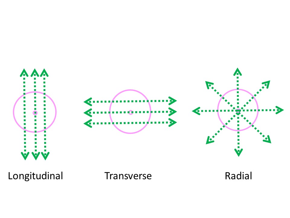
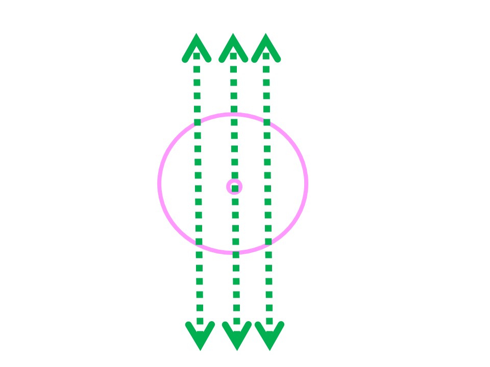
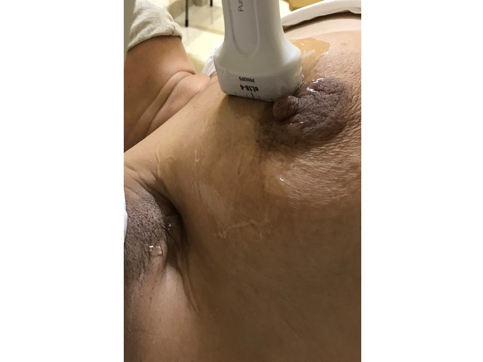
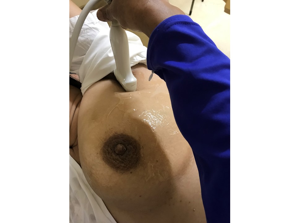
Longitudinal scan of the right breast in supine position


Transverse scan of the right breast in supine position


Radial scan of the left breast in right decubitus position


Scan of the left axilla in oblique or lateral decubitus position


Click on the pictures to magnify and display the legends


Click on this icon to display a case study

IARC, 150 Cours Albert Thomas, 69372 Lyon CEDEX 08, France - Tel: +33 (0)4 72 73 84 85 - Fax: +33 (0)4 72 73 85 75
© IARC 2024 - All Rights Reserved.