


Home / Training / Manuals / Atlas of breast cancer early detection / Learning
.png)
Click on the pictures to magnify and display the legends

Click on this icon to display a case study
Atlas of breast cancer early detection
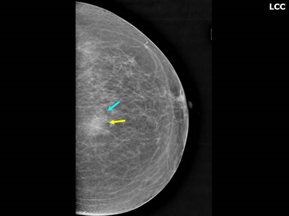
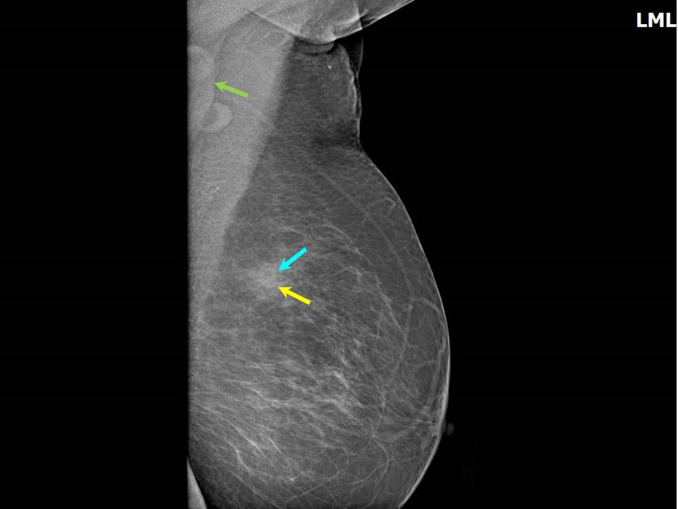
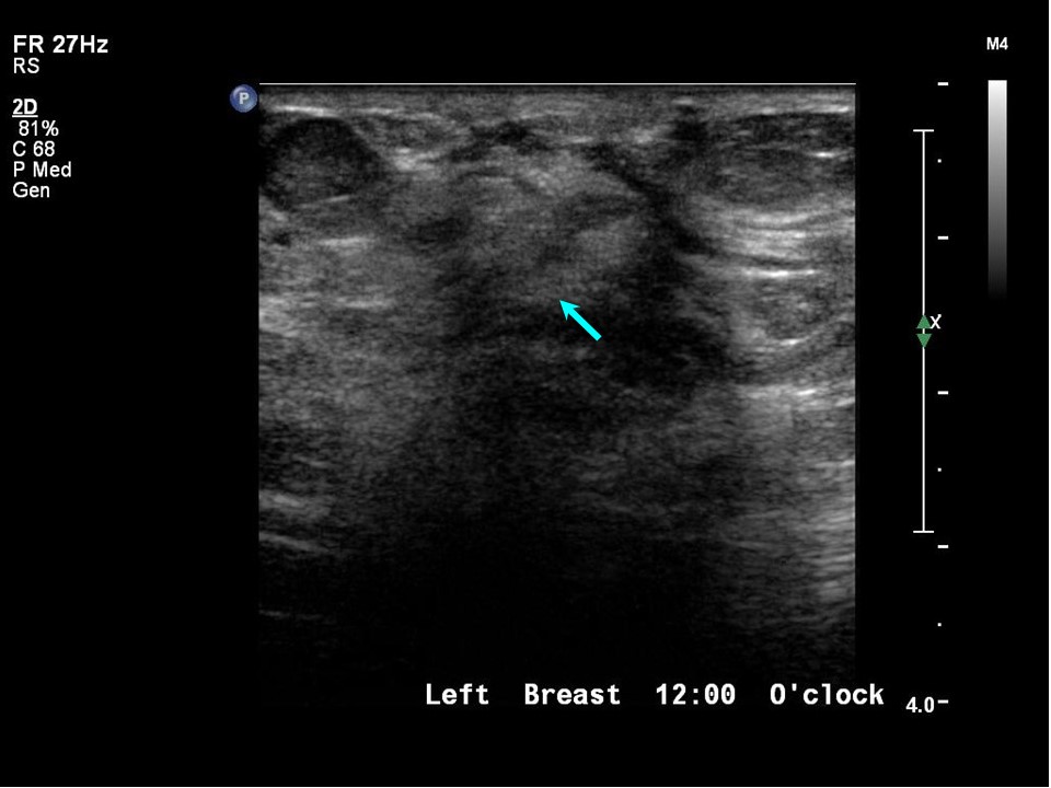
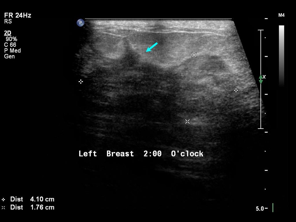
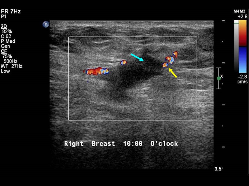
Filter by language: English / РусскийBreast imaging Breast ultrasound Ultrasound lexicon Breast masses Margin |
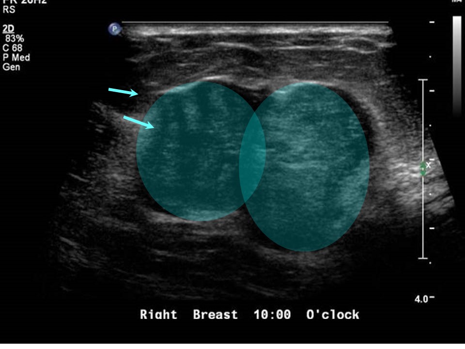
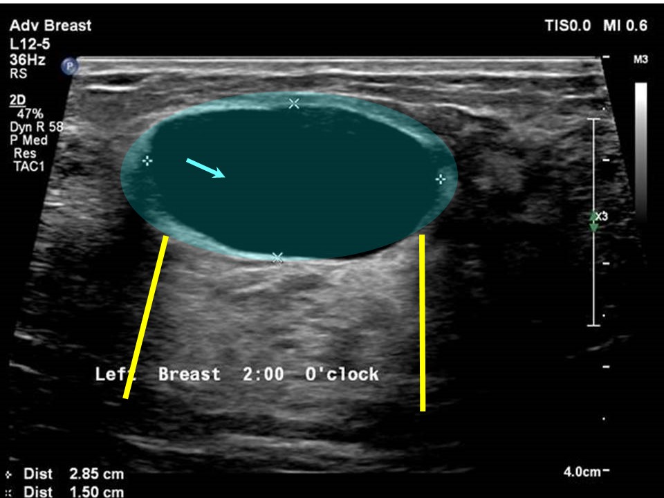
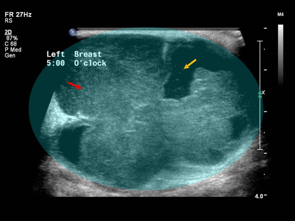
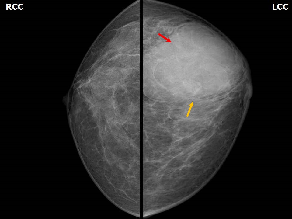
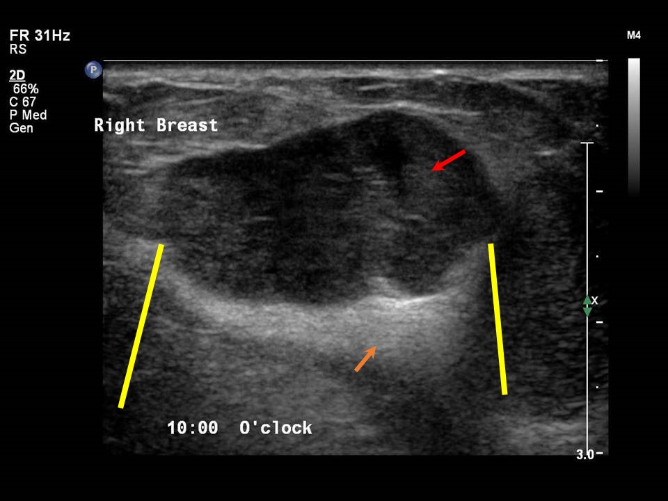
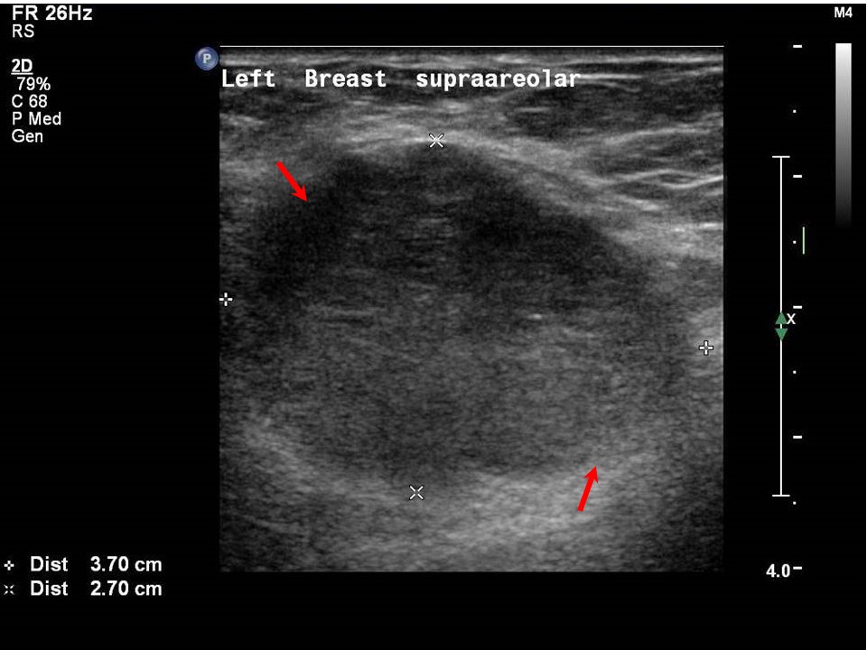
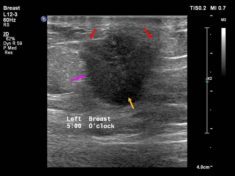
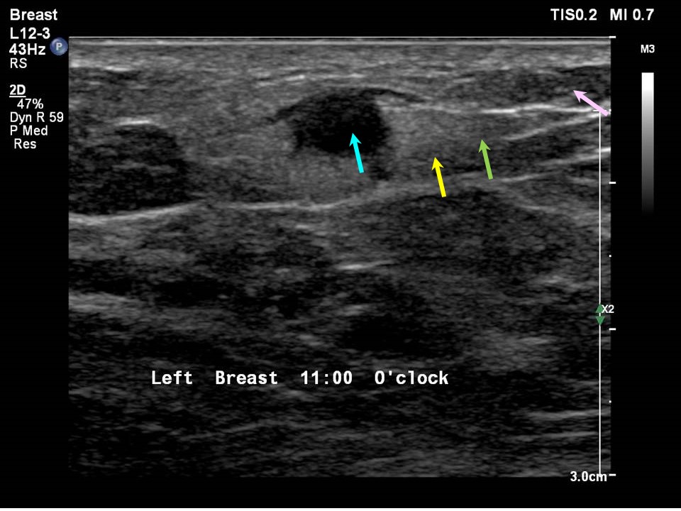
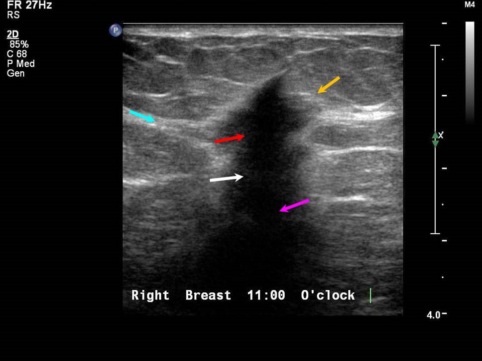
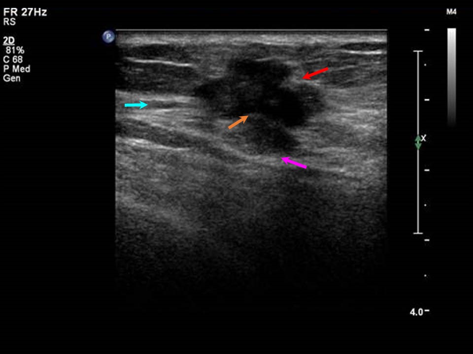
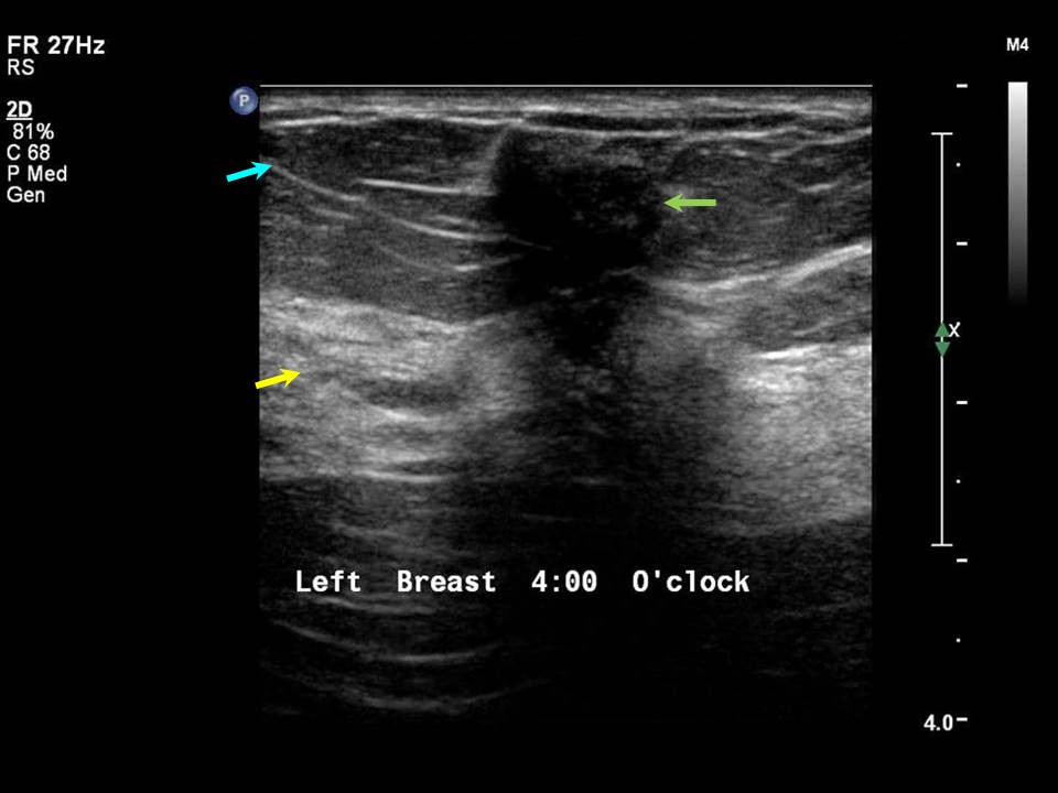
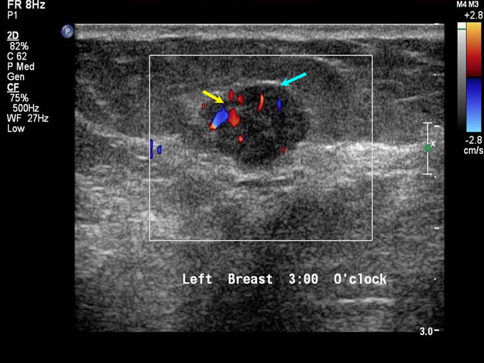
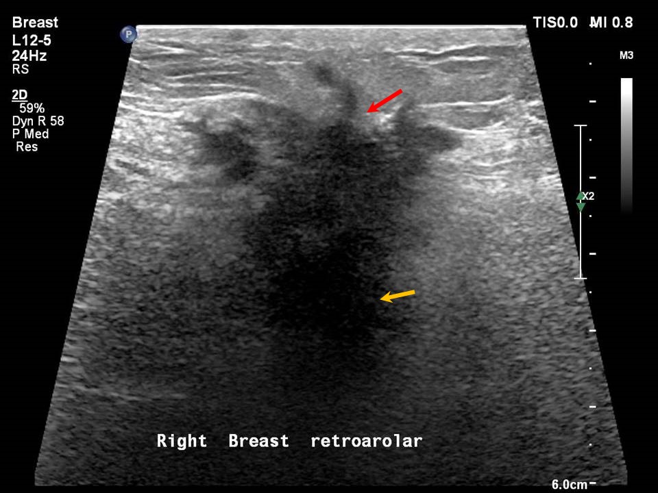
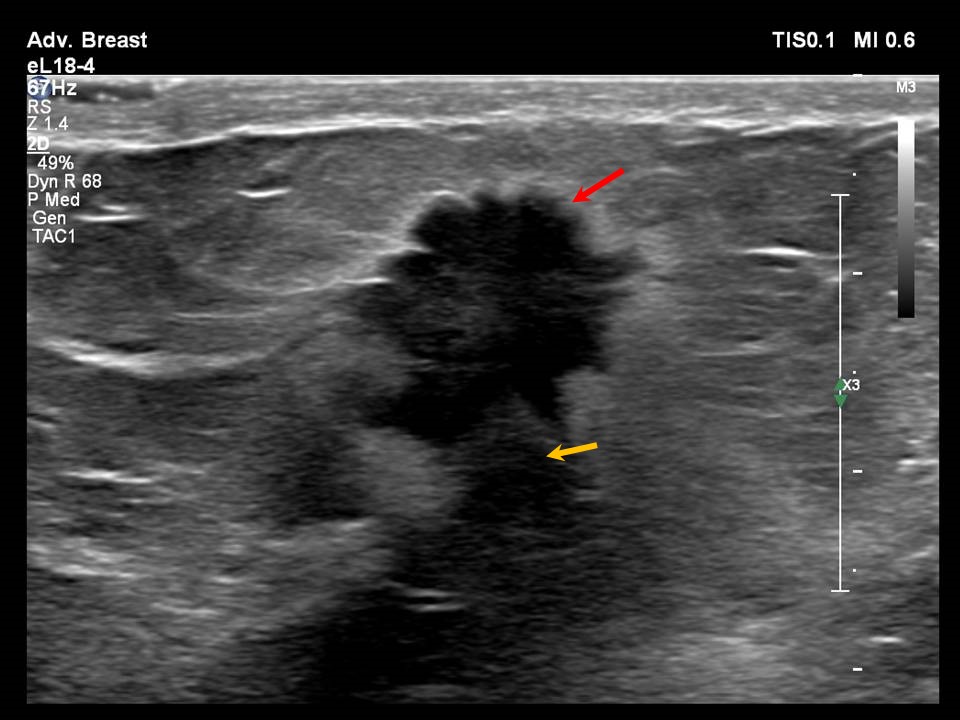
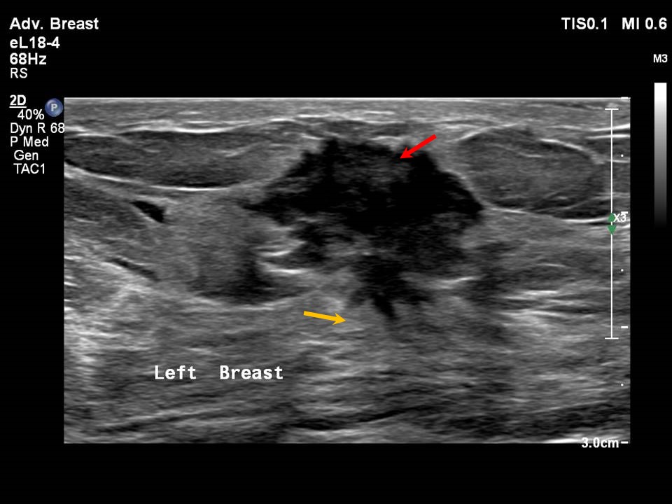
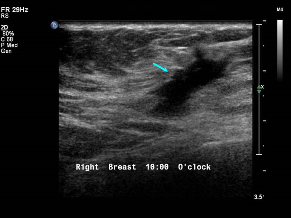
The margins of a mass may enable differentiation between benign and malignant. The margin is a feature that separates the mass from the adjacent breast parenchyma. Margins can be described as circumscribed or non-circumscribed.
Circumscribed margins A circumscribed margin indicates a probable benign finding .Non-circumscribed margins Non-circumscribed margins are seen in lesions caused by infection, inflammation, or neoplasia. They are also seen in lesions resulting from breast trauma and surgical scarring. Indistinct Indistinct margins are seen in malignant masses and also in lesions caused by infection, inflammation, breast trauma, and fat necrosis. A circumscribed mass that forms after interventions such as biopsy or FNAC will often have indistinct margins .Angular margins Angular margins are commonly seen in malignant masses. Atypical fibroadenomas or phyllodes may also have angular margins .Microlobulated margins Microlobulated margins are a feature of malignancy. Spiculated margins Spiculated margins are seen in malignant masses. They describe the extent and possible infiltration of the mass into the surrounding breast parenchyma . |


Click on the pictures to magnify and display the legends
Click on this icon to display a case study
25 avenue Tony Garnier CS 90627 69366, LYON CEDEX 07 France - Tel: +33 (0)4 72 73 84 85
© IARC 2025 - Terms of use - Privacy Policy.
© IARC 2025 - Terms of use - Privacy Policy.