


Home / Training / Manuals / Atlas of breast cancer early detection / Cases
Atlas of breast cancer early detection
Filter by language: English / Русский
Go back to the list of case studies
.png) Click on the pictures to magnify and display the legends
Click on the pictures to magnify and display the legends
| Case number: | 046 |
| Age: | 61 |
| Clinical presentation: | Postmenopausal woman with average risk of developing breast cancer presented with a lump in the upper quadrant of the left breast. Examination revealed a lump above the nippleareolar complex with an inverted nipple on the left side. |
Mammography:
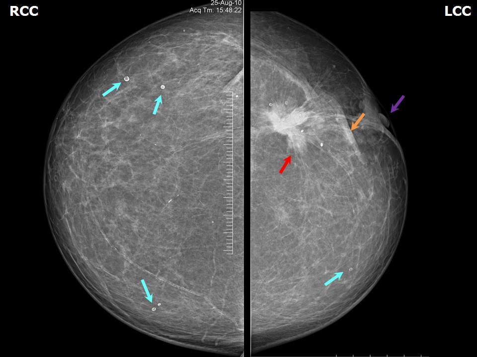 | 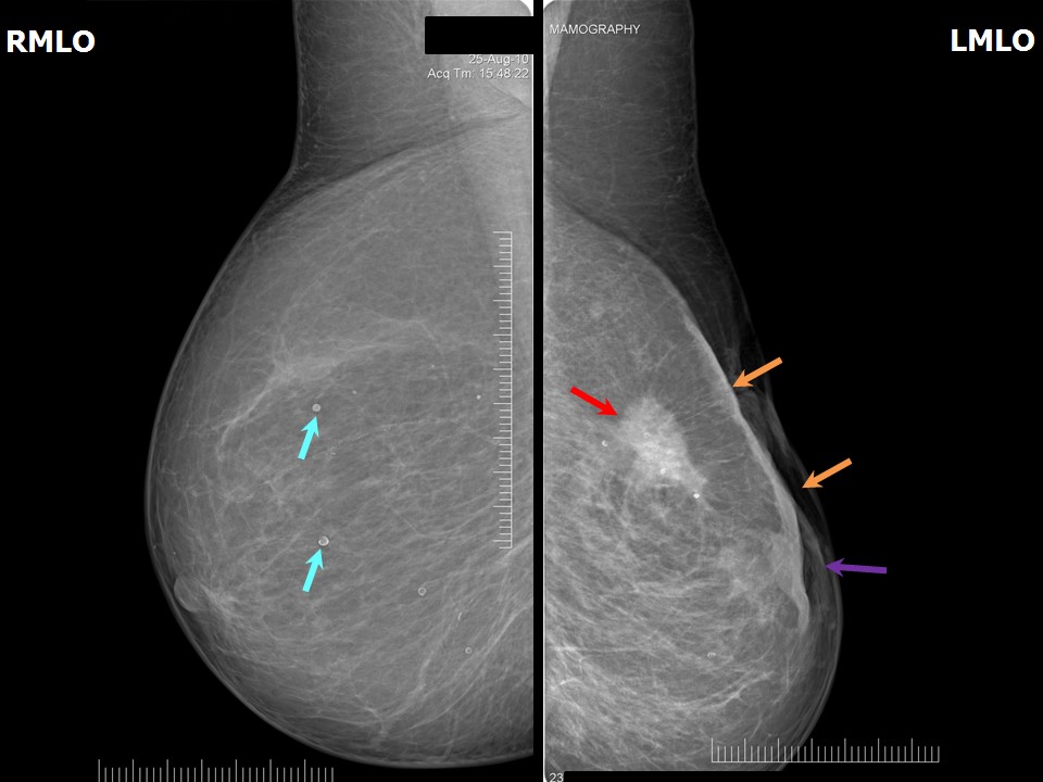 |
| Breast composition: | ACR category a (the breasts are almost entirely fatty) | Mammography features: |
| ‣ Location of the lesion: | Left breast, upper outer quadrant at 2 oclock, middle third |
| ‣ Mass: | |
| • Number: | 1 |
| • Size: | 3.7 cm in greatest dimension |
| • Shape: | Irregular |
| • Margins: | Spiculated |
| • Density: | High |
| ‣ Calcifications: | |
| • Typically benign: | None |
| • Suspicious: | None |
| • Distribution: | None |
| ‣ Architectural distortion: | Present |
| ‣ Asymmetry: | None |
| ‣ Intramammary node: | None |
| ‣ Skin lesion: | None |
| ‣ Solitary dilated duct: | None |
| ‣ Associated features: | Skin retraction, nipple retraction, skin thickening, trabecular thickening, and axillary adenopathy |
| Breast composition: | ACR category a (the breasts are almost entirely fatty) | Mammography features: |
| ‣ Location of the lesion: | Right breast, all quadrants, entire breast, middle third |
| ‣ Mass: | |
| • Number: | 0 |
| • Size: | No |
| • Shape: | None |
| • Margins: | None |
| • Density: | None |
| ‣ Calcifications: | |
| • Typically benign: | Round, rim |
| • Suspicious: | None |
| • Distribution: | Diffuse |
| ‣ Architectural distortion: | None |
| ‣ Asymmetry: | None |
| ‣ Intramammary node: | None |
| ‣ Skin lesion: | None |
| ‣ Solitary dilated duct: | None |
| ‣ Associated features: | None |
Ultrasound:
 | 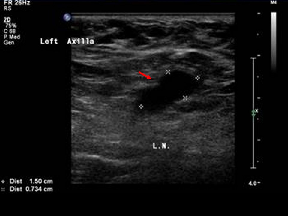 |
| Ultrasound features: Left breast, upper outer quadrant at 2 oclock | |
| ‣ Mass | |
| • Location: | Left breast, upper outer quadrant at 2 oclock |
| • Number: | 1 |
| • Size: | 3.8 × 2.7 cm |
| • Shape: | Irregular |
| • Orientation: | Not parallel |
| • Margins: | Angular |
| • Echo pattern: | Hypoechoic |
| • Posterior features: | Posterior shadowing |
| ‣ Calcifications: | None |
| ‣ Associated features: | Skin thickening, skin retraction, internal vascularity, and left axillary lymphadenopathy of altered morphology |
| ‣ Special cases: | None |
BI-RADS:
BI-RADS Category: 5 (highly suggestive of malignancy)Further assessment:
Further assessment advised: Referral for cytologyCytology:
 |
| Cytology features: | |
| ‣ Type of sample: | FNAC |
| ‣ Site of biopsy: | |
| • Laterality: | Left |
| • Quadrant: | Upper outer and another nodule in the areola |
| • Localization technique: | Palpation |
| • Nature of aspirate: | Whitish |
| ‣ Cytological description: | Smears from both areas reveal malignant cells arranged in sheets or isolated. Individual cells are pleomorphic with high N:C ratio, hyperchromatic nuclei, and prominent nucleoli |
| ‣ Reporting category: | Malignant |
| ‣ Diagnosis: | Carcinoma |
| ‣ Comments: | None |
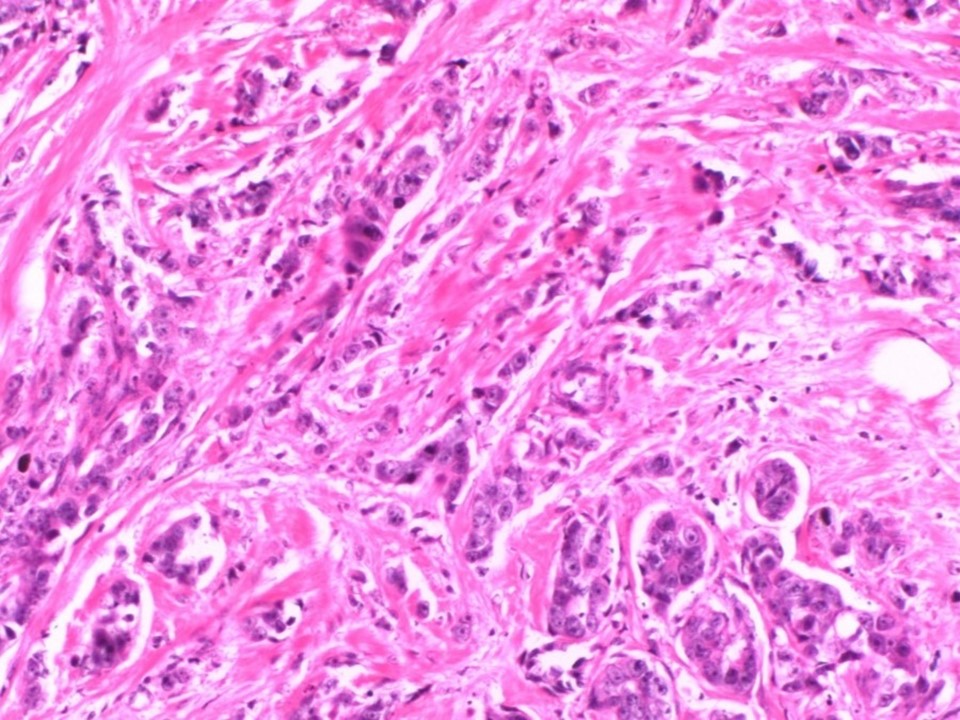
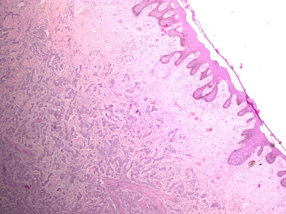
Histopathology:
MRM
 |  |
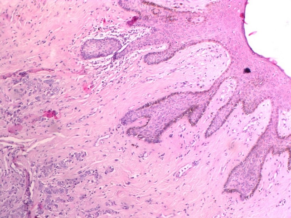 |
| Histopathology features: | |
| ‣ Specimen type: | MRM |
| ‣ Laterality: | Left |
| ‣ Macroscopy: | MRM specimen of left breast (28.0 × 20.0 × 7.0 cm) covered by a flap of skin (16.0 × 3.5 cm). Nipple is flattened and areola shows slightly elevated area just below the nipple. Cut surface shows a firm greyish white tumour (3.0 × 1.5 × 3.0 cm) with infiltrating margins seen in the upper outer quadrant. Base is 1.2 cm from the tumour. A second tumour nodule (2.0 × 1.2 × 2.0 cm) is seen below the areola. The rest of the breast is unremarkable |
| ‣ Histological type: | Invasive breast carcinoma of no special type |
| ‣ Histological grade: | Grade 3 (3 + 3 + 2 = 8) |
| ‣ Mitosis: | 14 |
| ‣ Maximum invasive tumour size: | 3.0 cm in greatest dimension |
| ‣ Lymph node status: | 9/17 |
| ‣ Peritumoural lymphovascular invasion: | Present |
| ‣ DCIS/EIC: | Cribriform DCIS of low grade; EIC absent |
| ‣ Margins: | Sections from nipple and areola are involved by the tumour. Sections from the base and separately sent muscle are free of tumour |
| ‣ Pathological stage: | pT2(2)N2 |
| ‣ Biomarkers: | |
| ‣ Comments: |
Case summary:
| Postmenopausal woman presented with left breast lump. Diagnosed as left breast carcinoma with skin thickening and retraction, and left nipple retraction, BI-RADS 5 on imaging, as left breast carcinoma on cytology, and as invasive breast carcinoma of no special type, pT2(2)N2 on histopathology. |
Learning points:
|