


Home / Training / Manuals / Atlas of breast cancer early detection / Learning
.png)
Click on the pictures to magnify and display the legends

Click on this icon to display a case study
Atlas of breast cancer early detection
Filter by language: English / РусскийBreast imaging Breast ultrasound Learning breast ultrasound Normal anatomy |
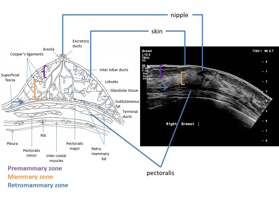
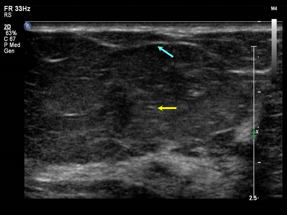
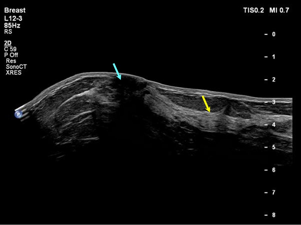
Breast ultrasound reveals breast parenchyma with varying proportion of fibrous, fatty, and glandular components at different stages of life as well as through the changing physiological status of the woman from thelarche to menopause.
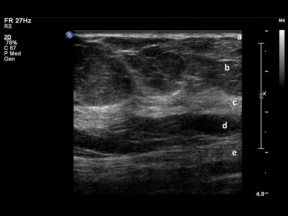
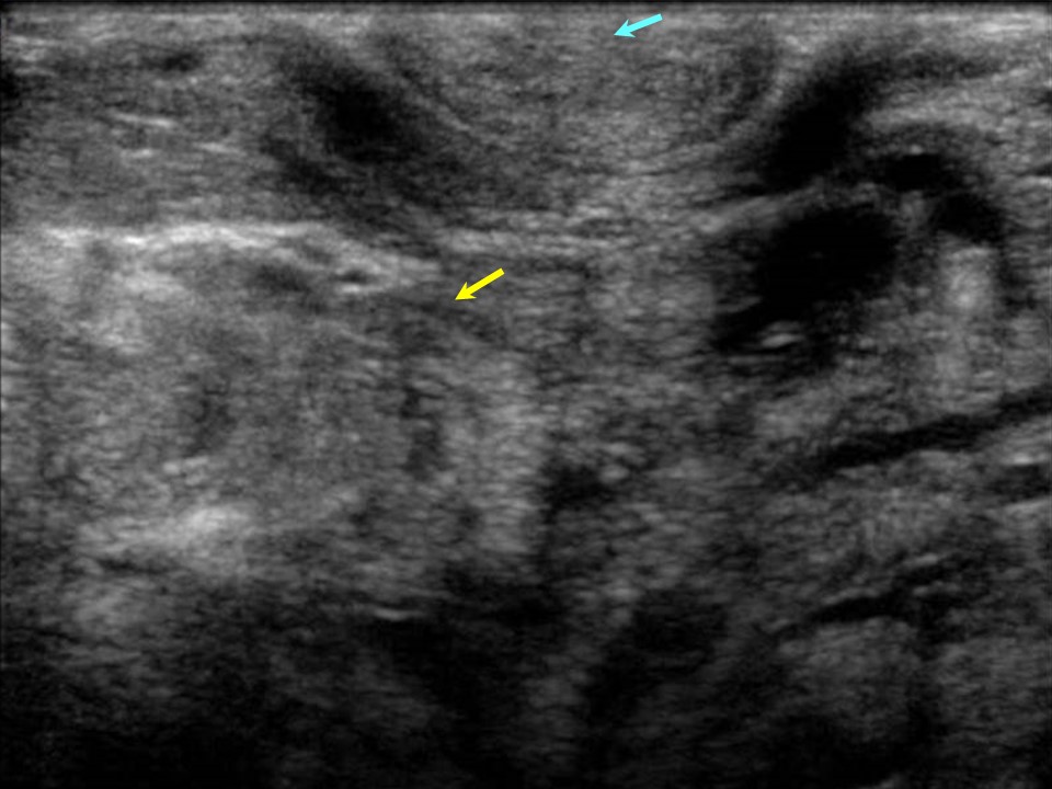
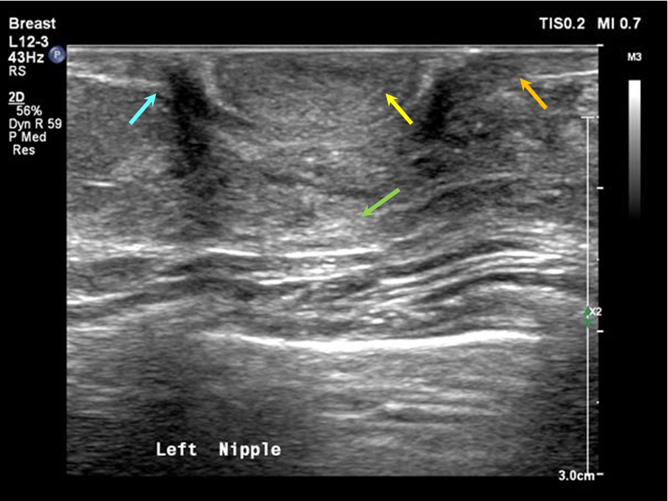
Breast skin (normal thickness 0.52.0 mm), subcutaneous fat, breast glandular parenchyma, pectoralis muscle, ribs, and the nippleareolar complex are detailed on ultrasound. The anatomy of the breast is divided into three zones: the premammary zone, the mammary zone, and the retromammary zone. The mammary glandular zone is made up of ducts and lobules and is the functional unit of the breast. Anterior suspensory ligaments, also known as ligaments of Cooper, formed from interlobular connective tissue are seen in the premammary zone. They insert into the dermis to stabilize the breast. |
Click on the pictures to magnify and display the legends
Click on this icon to display a case study
25 avenue Tony Garnier CS 90627 69366, LYON CEDEX 07 France - Tel: +33 (0)4 72 73 84 85
© IARC 2025 - Terms of use - Privacy Policy.
© IARC 2025 - Terms of use - Privacy Policy.