


Home / Training / Manuals / Atlas of breast cancer early detection / Learning
.png)
Click on the pictures to magnify and display the legends

Click on this icon to display a case study
Atlas of breast cancer early detection
Filter by language: English / РусскийBreast imaging Breast ultrasound Learning breast ultrasound Breast changes in special stages of life |
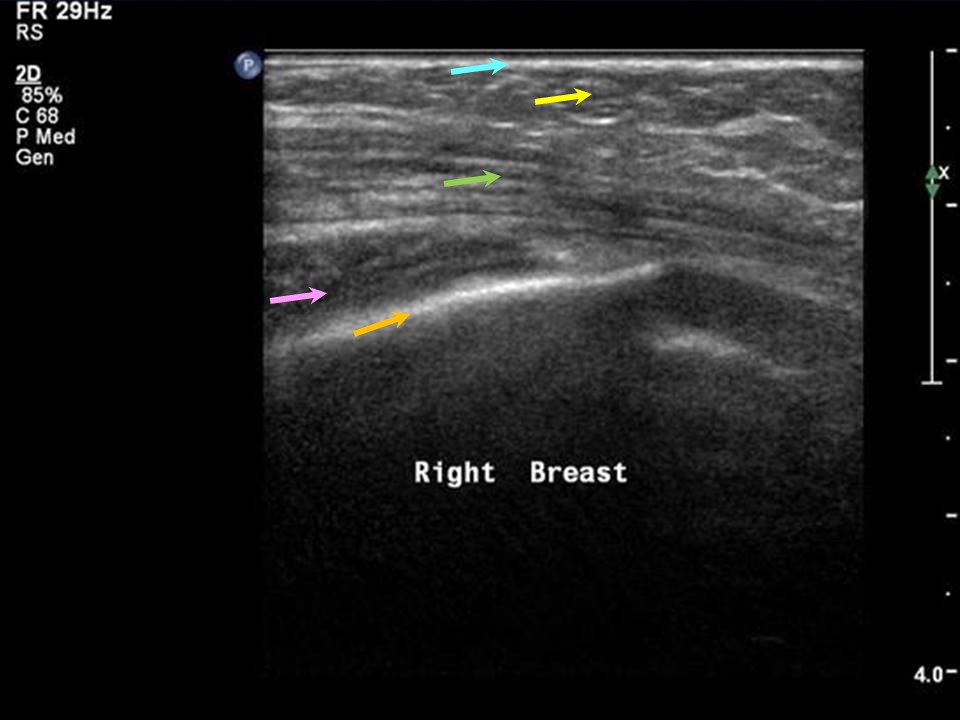
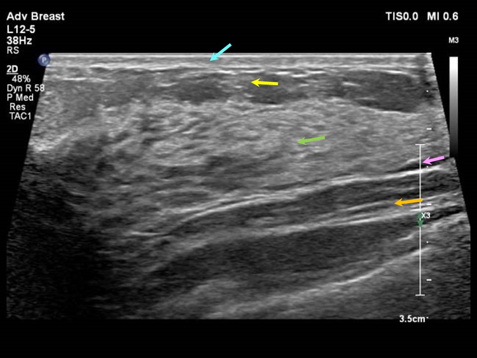
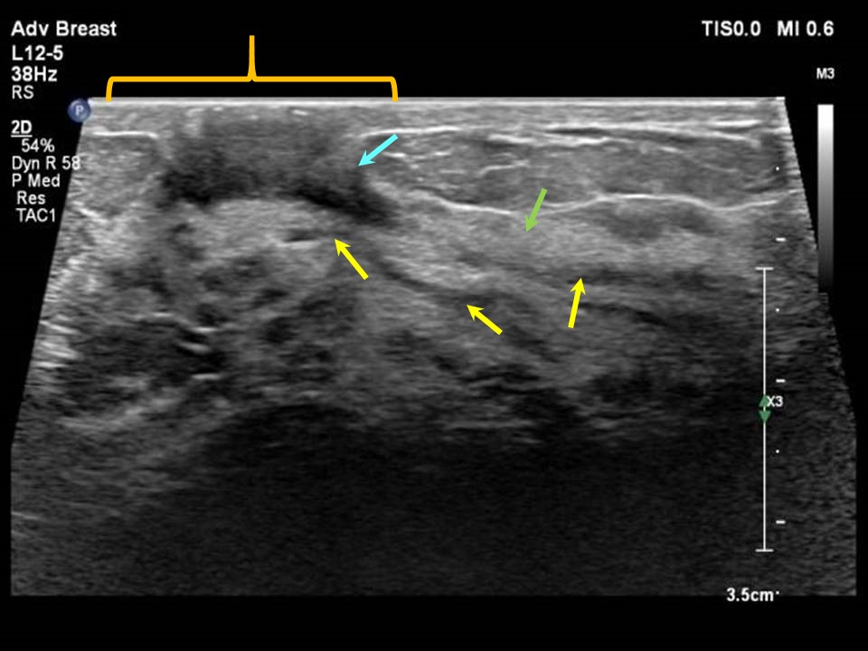
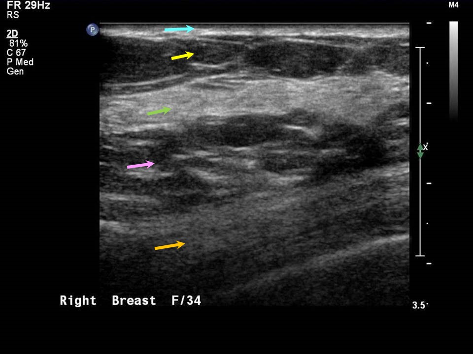
The breast shows significant morphological changes throughout life. Childhood (prethelarche) The nipple, surrounding skin, and retroareolar region all have homogeneous dermal and subcutaneous tissues. Morphologically similar appearances are seen in both sexes. Thelarche Breast buds develop before the onset of menarche and are seen on ultrasound as a subareolar area of proliferating elongating symmetrically arranged ducts. Puberty Rapid proliferation of the ducts and the lobules occur during puberty. Mammary zone differentiation can be seen on ultrasound as uniform, homogeneously echogenic glandular parenchyma with premammary and retromammary areas of fatty parenchyma and supporting connective tissues. Adult women, reproductive age group In adult women of reproductive age, ultrasound reveals homogeneous fibroglandular echotexture with normal distribution of the glandular tissues in the mammary zone and fatty parenchyma and connective tissues in the premammary and retromammary zones. This is seen from the stage of menarche in young girls and in postpubertal women until the stage of breast involution. Through this period of life, transient and revers/ible changes related to pregnancy and lactation will be seen. |


Click on the pictures to magnify and display the legends
Click on this icon to display a case study
25 avenue Tony Garnier CS 90627 69366, LYON CEDEX 07 France - Tel: +33 (0)4 72 73 84 85
© IARC 2025 - Terms of use - Privacy Policy.
© IARC 2025 - Terms of use - Privacy Policy.