


Home / Training / Manuals / Atlas of breast cancer early detection / Learning
.png)
Click on the pictures to magnify and display the legends

Click on this icon to display a case study
Atlas of breast cancer early detection
Filter by language: English / РусскийBreast imaging Techniques |
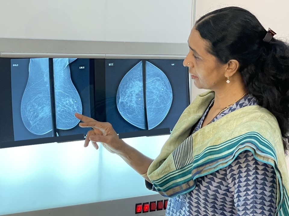
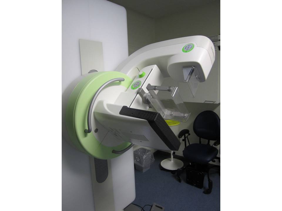
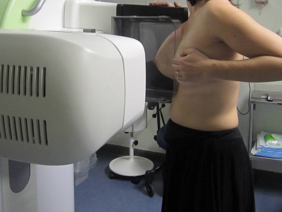
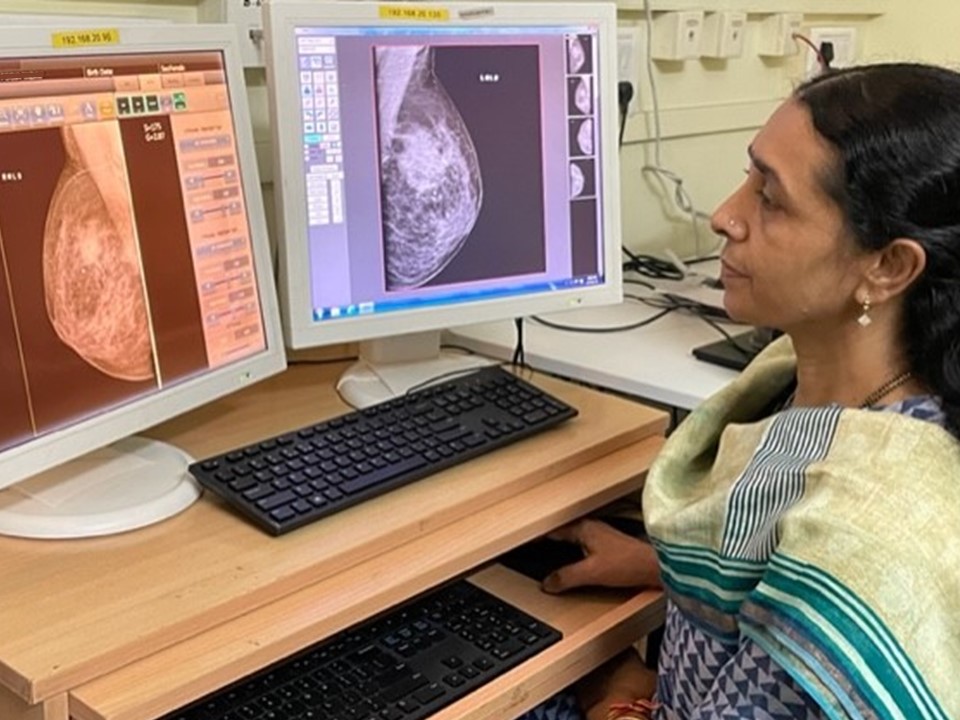
MammographyThe technology of mammography has evolved over time. It includes screen-film and digital techniques. Advantages of mammography
Screen-film mammographyScreen-film mammography uses a molybdenum anode X-ray unit with a uniform-thickness breast-compression paddle to enable proper exposure of the breast tissue from chest wall to nipple. The image obtained on the single-emulsion film is developed and processed to get a visible image of the breast of optimal contrast and density. Full-field digital mammography (FFDM)FFDM, more commonly known as digital mammography, has better contrast resolution than conventional mammography and enables optimization of both image acquisition and display. Screen-film mammography has better spatial resolution than FFDM, but FFDM has better contrast resolution, which overcomes the decreased spatial resolution. FFDM uses digital detectors that absorb X-ray photons and convert them into an electric charge, which is converted to an image. A computer processes the signal and displays the image on a high-resolution monitor, which can be viewed directly for interpretation. Digital images can be modified on the monitor to lighten or darken the image and to zoom in on certain sections to get a magnified view. Digital mammography has become the standard of care and has replaced screen-film mammography. Computed radiography digital mammography (CR FFDM)CR FFDM differs from FFDM in its image acquisition technology. Computed radiography is an indirect digital mammography technique that captures the image on a reusable plate. This reusable plate is then scanned by a reader to produce the digital image. CR FFDM and FFDM images can be either viewed on a monitor or printed and viewed for interpretation. Studies did not observe any advantages of CR FFDM over FFDM. Digital breast tomosynthesis (DBT)DBT represents a significant advance in technology and overcomes the inability of mammography to detect lesions hidden by dense overlapping fibroglandular breast tissues. DBT involves volumetric reconstruction of the whole breast from a finite number of low-dose two-dimensional projections obtained by different X-ray tube angles. The reconstructed images are presented on a high-resolution monitor using 0.51 mm slice separation. In DBT, morphological features for the detection of breast masses and breast calcifications are better defined, which makes diagnosis more accurate. The process is similar to computed tomography (CT) scan images of structures inside the body. Sequential visualization of thin images (slices) of the breast minimizes the masking effect of overlapping fibroglandular tissue and improves detection of a lesion or abnormality, particularly in dense breasts. DBT improves the accuracy of reporting, minimizes the number of unnecessary biopsies, and ensures that fewer women need to come back for a repeat mammogram. Advantages of DBT
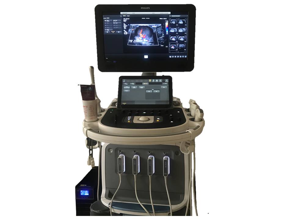
Breast ultrasound (hand-held)Ultrasound is an imaging technique used to detect breast abnormalities using a high-frequency linear transducer on a high-resolution ultrasound machine. Introduced in the 1980s, breast ultrasound is commonly used as an adjunct to mammography, particularly to further evaluate women with dense breast tissues. Advantages of breast ultrasound
Automated breast ultrasound (ABUS)ABUS is a breast ultrasound technique in which image acquisition is separated from interpretation. A trained nurse or technician applies the transducers in the right positions and keeps gentle pressure on the breast tissue while the breast is scanned craniocaudally and mediolaterally. The radiologist reviews the entire set of images, which are stored digitally on a dedicated workstation. Because breast image acquisition is minimally operator-dependent, consistency in identifying suspicious lesions is improved. Several types of ABUS are available, with different designs, image acquisition approaches, and workstation features. Dedicated computer-aided detection software for ABUS has the potential to create three-dimensional images. Conventional hand-held breast ultrasound is very operator-dependent and requires considerable time for the radiologist to assess both breasts. In contrast, ABUS is less operator-dependent, requires less time from a radiologist, and has more reproducible results. In September 2012, the United States Food and Drug Administration approved ABUS for screening women with dense breasts after negative findings on mammography. One multicentre cross-sectional study was carried out in China in 2016 to compare the diagnostic performance of hand-held breast ultrasound, ABUS, and mammography. A total of 1974 women aged 3069 years were recruited. The results suggested that the clinical performance of ABUS was comparable with that of hand-held ultrasound for breast cancer detection, and that both traditional ultrasound and ABUS have better performance than mammography, especially among women with high-density breasts. Advantages of ABUS
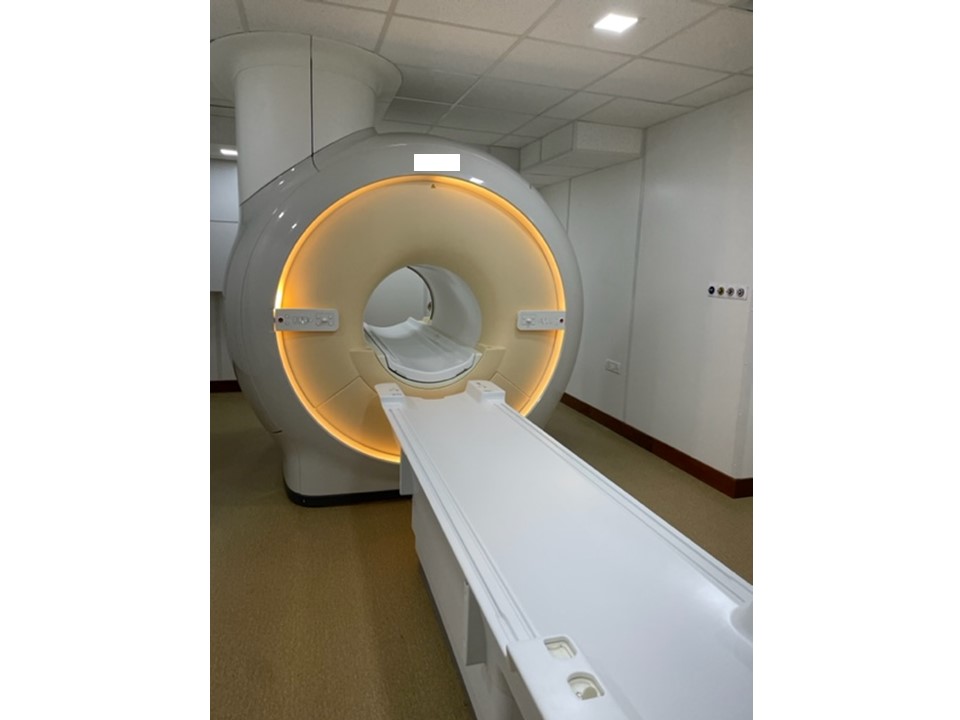
Breast MRIBreast MRI is primarily a supplemental tool for breast imaging used along with mammography and/or breast ultrasound. The sensitivity of contrast-enhanced breast MRI in the detection of cancer is considerably higher than that of either mammography or ultrasound. MRI is the screening modality of choice for women at high lifetime risk of breast cancer, such as those with mutations in the BRCA1 and/or BRCA2 genes. Advantages of breast MRI
|


Click on the pictures to magnify and display the legends
Click on this icon to display a case study
25 avenue Tony Garnier CS 90627 69366, LYON CEDEX 07 France - Tel: +33 (0)4 72 73 84 85
© IARC 2025 - Terms of use - Privacy Policy.
© IARC 2025 - Terms of use - Privacy Policy.