Home / Training / Manuals / Atlas of breast cancer early detection / Learning

Atlas of breast cancer early detection

Breast imaging – Breast ultrasound – Ultrasound-guided interventional procedures – Fine-needle aspiration cytology (FNAC)


Indications for FNAC

Diagnostic
  • An abnormality that is suspicious for malignancy detected on clinical examination.
  • An abnormality that is suspicious for malignancy detected on diagnostic breast imaging performed to investigate any breast problem and/or abnormal clinical findings.
  • A non-palpable breast lesion detected on imaging during routine screening of an asymptomatic woman.
  • Inflammatory diseases (uncommon).
  • Suspected secondary or metastatic tumours in the breast (including haematological or lymphoid malignancies).
  • Suspected recurrence of malignancy after treatment.


Therapeutic
  • Evacuation of simple or inflammatory cysts.


Steps
  1. Counsel the patient and obtain an informed consent.
  2. Perform diagnostic breast imaging (mammography or ultrasound) to localize the lesion. Mark the suspicious finding for ultrasound-guided intervention.
  3. Position the patient in dorsal decubitus and ask her to raise the arm ipsilateral to the lesion and rest it behind her head. An interior oblique position may be more suitable for a patient with large breasts or if the lesion is in an extremely lateral location. Patient position for ultrasound-guided FNAC is similar to that for diagnostic breast ultrasound.
  4. The FNAC procedure and preparation of cytology smears are described in the section on Laboratory processing of breast specimens for cytology and histology.
  5. Clean and drape the area of breast with a suspicious finding.
  6. Clean and cover the ultrasound probe with a sterile probe drape.
  7. Localize the non-palpable lesion and measure the skin depth to be traversed.
  8. Stabilize the lesion with firm pressure of the ultrasound probe.
  9. Select a 23G or 24G needle that is long enough to reach the centre of the lesion.
  10. Introduce the needle under guidance parallel to the length of the ultrasound probe and advance it into the breast tissue up to the lesion.
  11. View the needle in its entire length throughout the procedure and ensure that the lesion is hit and pierced towards its centre.
  12. Document the position of the needle in the breast (mark the quadrant and the position of the transducer on the image). Save the first screenshot, and with the needle in the tumour, save the second image.
  13. Pull the plunger back to create suction while holding the needle steady throughout the sampling procedure.
  14. Move the needle backwards and forwards inside the lesion 10–20 times.
  15. Release suction by letting go of the plunger.
  16. Withdraw the needle.
  17. Transfer the material onto the prepared slides. Ensure they are labelled with the patient’s unique identification details.
  18. Keep compression on the area with a gauze pad to prevent the formation of haematoma.
  19. Further processing of slides is discussed in detail in the section Breast Pathology.


Further details about the advantages and disadvantages of FNAC are given later.


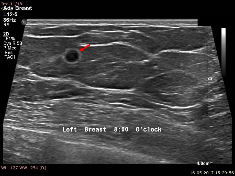
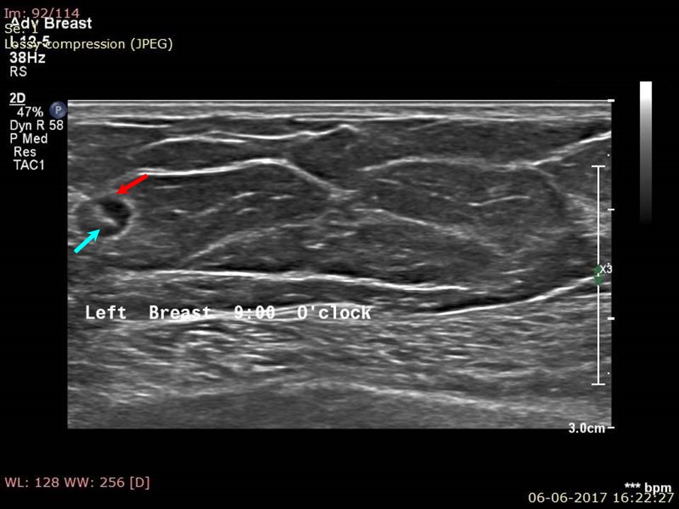
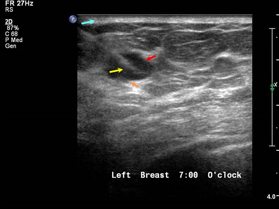
Click on the pictures to magnify and display the legends


Click on this icon to display a case study

IARC, 150 Cours Albert Thomas, 69372 Lyon CEDEX 08, France - Tel: +33 (0)4 72 73 84 85 - Fax: +33 (0)4 72 73 85 75
© IARC 2024 - All Rights Reserved.