Home / Training / Manuals / Atlas of breast cancer early detection / Learning
.png)
Click on the pictures to magnify and display the legends

Click on this icon to display a case study
Atlas of breast cancer early detection
Filter by language: English / РусскийBreast imaging – Breast ultrasound – Getting ready |
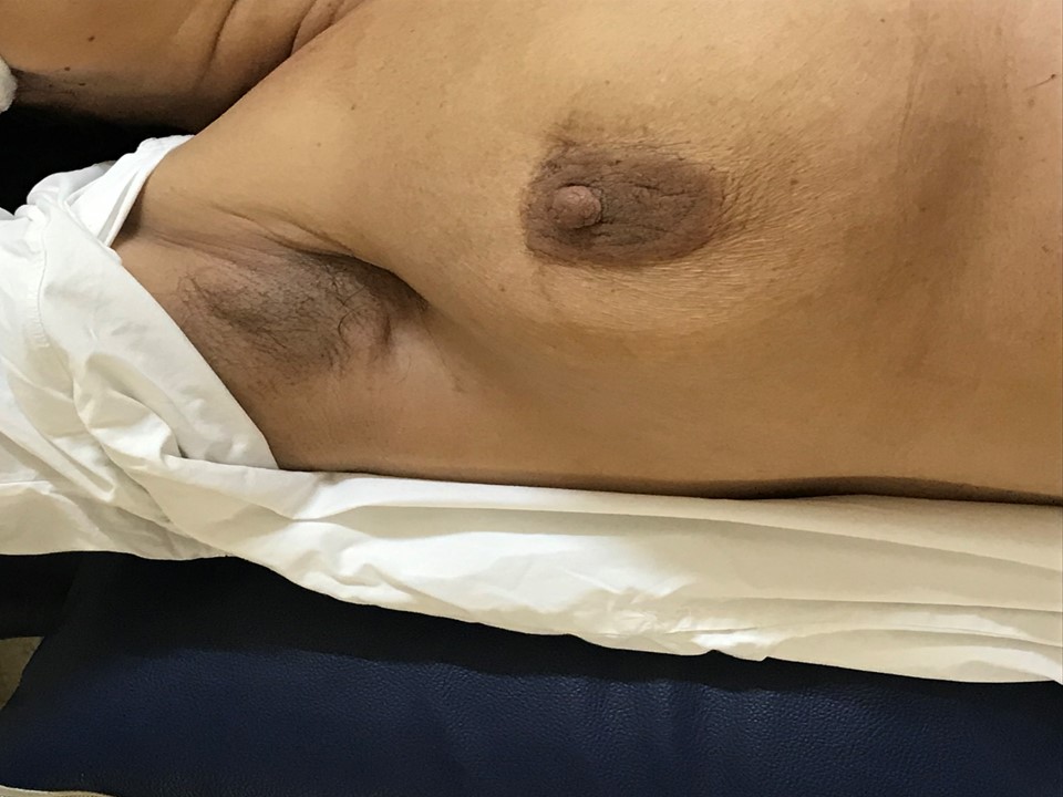
Patient in supine position for bilateral breast ultrasound If both breasts are to be scanned, the right breast is usually scanned first, then the left. Raise the patient’s arm above her head and place a soft support beneath the shoulder to minimize movement of the breast during the examination. Move the patient’s gown to uncover the side under examination up to the axilla. Cover the other breast for comfort. Ask the patient to turn her head to the opposite side. Patient in supine position for ultrasound of right breast Patient in supine position for ultrasound of left breast Patient in right decubitus position for ultrasound of the far lateral tissues of the left breast The step-by-step breast ultrasound examination checklist acts as a useful reminder of the steps to be taken in the clinic. The checklist can also be used to assess the trainee’s skill during a training programme. |
Click on the pictures to magnify and display the legends
Click on this icon to display a case study
25 avenue Tony Garnier CS 90627 69366, LYON CEDEX 07 France - Tel: +33 (0)4 72 73 84 85
© IARC 2025 - Terms of use - Privacy Policy.
© IARC 2025 - Terms of use - Privacy Policy.


