Home / Training / Manuals / Atlas of breast cancer early detection / Learning

Atlas of breast cancer early detection

Breast imaging – Mammography technique – Mammography procedure – Additional mammographic views – Spot compression magnification view


Indication:
The spot compression magnification view may be advised to delineate the detailed morphology of a mass palpable on clinical examination or an abnormality seen on screening mammography in the absence of any palpable lump.

Advantages
  • The spot compression magnification view increases the spatial resolution because the distance between the focal spot and the breast is reduced; this enhances the geometric sharpness, which differentiates a real lesion from overlapping summation shadows.
  • A magnified view of the microcalcifications seen on CC and MLO projections helps the radiologist to understand the morphological appearance of the calcifications and to differentiate them into typically benign, suspicious, or malignant calcifications.
  • The spot compression magnification view is very helpful to delineate suspicious microcalcifications and to plan an image-guided biopsy.

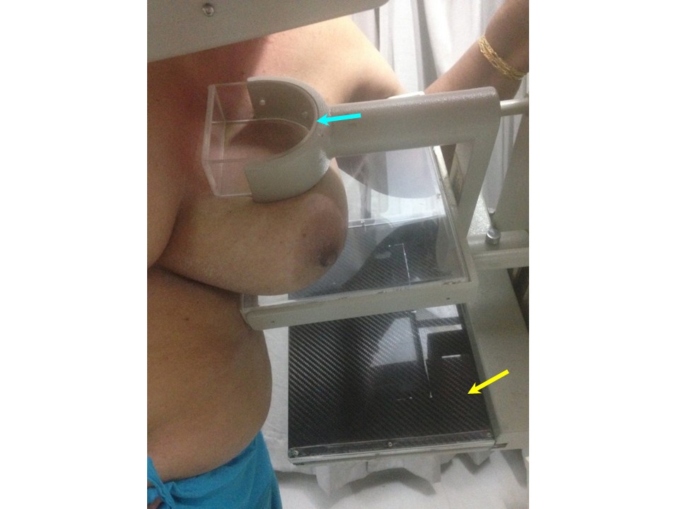
Steps to obtain a spot compression magnification view
  1. Survey the CC and MLO views in detail for the suspicious finding and mark it. Mark the distance between this finding and the nipple. Use this to position the breast accordingly for spot compression magnification views.
  2. Talk to the patient and explain the need for an additional view. This helps to reduce anxiety.
  3. Detach the mammography unit compression device used for standard positioning.
  4. Attach the spot compression magnification breast support with a smaller compression device in the slots provided. The focal spot focus distance is shorter and the breast under examination is positioned at a higher level, away from the detector.
  5. Place the image receptor in the cassette holder with the detector at the same level as for the conventional mammography position.
  6. Position the patient with the marked area of the suspicious finding under the spot compression paddle. Apply gentle compression. Take care to move the abdomen out of the edge of the image detector to avoid artefacts from overlapping body parts.
  7. After adequate compression is achieved, step behind the protective lead shield and take the X-ray.


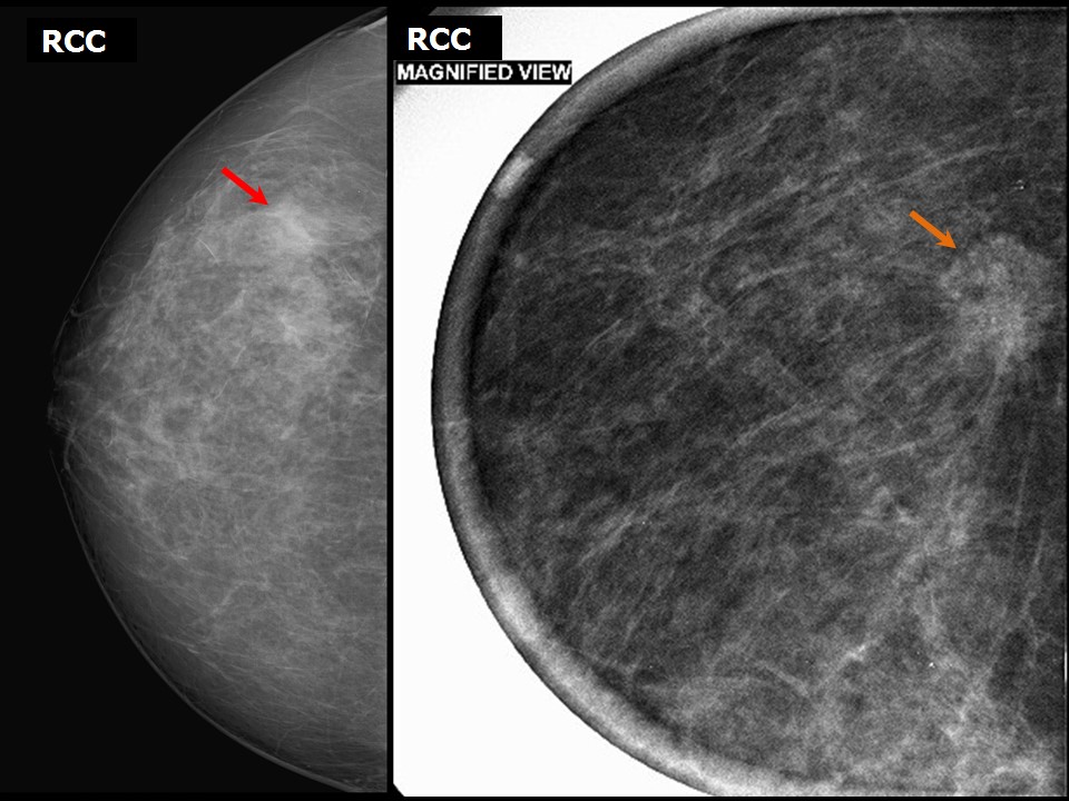
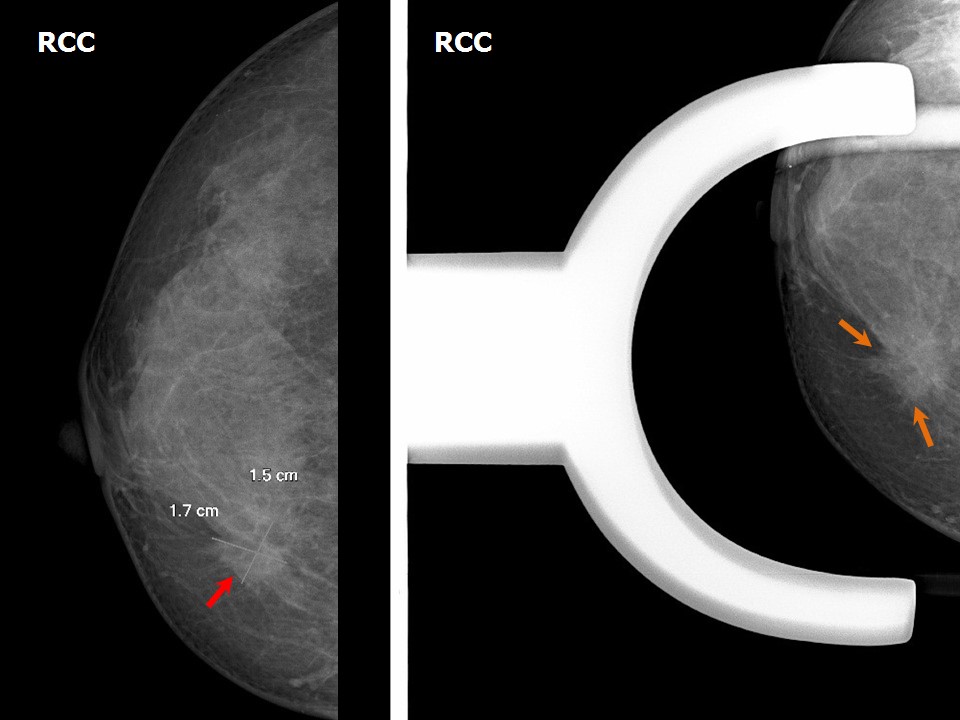
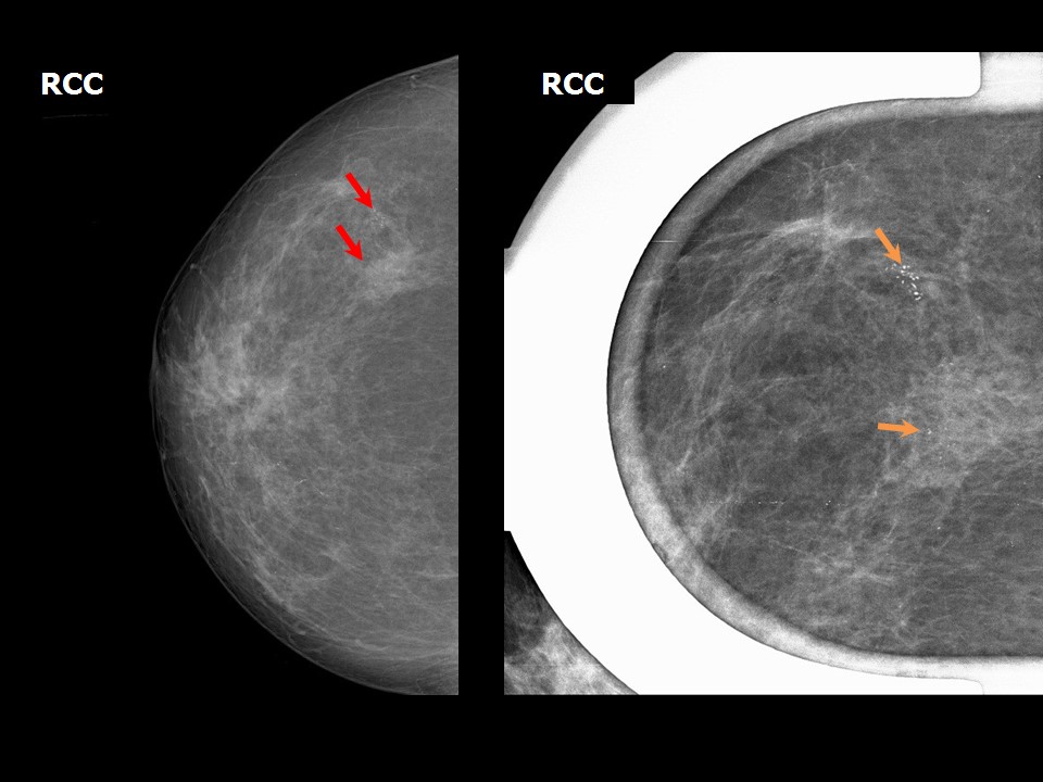
Spot compression magnification view to visualize the borders of an abnormality or a suspicious developing asymmetry


Spot compression magnification view to differentiate between a true lesion and a summation shadow


Spot compression magnification view to enhance the geometric sharpness and margins of a suspicious mass


Spot compression magnification view to enhance geometric sharpness and the margins of a focal edge retraction


Spot compression magnification view to enhance geometric sharpness and delineation of microcalcifications in a suspicious mass


Spot compression magnification view to enhance geometric sharpness of microcalcifications


Click on the pictures to magnify and display the legends


Click on this icon to display a case study

IARC, 150 Cours Albert Thomas, 69372 Lyon CEDEX 08, France - Tel: +33 (0)4 72 73 84 85 - Fax: +33 (0)4 72 73 85 75
© IARC 2024 - All Rights Reserved.