Home / Training / Manuals / Atlas of breast cancer early detection / Learning

Atlas of breast cancer early detection

Breast imaging – Mammography interpretation – Interpreting the abnormal mammogram


The primary role of mammography is to detect cancer at an early stage when the lesion is small. The steps used to interpret an abnormality detected on mammography are outlined in the following sections.

1. Identify any suspicious features
An abnormality is suspected if any of the following features are seen:
  • a developing asymmetry or new lesion that was not visible on earlier mammograms;
  • a mass lesion on the mammogram;
  • calcifications with or without a mass;
  • focal or global asymmetry of the breast;
  • architectural distortion;
  • skin changes as an isolated finding or associated with a mass;
  • nipple retraction; or
  • enlarged axillary nodes of high density, with loss of normal uniform shape and loss of fatty hilum. These are features suspicious for metastatic nodes.


2. Differentiate true abnormalities from apparent ones
  • Additional mammographic views on DBT and contrast-enhanced digital mammography (spot compression magnification view is the most common) or adjuvant imaging techniques (breast ultrasound is the most common) will help to differentiate true abnormalities from apparent ones.
  • Overlapping breast tissues called summation shadows are seen on only one mammographic view. A true abnormality is seen on more than one mammographic view and needs further assessment or biopsy.


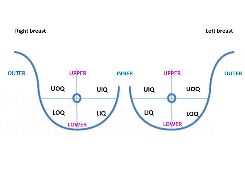
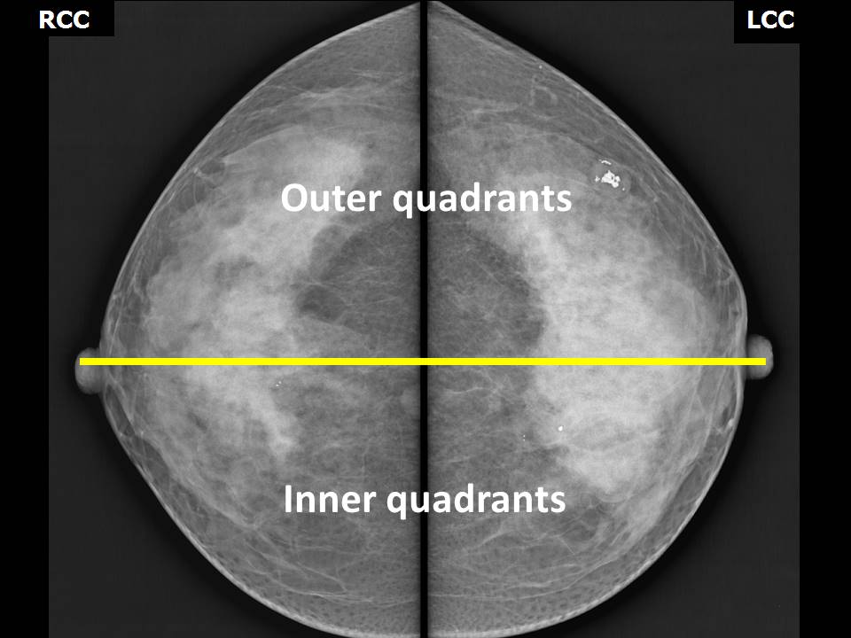
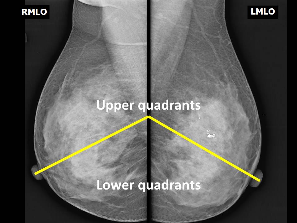
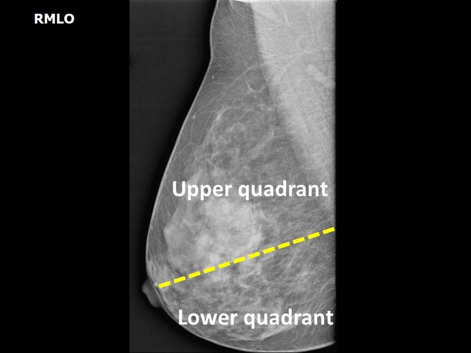
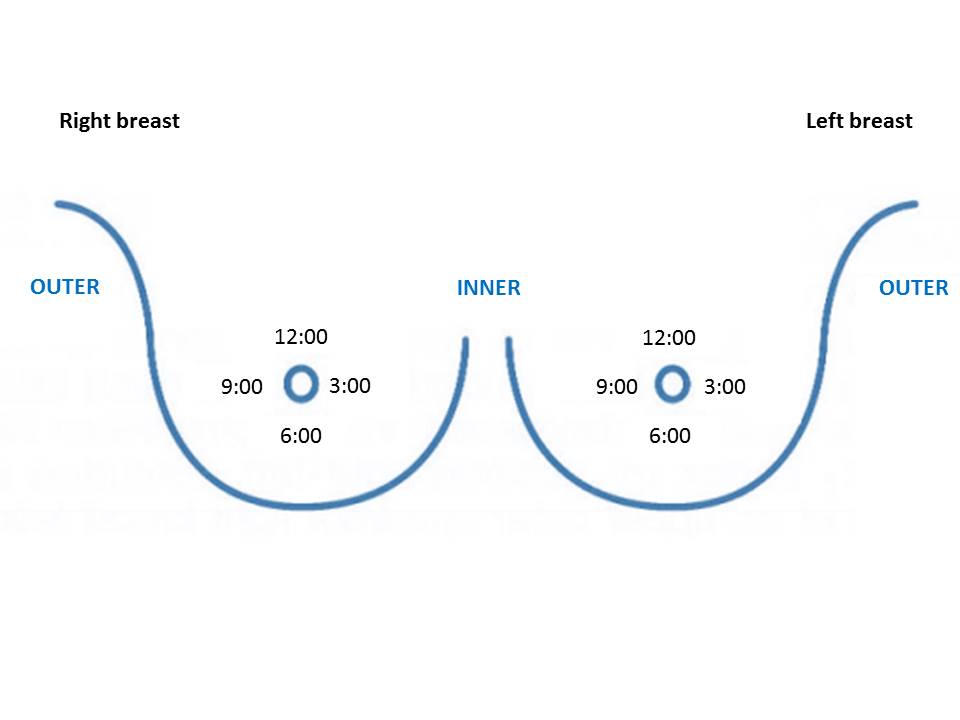
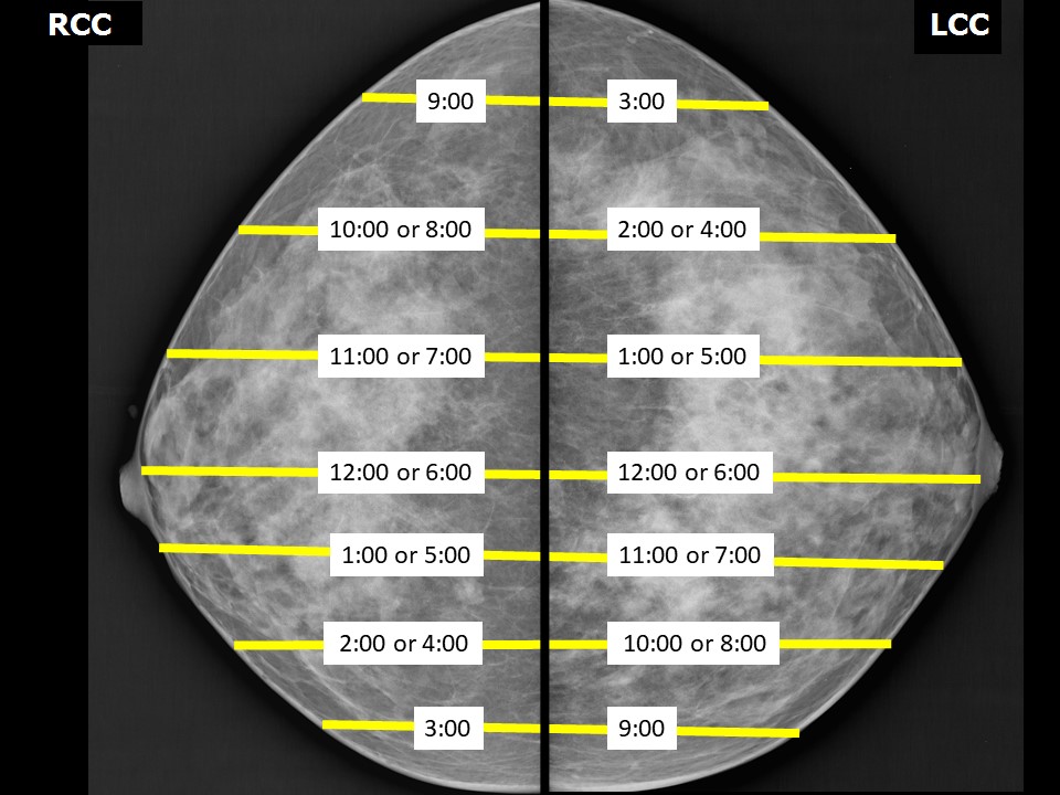
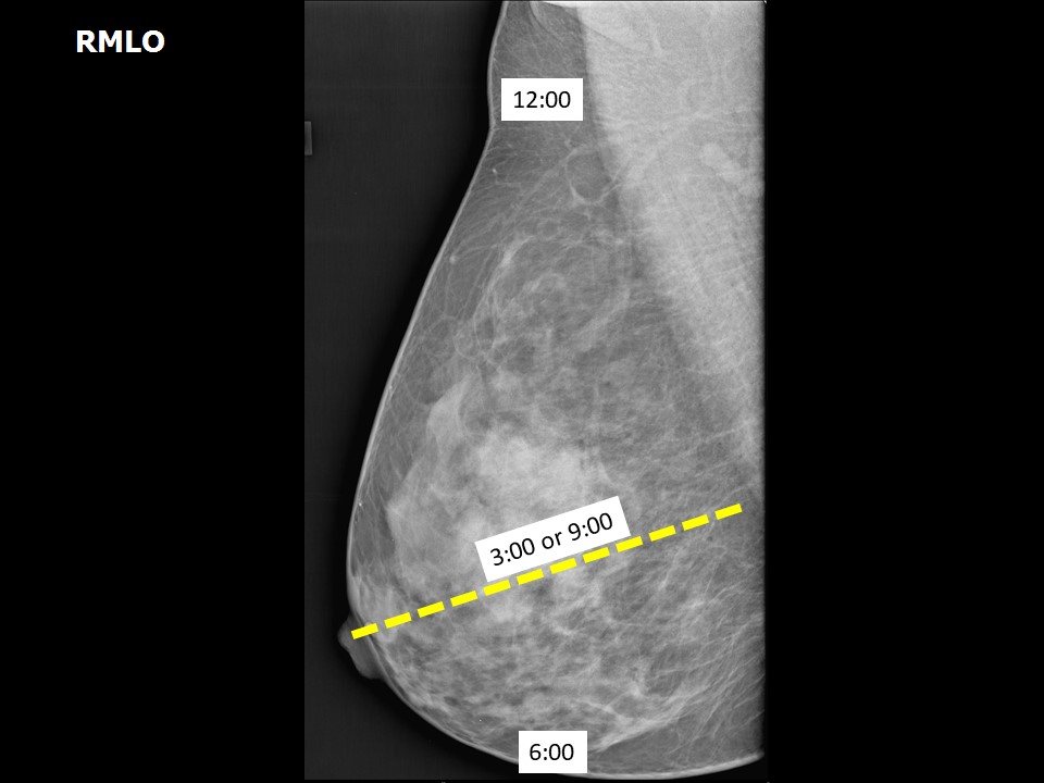
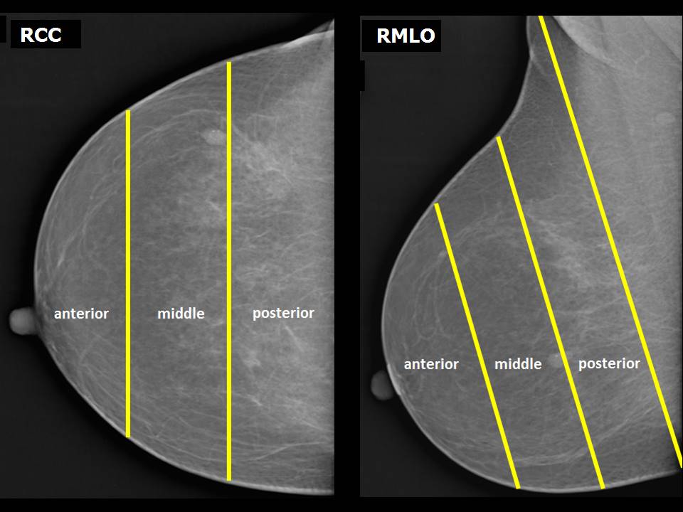
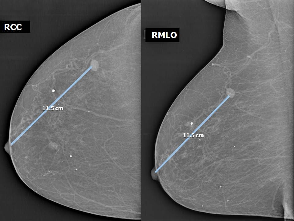
3. Categorize the abnormality using a common lexicon
The categorization of abnormalities detected on mammography is observer-dependent and can vary widely. To maintain uniformity of interpretation, the descriptors given in the BI-RADS lexicon should be used to document the findings, to make a provisional diagnosis, and to suggest further assessment.


4. Localize the abnormality
The abnormality should be localized according to the following:
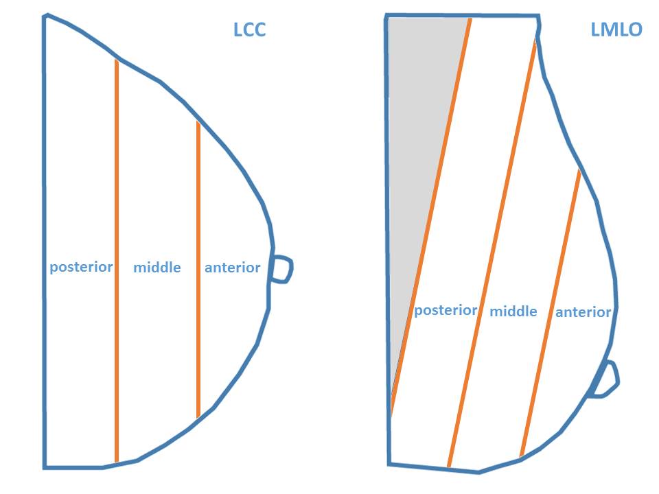
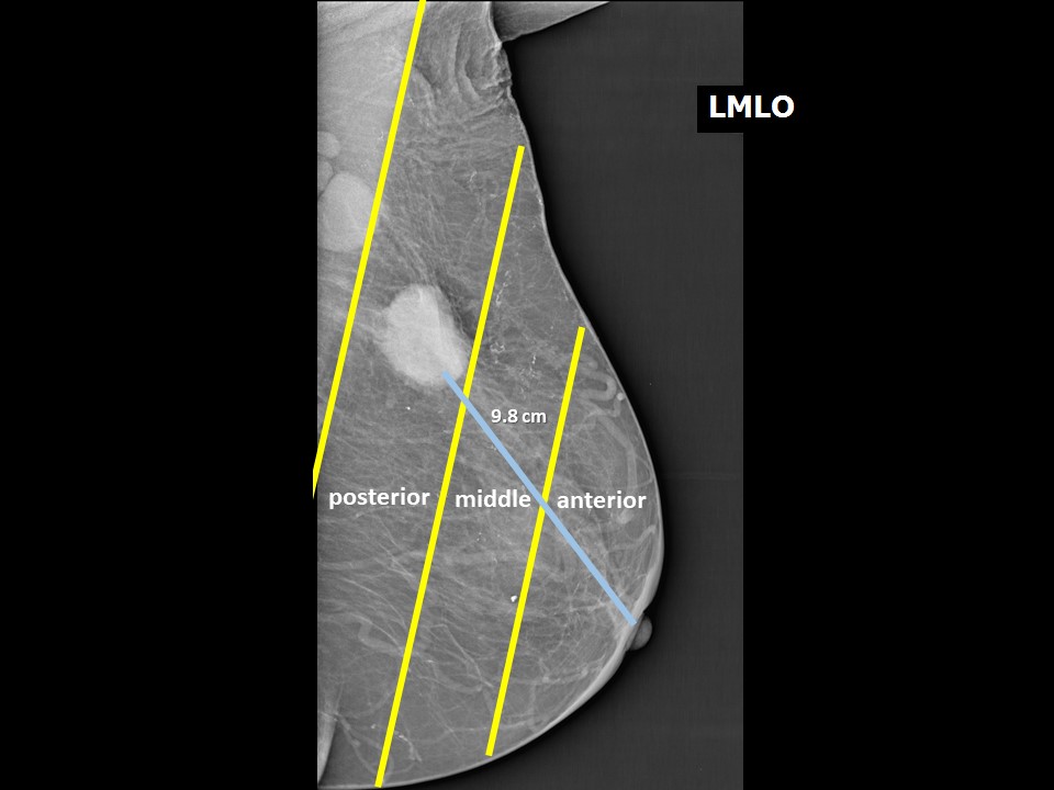
  • Laterality: Left breast or right breast.
  • Quadrant of the breast and o’clock position: The CC view on mammography localizes the lesion by the o’clock position. The MLO view is then used to determine whether the mass is in the upper (above the nipple) or lower (below the nipple) quadrant of the breast.
  • Depth: A lesion is localized either in the anterior third, middle third, or posterior third of the breast, depending on its depth within the breast.
  • Distance from nipple: The distance of the mass from the nipple is measured, the nipple being the fixed reference point.

Quadrant localization


O’clock position


Depth of lesion (anterior, middle, and posterior thirds)


Distance from nipple


Summary
To summarize, the following location descriptors are used in mammography:
  • Laterality: Right breast or left breast.
  • Quadrant and o’clock location: Understand that MLO projections distinguish between superior (upper) and inferior (lower) quadrants. CC projections distinguish between lateral (outer) and medial (inner) quadrants. The four quadrants are used in mammography; a clock face is used in ultrasound. Understand that the clock face is a mirror image for lateral (outer) and medial (inner) quadrants, so that 3 o’clock on the left is 9 o’clock on the right, 2 o’clock on the left is 10 o’clock on the right and so forth.
  • Depth of the lesion in the breast – anterior, middle, or posterior thirds on the mammogram.
  • Distance from the nipple.


5. Determine whether multifocal or multicentric
Survey the rest of the breast and the other breast to detect further lesions. Breast imaging may detect additional findings that are benign lesions, precursor lesions, or malignant neoplasms. The presence of two or more foci of cancer within the same breast quadrant is defined as multifocal , whereas the presence of two or more foci of cancer in different quadrants of the same breast is defined as multicentric .
Thus, an abnormal mammogram may detect:
  • unifocal breast cancer, where there is only one tumour in the breast and no significant findings in the other breast ;
  • multifocal breast cancer, where two or more foci of cancer are seen in the same quadrant or area of the breast ;
  • multicentric breast cancer, where two or more foci of cancer are seen separately in different quadrants or areas of the breast;
  • bilateral breast cancers in a patient with abnormal CBE in a single breast or palpable lumps in both breasts ; or
  • unifocal breast cancer with a benign finding elsewhere in the breast or in the other breast .
Mammography and breast ultrasound are complementary imaging modalities used to evaluate the presence of multifocal or multicentric breast cancers or bilateral breast cancers. Tumours are considered to be multifocal when the distance between the tumour masses is ≤ 5 cm and multicentric when this distance is > 5 cm .

6. Suggest further assessment if necessary to confirm the diagnosis
If necessary, further assessments can be suggested to confirm the diagnosis:
  • Additional mammographic views for better visualization of a finding seen on standard mammographic views.
  • Breast ultrasound as a supplementary tool.
  • In some cases, breast MRI or image-guided biopsy may be needed.


Click on the pictures to magnify and display the legends


Click on this icon to display a case study

IARC, 150 Cours Albert Thomas, 69372 Lyon CEDEX 08, France - Tel: +33 (0)4 72 73 84 85 - Fax: +33 (0)4 72 73 85 75
© IARC 2024 - All Rights Reserved.