


Home / Training / Manuals / Atlas of breast cancer early detection / Cases
Atlas of breast cancer early detection
Filter by language: English / Русский
Go back to the list of case studies
.png) Click on the pictures to magnify and display the legends
Click on the pictures to magnify and display the legends
| Case number: | 072 |
| Age: | 58 |
| Clinical presentation: | Postmenopausal woman with average risk of developing breast cancer presented with a lump in her right breast. On clinical examination, she had a hard lump in the upper quadrant of the right breast. |
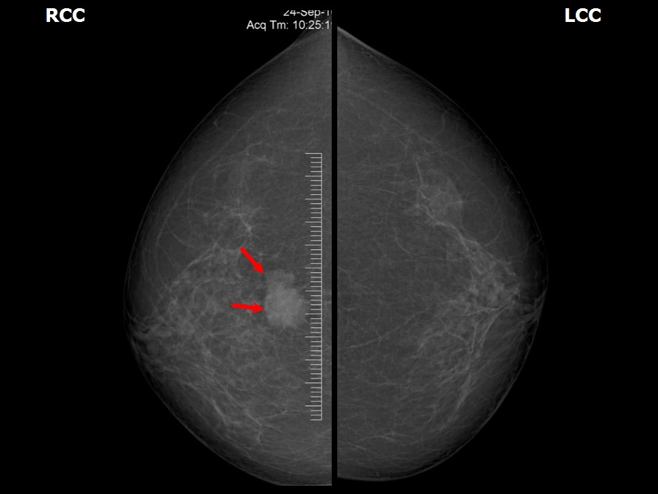
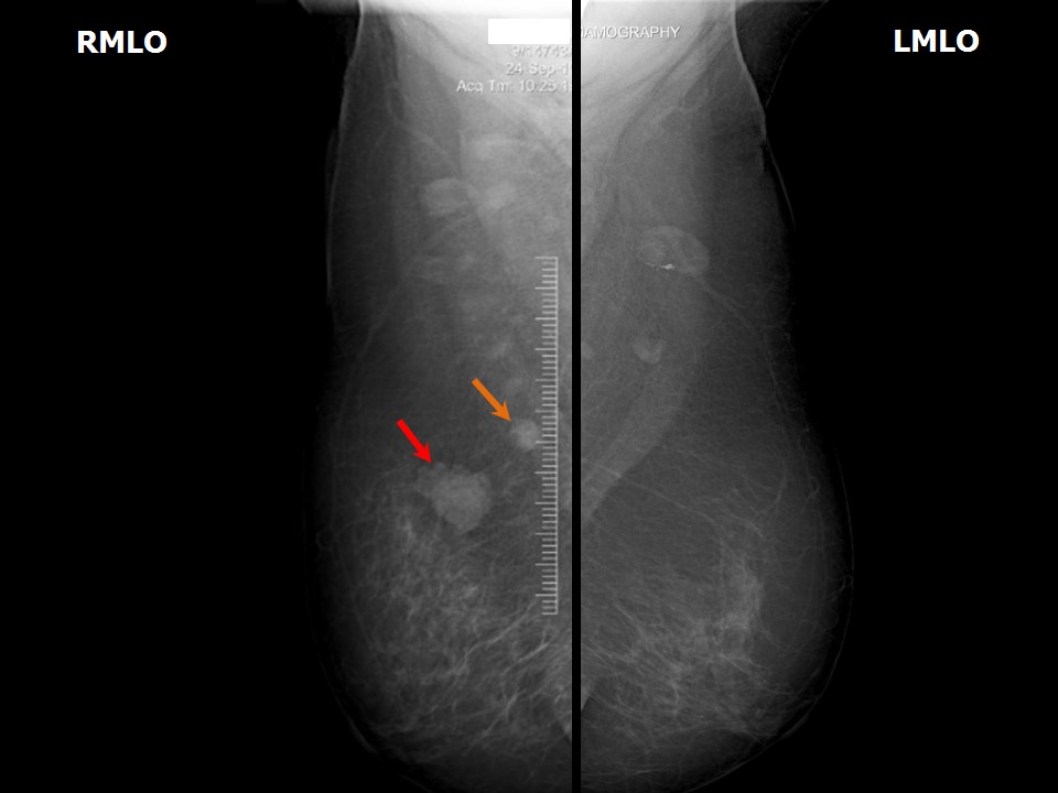
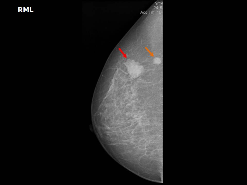
Mammography:
 |  |
 |  |
| Breast composition: | ACR category a (the breasts are almost entirely fatty) | Mammography features: |
| ‣ Location of the lesion: | Right breast, upper inner quadrant at 12-1 oclock, posterior third |
| ‣ Mass: | |
| • Number: | 2 |
| • Size: | 2.0 × 1.4 cm and 0.8 × 0.5 cm |
| • Shape: | Irregular |
| • Margins: | Microlobulated |
| • Density: | High |
| ‣ Calcifications: | |
| • Typically benign: | None |
| • Suspicious: | None |
| • Distribution: | None |
| ‣ Architectural distortion: | None |
| ‣ Asymmetry: | None |
| ‣ Intramammary node: | None |
| ‣ Skin lesion: | None |
| ‣ Solitary dilated duct: | None |
| ‣ Associated features: | None |
Ultrasound:
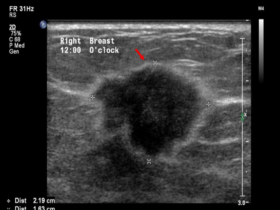 |
| Ultrasound features: Right breast, upper quadrants at 12 oclock | |
| ‣ Mass | |
| • Location: | Right breast, upper quadrants at 12 oclock |
| • Number: | 1 |
| • Size: | 2.2 × 1.6 cm |
| • Shape: | Irregular |
| • Orientation: | Not parallel |
| • Margins: | Angular |
| • Echo pattern: | Hypoechoic |
| • Posterior features: | No posterior features |
| ‣ Calcifications: | None |
| ‣ Associated features: | Internal vascularity |
| ‣ Special cases: | None |
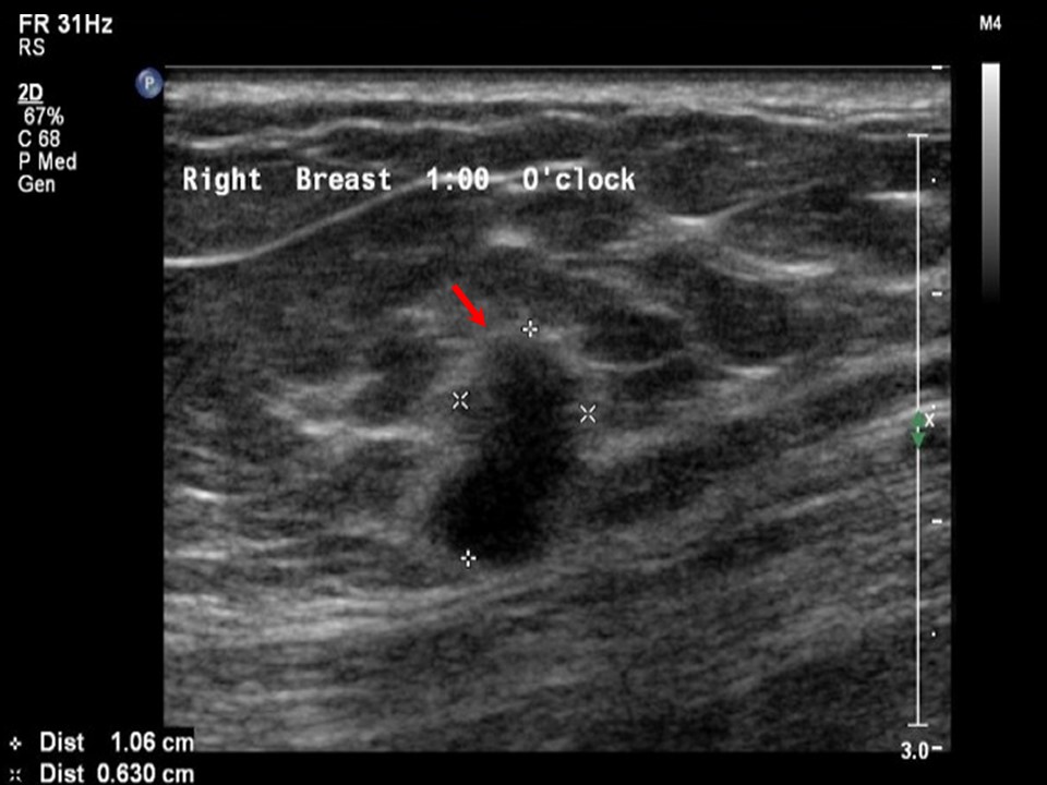 |
| Ultrasound features: Right breast, upper inner quadrant at 1 oclock | |
| ‣ Mass | |
| • Location: | Right breast, upper inner quadrant at 1 oclock |
| • Number: | 1 |
| • Size: | 1.0 × 0.6 cm |
| • Shape: | Irregular |
| • Orientation: | Not parallel |
| • Margins: | Angular |
| • Echo pattern: | Hypoechoic |
| • Posterior features: | No posterior features |
| ‣ Calcifications: | None |
| ‣ Associated features: | Internal vascularity |
| ‣ Special cases: | None |
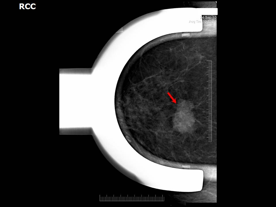
BI-RADS:
BI-RADS Category: 5 (highly suggestive of malignancy)Further assessment:
Further assessment advised: Referral for cytologyCytology:
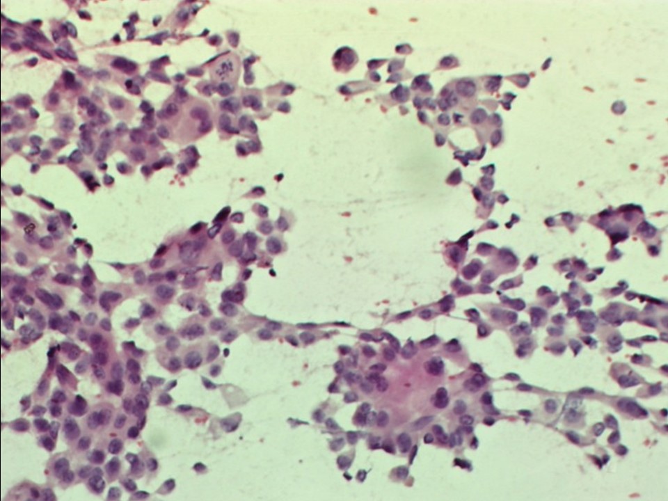 |
| Cytology features: | |
| ‣ Type of sample: | FNAC (solid lesion) |
| ‣ Site of biopsy: | |
| • Laterality: | Right |
| • Quadrant: | Upper inner |
| • Localization technique: | Palpation |
| • Nature of aspirate: | Whitish |
| ‣ Cytological description: | Smears are very cellular and show loosely cohesive sheets of larger ductal epithelial cells with high N:C ratio, pleomorphism, and hyperchromatic nuclei |
| ‣ Reporting category: | Malignant |
| ‣ Diagnosis: | Carcinoma |
| ‣ Comments: | None |
Histopathology:
MRM
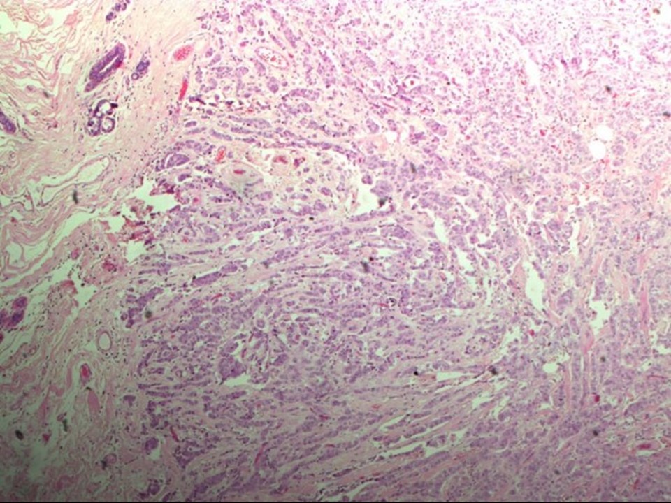 | 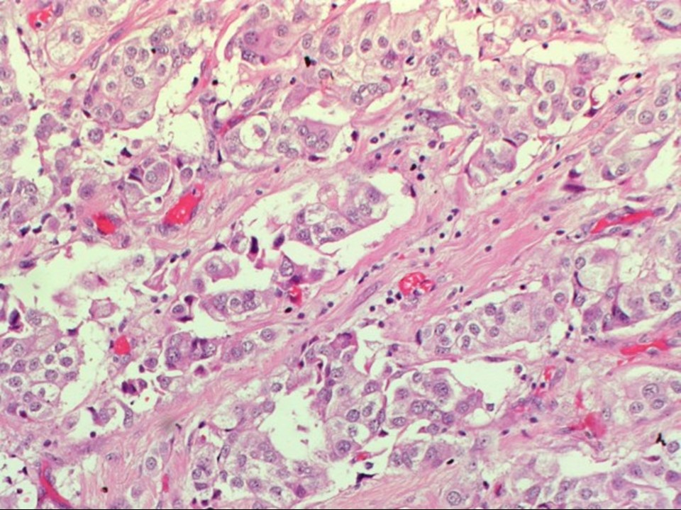 |
| Histopathology features: | |
| ‣ Specimen type: | MRM |
| ‣ Laterality: | Right |
| ‣ Macroscopy: | On serial sectioning, two nodules 2.2 × 1.6 × 1.0 cm and 1.0 × 0.8 × 0.4 cm were seen 4.0 cm apart. Both were well circumscribed and firm in consistency |
| ‣ Histological type: | Invasive breast carcinoma of no special type |
| ‣ Histological grade: | Grade 3 (3 + 3 + 2 = 8) |
| ‣ Mitosis: | 14 |
| ‣ Maximum invasive tumour size: | Largest 2.2 cm (another smaller at 1.0 cm) |
| ‣ Lymph node status: | 0/26 |
| ‣ Peritumoural lymphovascular invasion: | Not identified |
| ‣ DCIS/EIC: | Solid and cribriform intermediate grade |
| ‣ Margins: | Free of tumour |
| ‣ Pathological stage: | pT2(2)N0 |
| ‣ Biomarkers: | |
| ‣ Comments: |
Case summary:
| Postmenopausal woman presented with lump in the right breast. Diagnosed as right breast carcinoma (multifocal), BI-RADS 5 on imaging, as breast carcinoma on cytology, and as invasive breast carcinoma of no special type, pT2(2)N0 on histopathology. |
Learning points:
|