


Home / Training / Manuals / Atlas of breast cancer early detection / Learning
.png)
Click on the pictures to magnify and display the legends

Click on this icon to display a case study
Atlas of breast cancer early detection
Filter by language: English / РусскийBreast imaging Mammography interpretation Mammography lexicon Asymmetry |
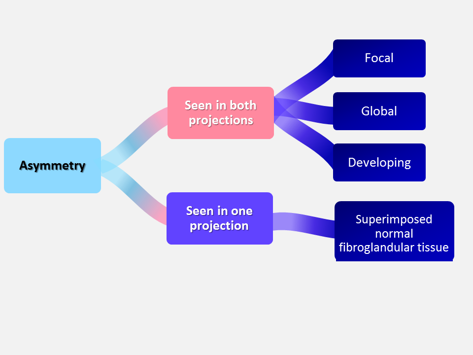
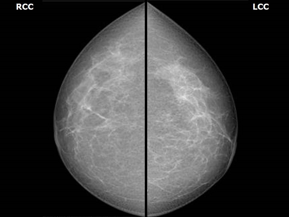
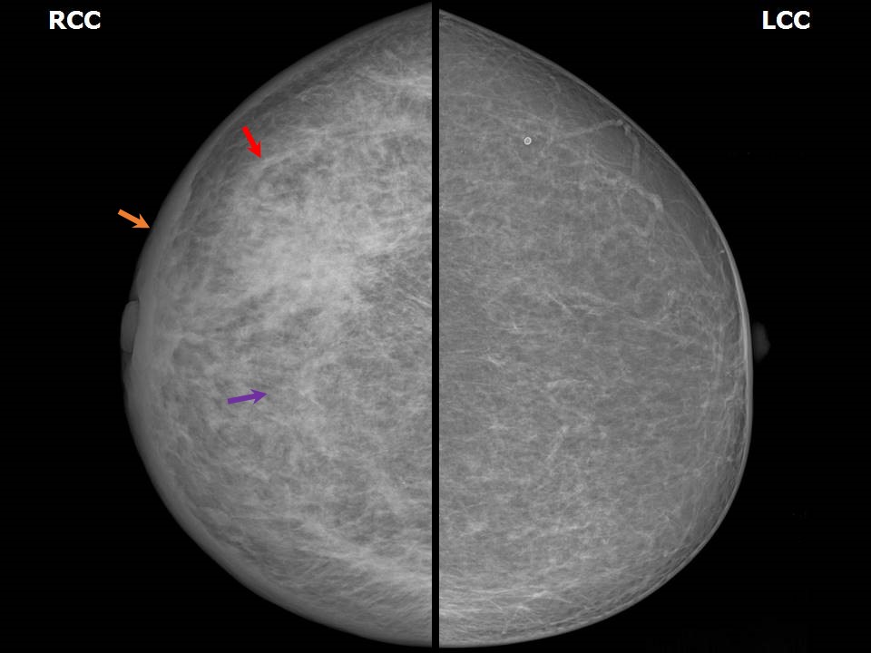
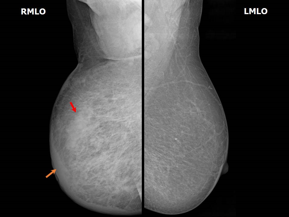
The breast parenchyma shows bilaterally symmetrical density on the mammogram. Although the breast parenchymal density seen on mammography is variable, as described in earlier sections, the symmetry is maintained. Normal mammogram: Symmetrical glandular parenchyma Asymmetry Asymmetry is defined as the loss of the normal symmetrical glandular parenchymal pattern seen on mammography. It may be caused by a constitutional difference between the two breasts, which is a developmental change where there is no underlying abnormality to cause the asymmetry. It may also be caused by variations in the compression and positioning of the breasts during mammography. Constitutional asymmetry Constitutionally asymmetrical breast tissue:
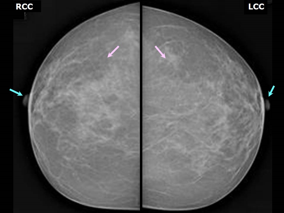
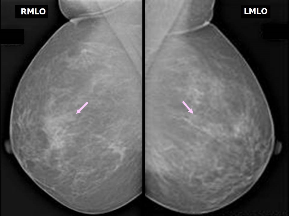
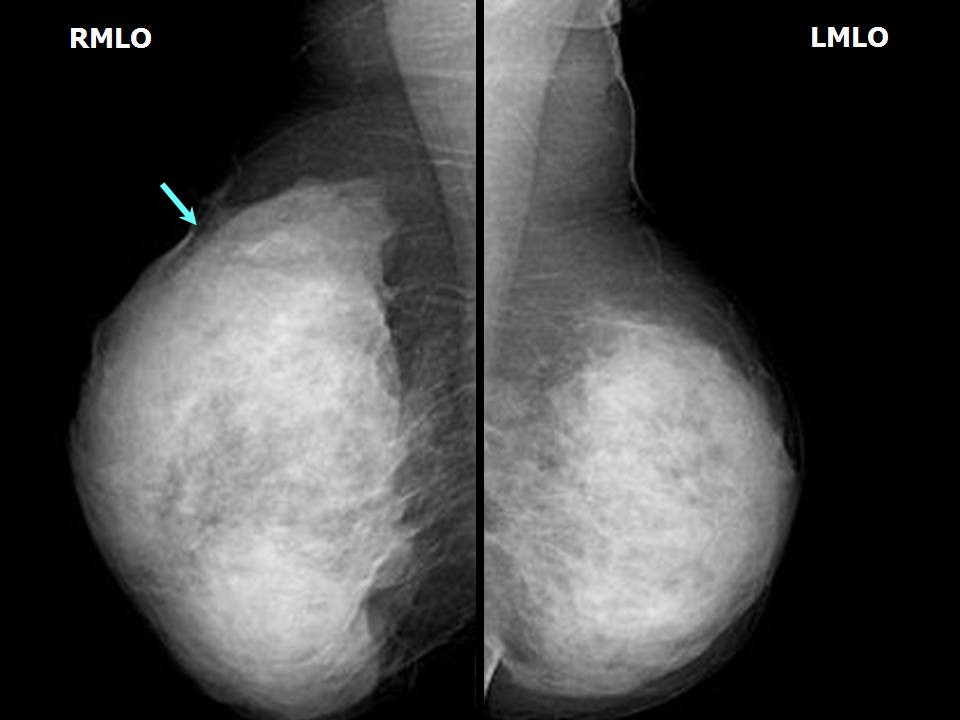
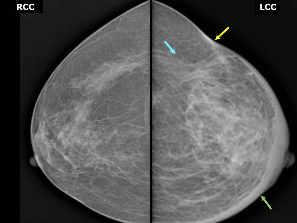
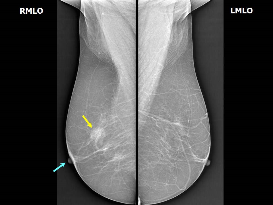
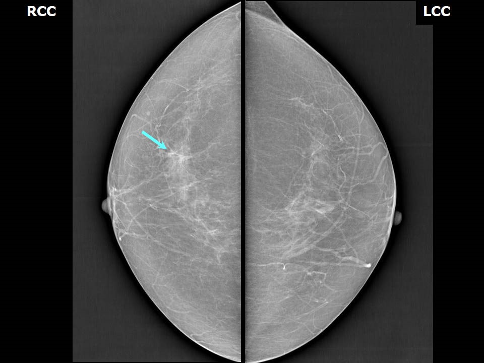
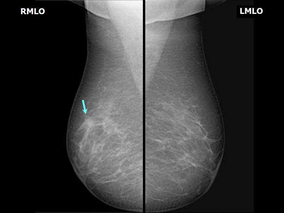
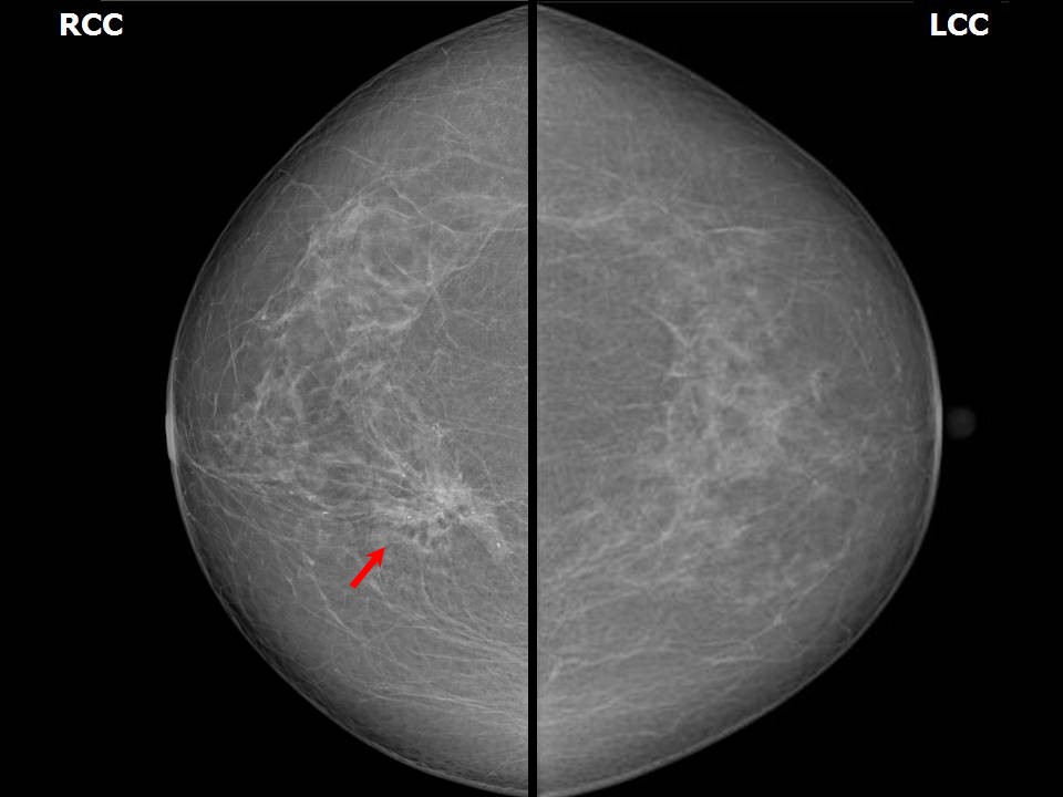
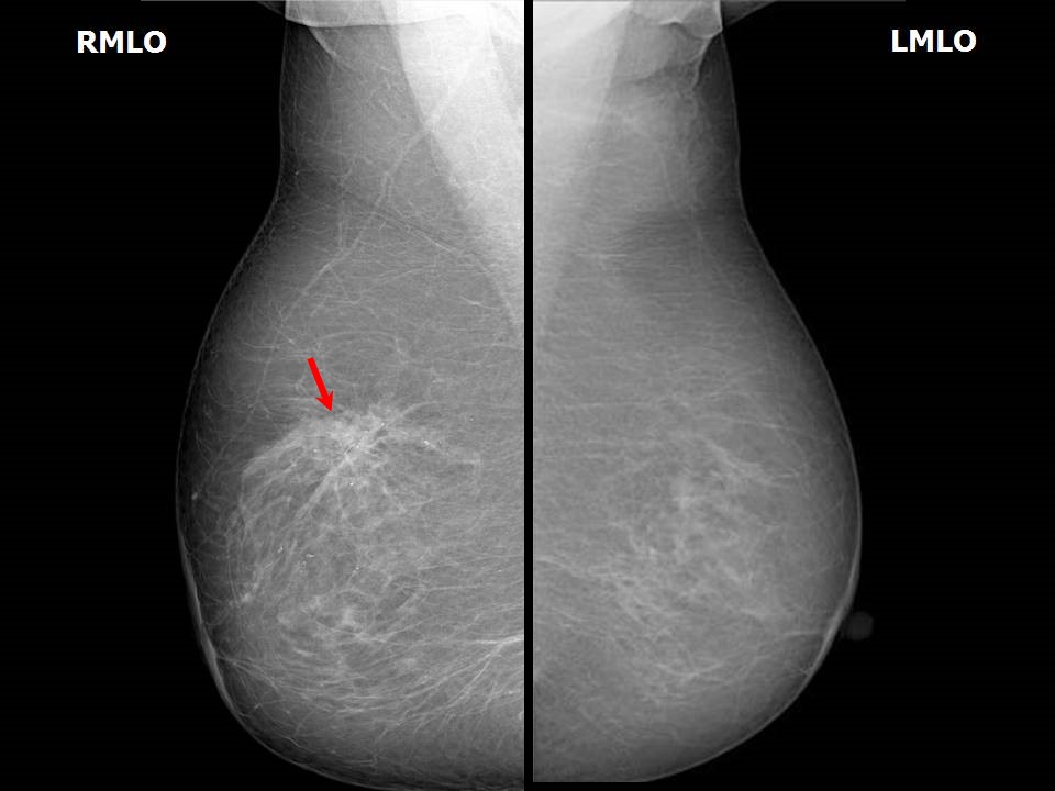
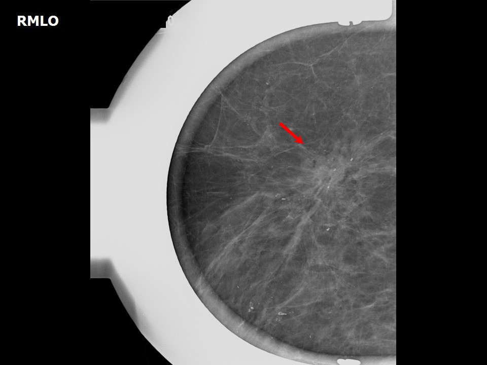
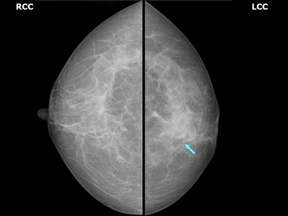
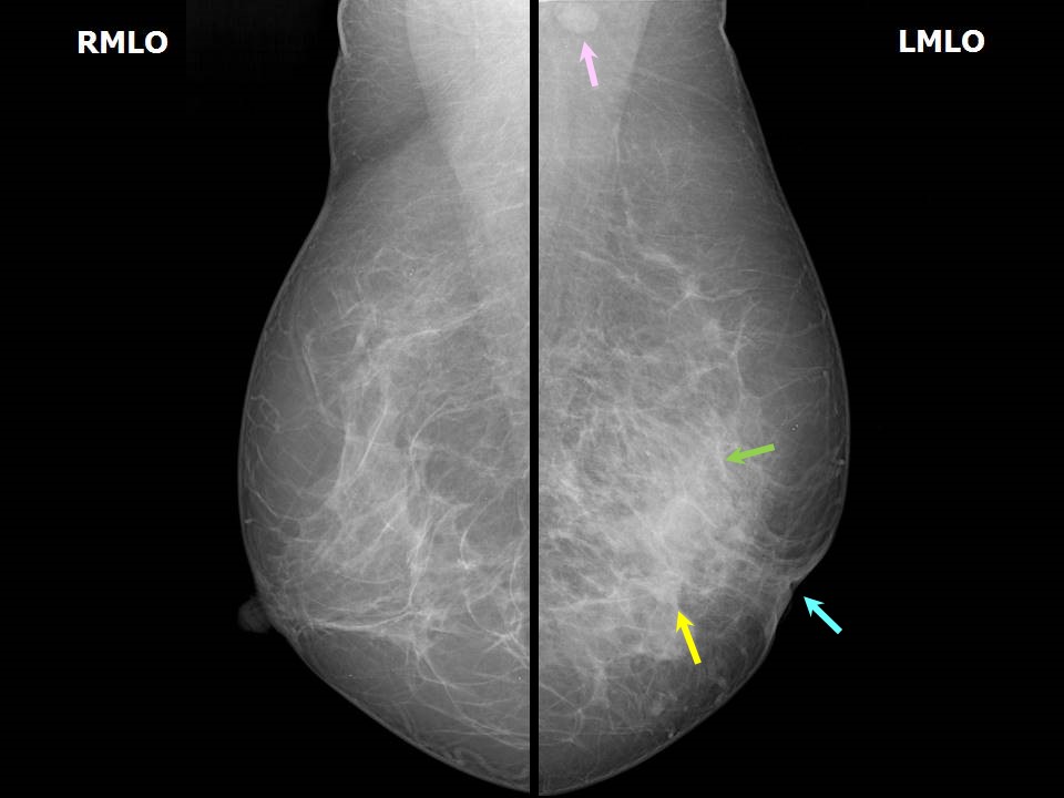
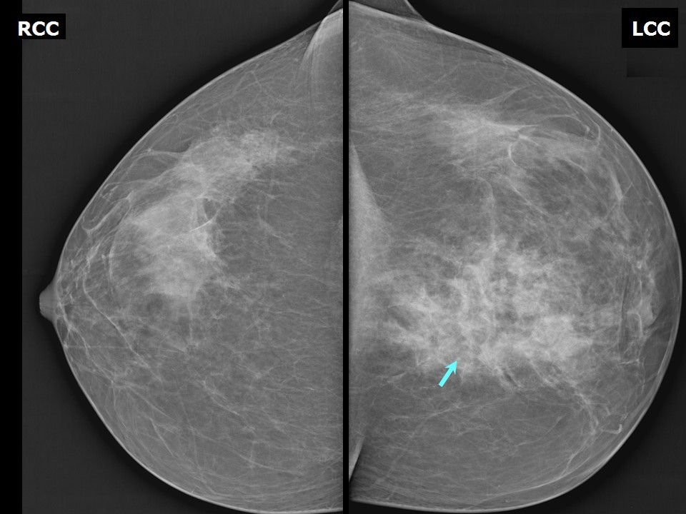
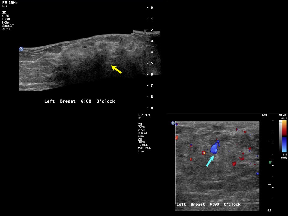
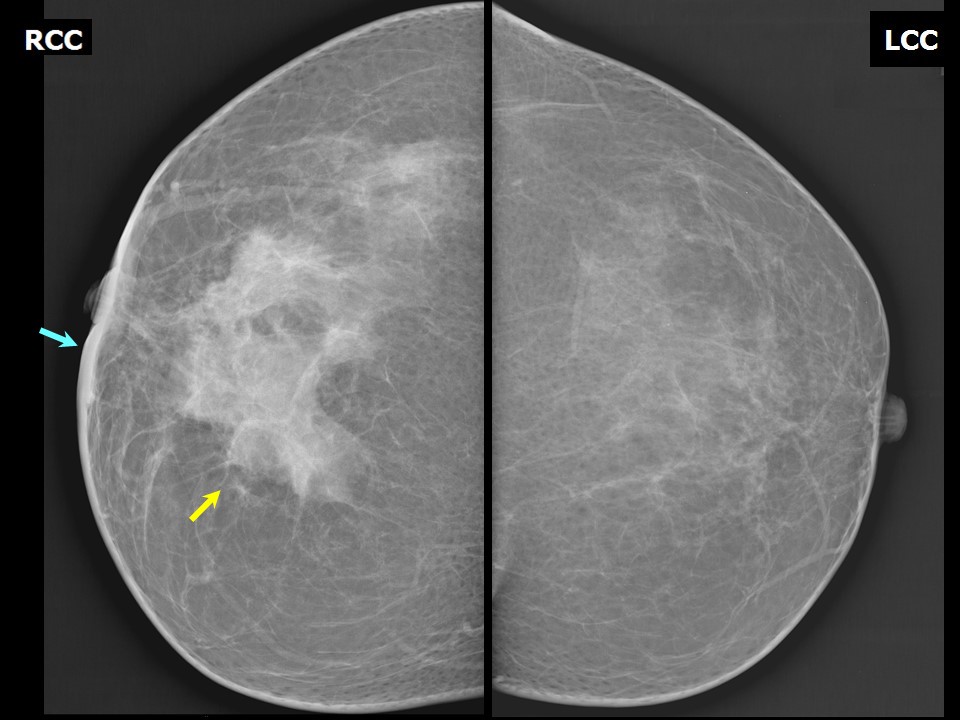
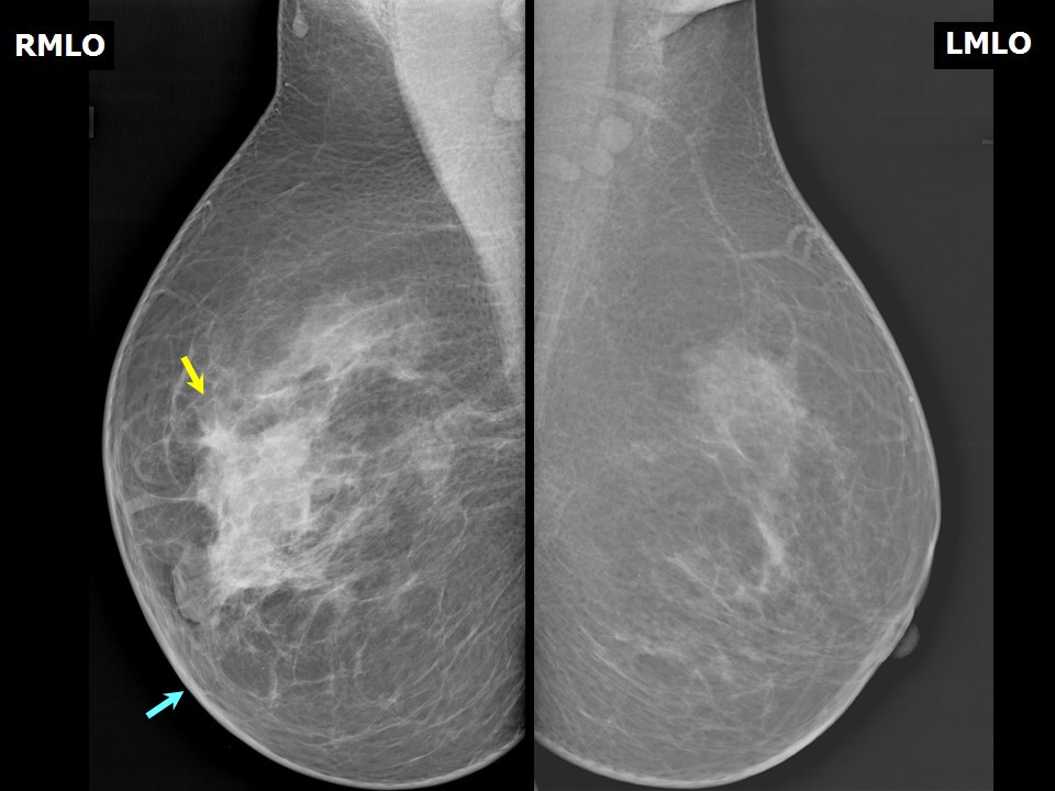
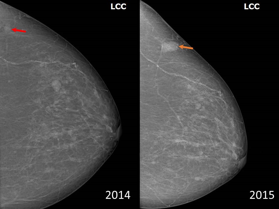
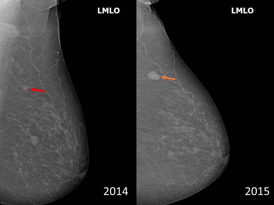
Postsurgical asymmetry Asymmetry may be caused by earlier interventions such as surgical excision or biopsy. Evaluation of asymmetry Asymmetry visible on one projection If the area of fibroglandular tissue is visible on only one mammographic projection, the cause may be summation of normal breast tissue as discussed earlier. A detailed history and the absence of positive findings on thorough clinical examination and ultrasound of the breasts will help to confirm that the asymmetry is a simple variant of normal. A short-interval follow-up to further ascertain the absence of pathology in the region of asymmetry should be followed by regular follow-up for 3 years to document stability. Asymmetry on two projections Focal asymmetry visible on two projections is a real finding and not a summation shadow. This needs to be differentiated from a mass. Further evaluation of a focal asymmetry is done in a similar manner to that for a mass lesion. Spot compression magnification views or views obtained by changing the angle of the gantry can be used to confirm the morphology. Global asymmetry Global asymmetry consists of asymmetry involving at least one quarter of the breast. It is commonly seen in women who have symptoms of breast inflammation, puerperal mastitis, or breast trauma. Breast oedema as a result of infection or inflammation or inflammatory carcinoma causes global asymmetry. These patients are usually symptomatic with a positive CBE. Asymmetry may also be caused by earlier interventions such as surgical excision or biopsy. If the area of asymmetry is visible on only one mammographic projection, the cause may be summation of normal breast tissue as discussed earlier. Global asymmetry with breast inflammation .Global asymmetry with non-puerperal mastitis .Global asymmetry with history of breast trauma .Global asymmetry with inflammatory carcinoma .A new asymmetry which was not seen on earlier mammogram or an increase in size and density of the asymmetry seen on previous mammogram, is labelled as developing asymmetry.
This asymmetry includes a finding that has changed over time and thus is of high suspicion. This term is unique to mammography and the finding can be extremely subtle. Thus all prior mammograms arranged in year follow-up sequence and eye-balling each view helps identify the new asymmetry. Change overtime in the same patient Abnormalities identified on breast ultrasonography (US) or magnetic resonance (MRI) are not called as developing asymmetries. |


Click on the pictures to magnify and display the legends
Click on this icon to display a case study
25 avenue Tony Garnier CS 90627 69366, LYON CEDEX 07 France - Tel: +33 (0)4 72 73 84 85
© IARC 2025 - Terms of use - Privacy Policy.
© IARC 2025 - Terms of use - Privacy Policy.