


Home / Training / Manuals / Atlas of breast cancer early detection / Cases
Atlas of breast cancer early detection
Filter by language: English / Русский
Go back to the list of case studies
.png) Click on the pictures to magnify and display the legends
Click on the pictures to magnify and display the legends
| Case number: | 117 |
| Age: | 60 |
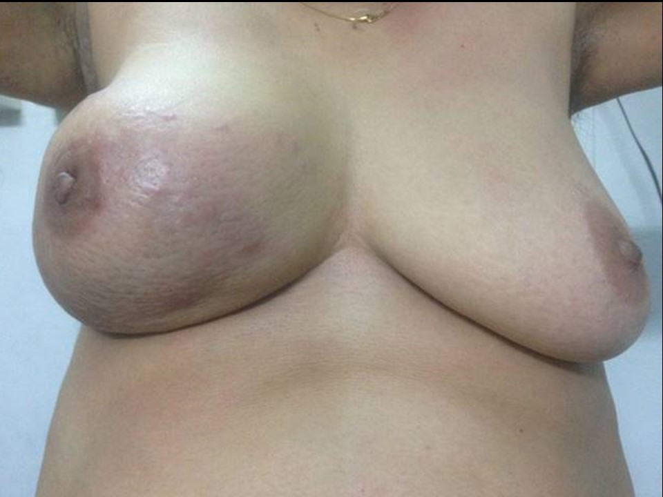
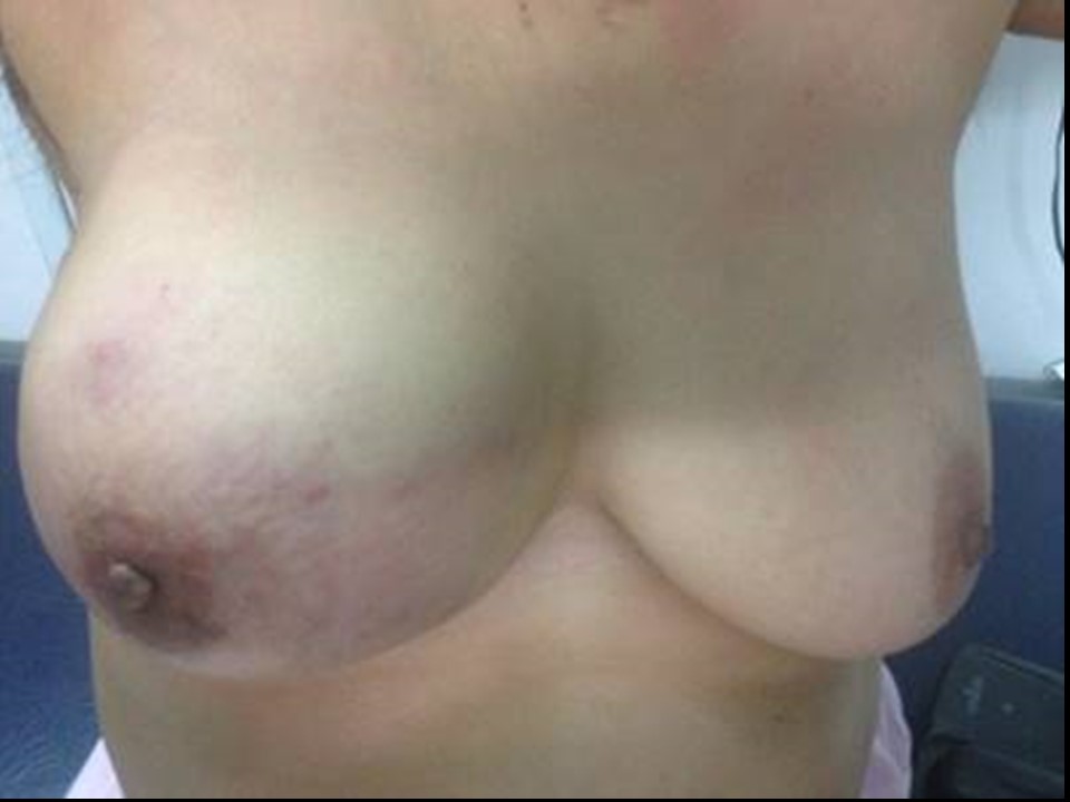
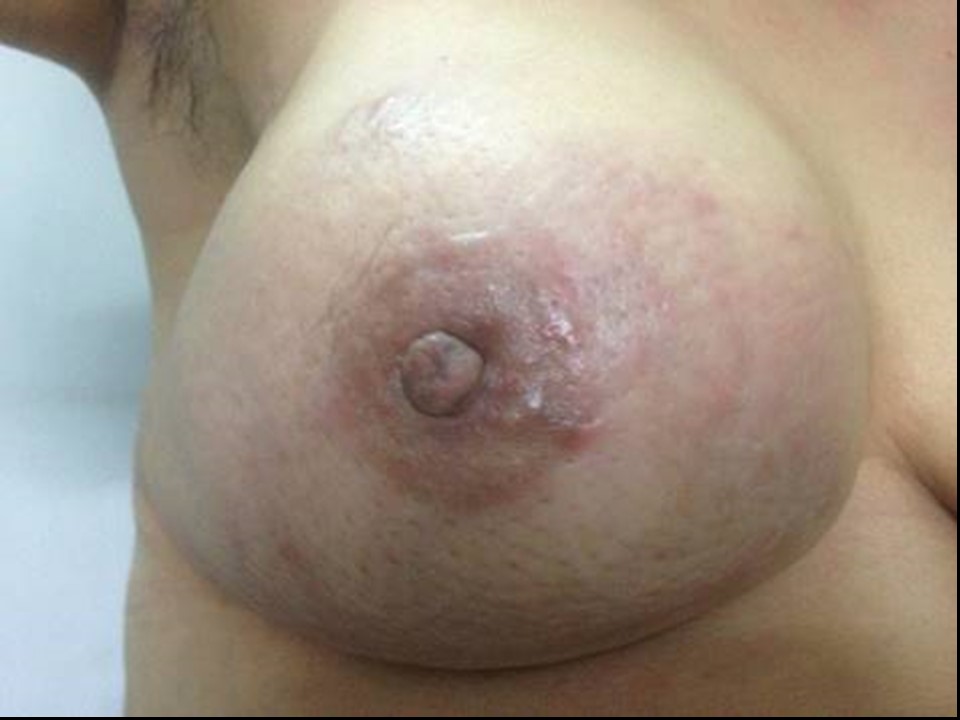
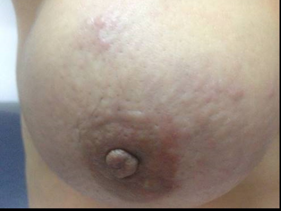
| Clinical presentation: | Postmenopausal woman with increased risk because of a family history of breast cancer presented with increased size of the breast with pain. Examination revealed warmth and redness over the right breast with a large hard lump 8 cm in diameter. The lump was not fixed to the skin or the chest wall but she had nipple retraction on the same side. |
 |  |
 |  |
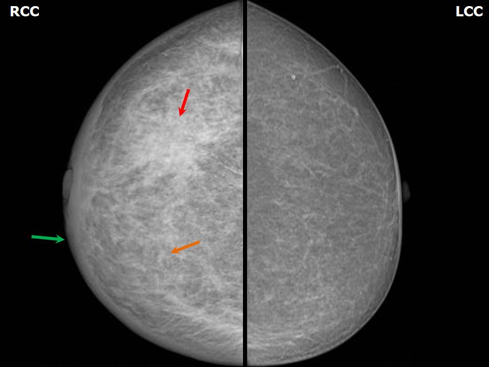
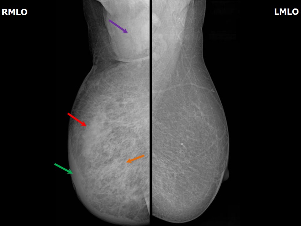
Mammography:
 |  |
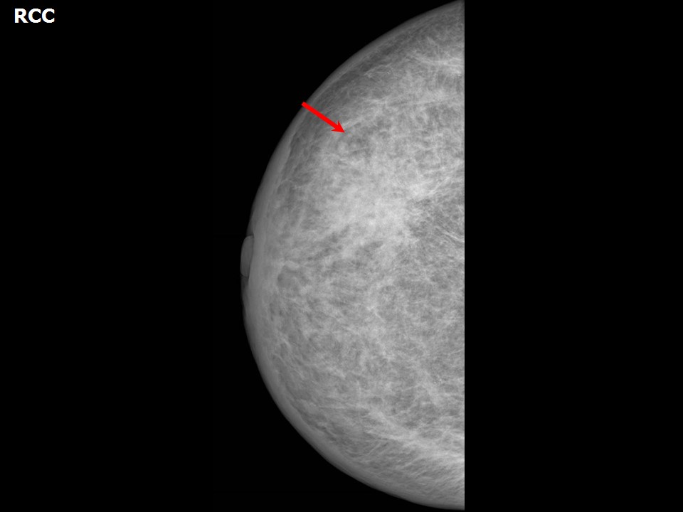 | 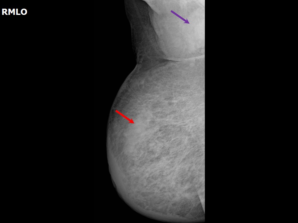 |
| Breast composition: | ACR category b (there are scattered areas of fibroglandular density) | Mammography features: |
| ‣ Location of the lesion: | Right breast, upper outer quadrant at 10 oclock, middle third |
| ‣ Mass: | |
| • Number: | 1 |
| • Size: | 4.8 × 4.4 cm |
| • Shape: | Irregular |
| • Margins: | Spiculated |
| • Density: | High |
| ‣ Calcifications: | |
| • Typically benign: | None |
| • Suspicious: | None |
| • Distribution: | None |
| ‣ Architectural distortion: | None |
| ‣ Asymmetry: | Focal |
| ‣ Intramammary node: | None |
| ‣ Skin lesion: | None |
| ‣ Solitary dilated duct: | None |
| ‣ Associated features: | Skin thickening, trabecular thickening, metastatic, axillary lymphadenopathy, and architectural distortion |
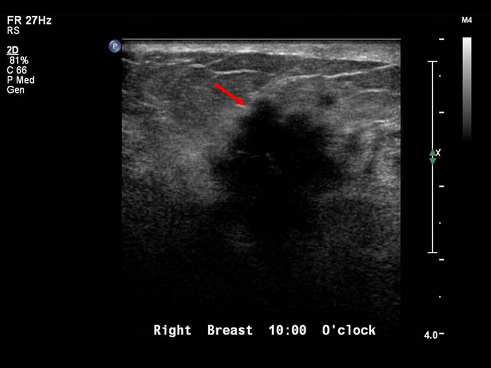
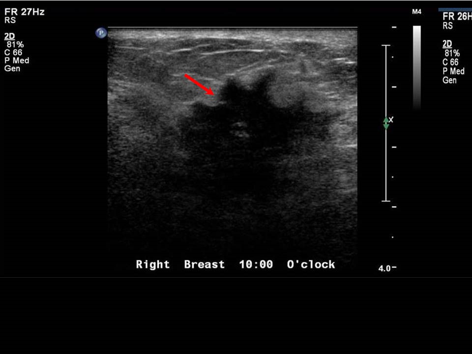
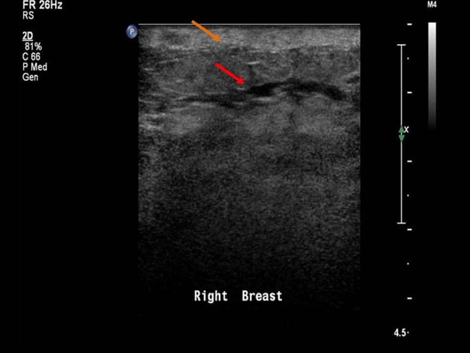
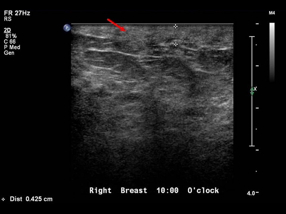
Ultrasound:
 |  |
 |  |
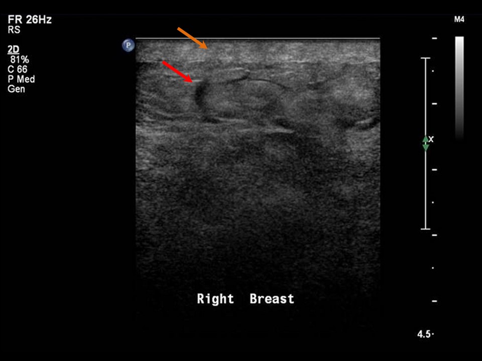 | 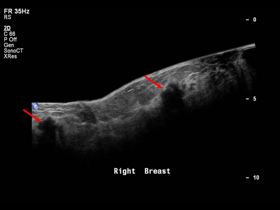 |
| Ultrasound features: Right breast, upper outer quadrant at 10 oclock | |
| ‣ Mass | |
| • Location: | Right breast, upper outer quadrant at 10 oclock |
| • Number: | 1 |
| • Size: | 4.0 × 3.5 cm |
| • Shape: | Irregular |
| • Orientation: | Not parallel |
| • Margins: | Spiculated |
| • Echo pattern: | Hypoechoic |
| • Posterior features: | Posterior shadowing |
| ‣ Calcifications: | None |
| ‣ Associated features: | Architectural distortion, skin thickening (5 mm), oedema, internal vascularity, and enlarged right axillary lymph nodes with thickened cortex |
| ‣ Special cases: | None |
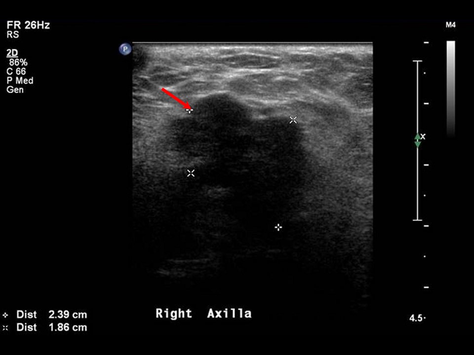 | 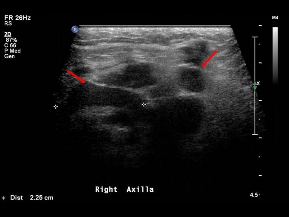 |
| Ultrasound features: Right axillary tail | |
| ‣ Mass | |
| • Location: | Right axillary tail |
| • Number: | 1 |
| • Size: | 2.4 × 1.9 cm |
| • Shape: | Irregular |
| • Orientation: | Not parallel |
| • Margins: | Spiculated |
| • Echo pattern: | Hypoechoic |
| • Posterior features: | Posterior shadowing |
| ‣ Calcifications: | None |
| ‣ Associated features: | Internal vascularity and enlarged right axillary lymph nodes |
| ‣ Special cases: | None |
BI-RADS:
BI-RADS Category: 5 (highly suggestive of malignancy)Further assessment:
Further assessment advised: Referral for cytologyCytology:
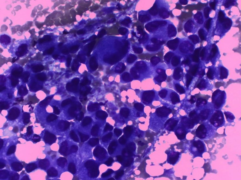 |
| Cytology features: | |
| ‣ Type of sample: | FNAC |
| ‣ Site of biopsy: | |
| • Laterality: | Right |
| • Quadrant: | Upper outer |
| • Localization technique: | Palpation |
| • Nature of aspirate: | whitish |
| ‣ Cytological description: | Smears are very cellular and have loosely cohesive clusters of malignant ductal cells. These cells have large irregular nuclei with prominent nucleoli |
| ‣ Reporting category: | Malignant |
| ‣ Diagnosis: | Carcinoma |
| ‣ Comments: | None |
Case summary:
| Postmenopausal woman presented with a rapidly increasing painful right breast lump. Diagnosed as inflammatory right breast carcinoma with breast oedema and skin thickening, BI-RADS 5 on imaging and as breast carcinoma on cytology. |
Learning points:
|