


Home / Training / Manuals / Atlas of breast cancer early detection / Cases
Developing asymmetries Go back to the list of case studies
Total record number: 12
Atlas of breast cancer early detection
Filter by language: English / РусскийDeveloping asymmetries Go back to the list of case studies
Case:097 | 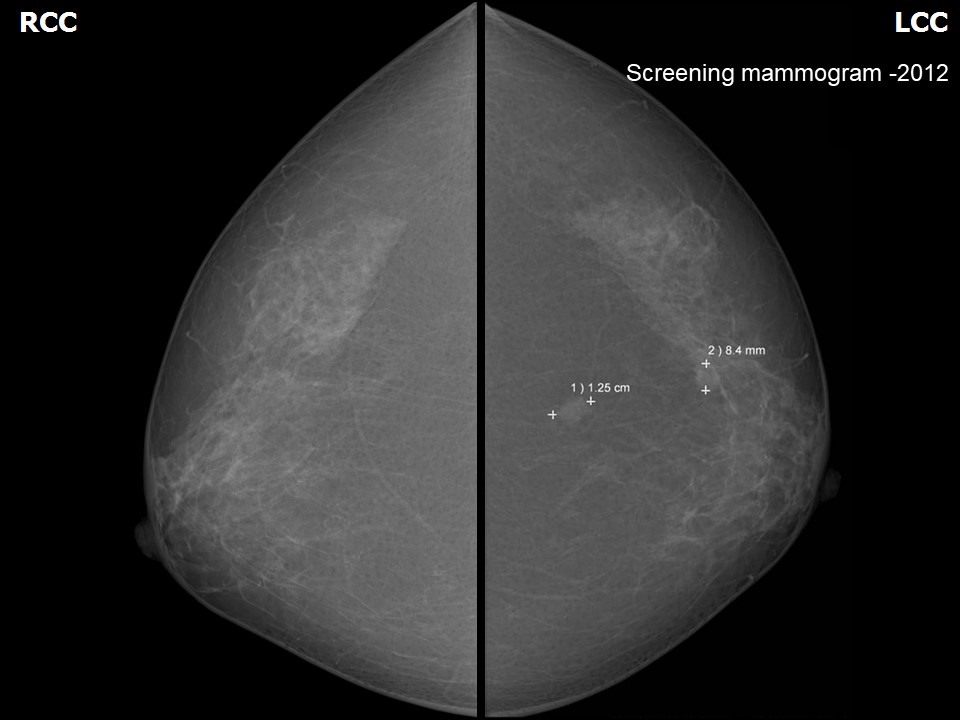 | 2012: 4C High suspicion for malignancy | ||
Case:098 | 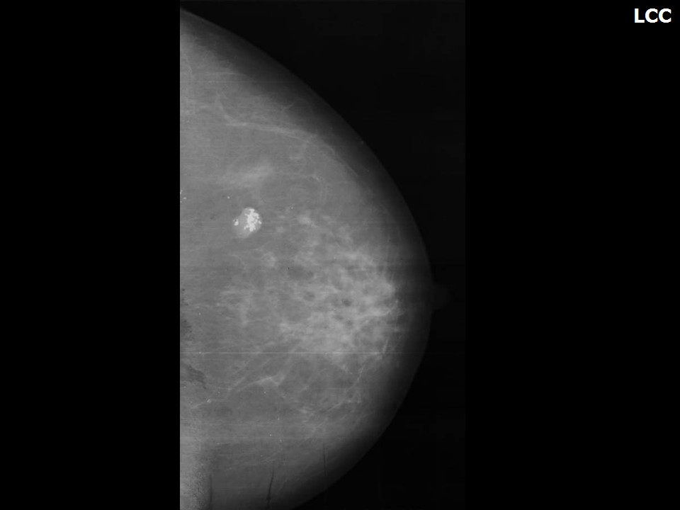 | 2013: 5 (Highly Suggestive of Malignancy) | ||
Case:099 | 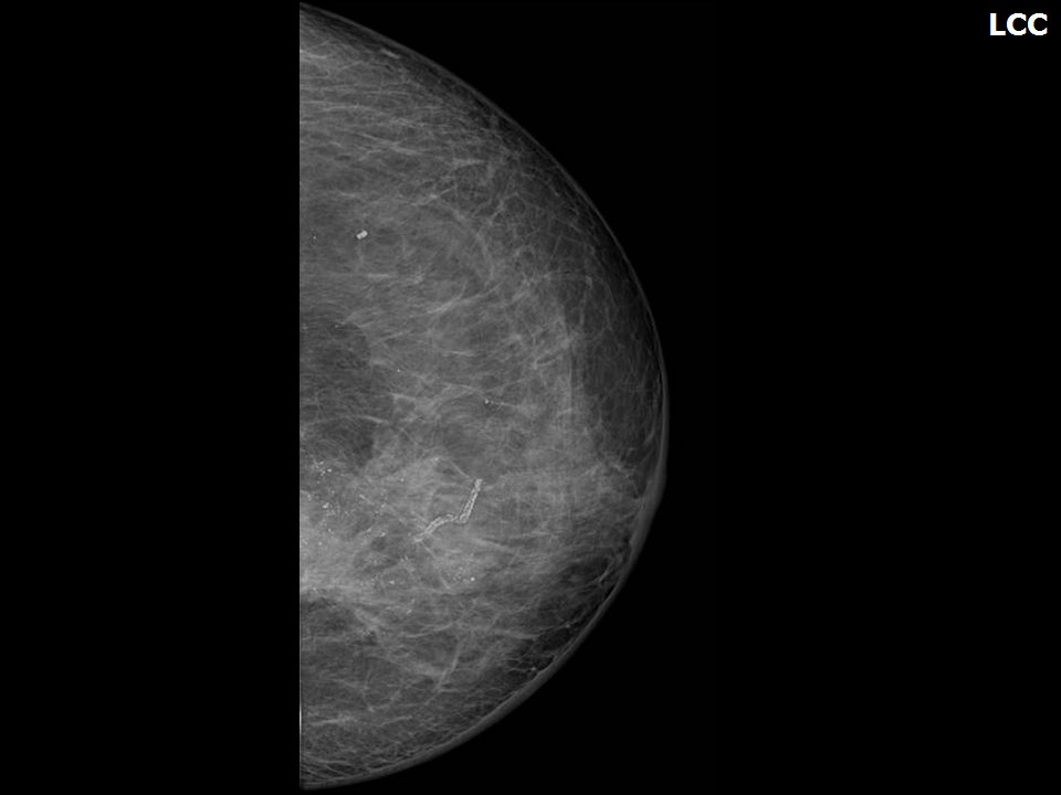 | |||
Case:126 | 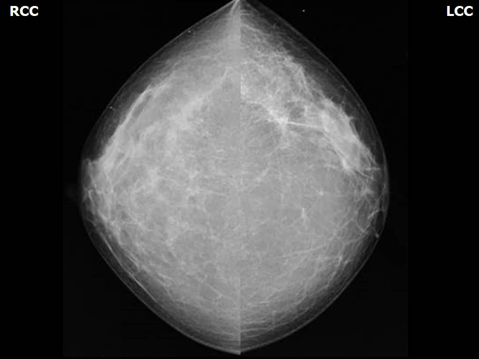 | 2018: Left BCS, post operative breast: 4C High suspicion for malignancy | ||
Case:100 | 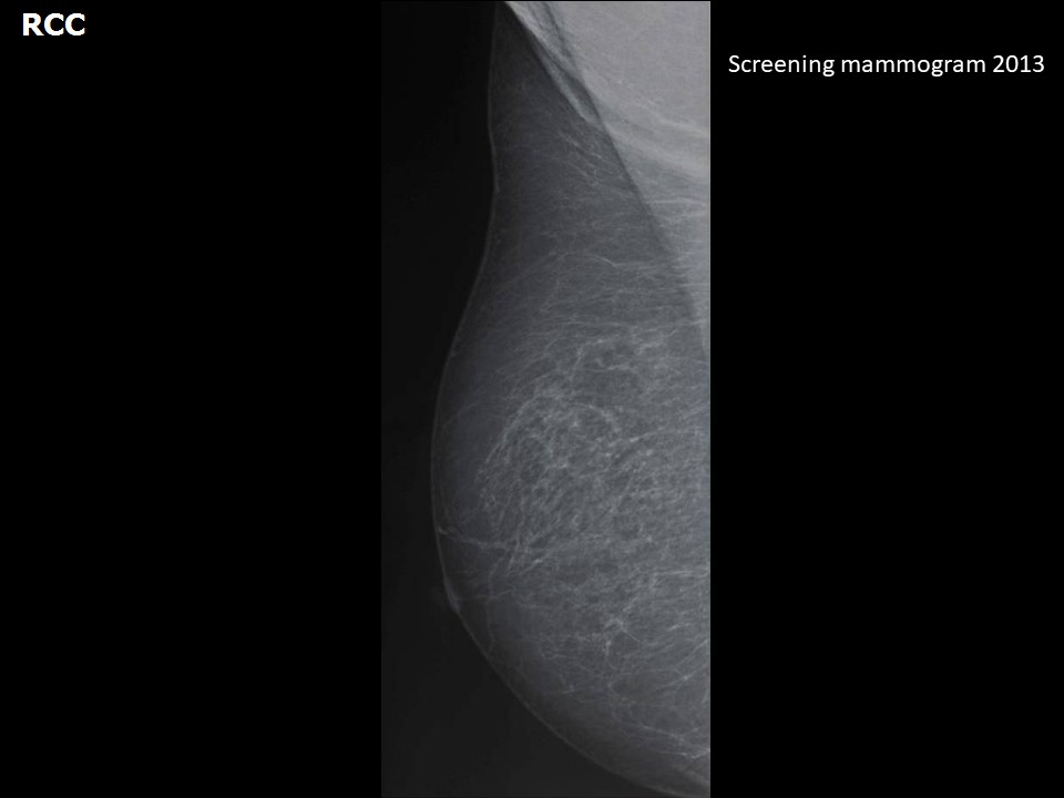 | 2014: 5 (Highly Suggestive of Malignancy) | ||
Case:102 | 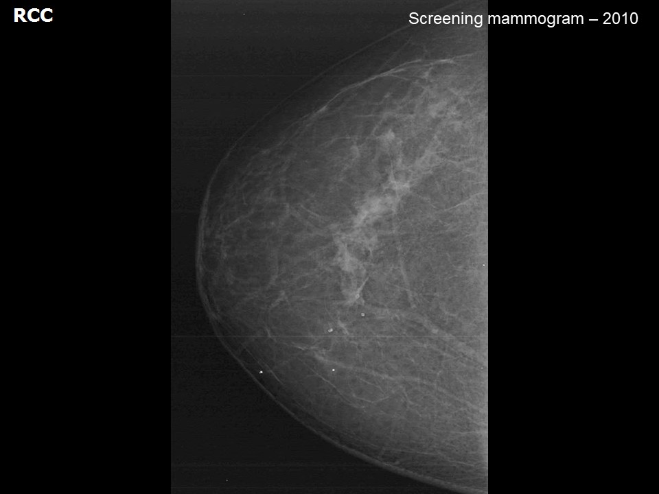 | 2012: 5 (Highly Suggestive of Malignancy) | ||
Case:103 | 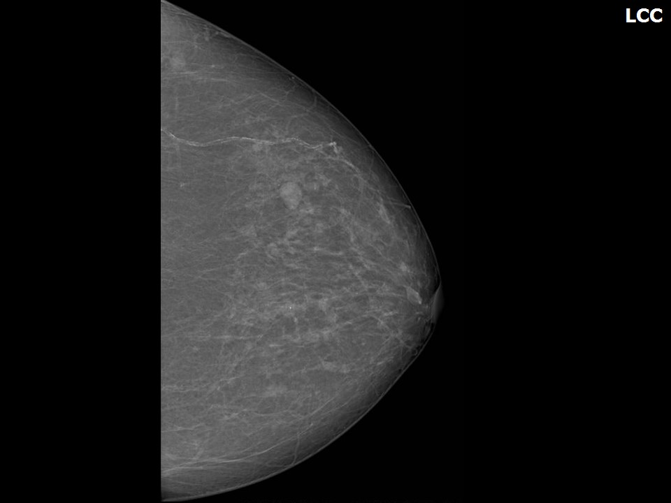 | 2015: 4C High suspicion for malignancy | ||
Case:104 | 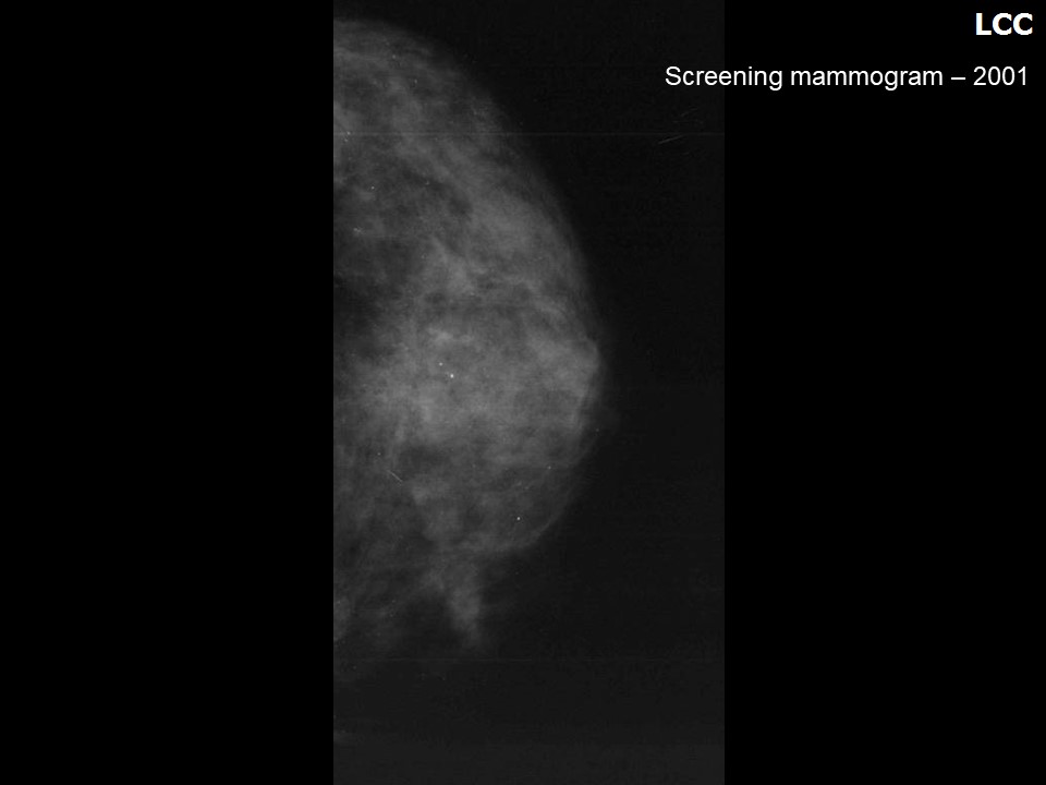 | 2002: 2 (Benign) 2010: 5 (Highly Suggestive of Malignancy) | ||
Case:105 | 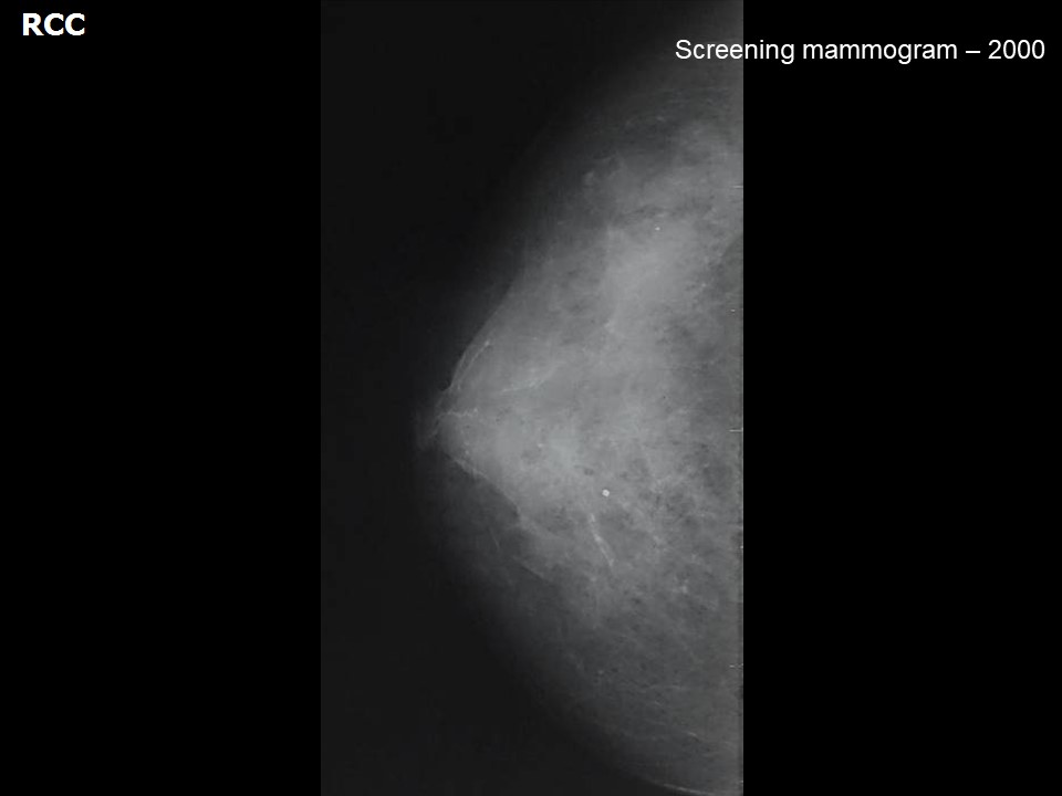 | 2014: 4B Moderate suspicion for malignancy | ||
Case:113 | 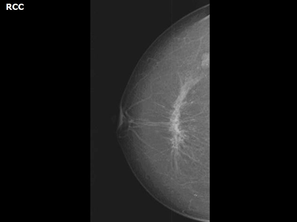 | 2016: 4C High suspicion for malignancy | ||
Case:115 | 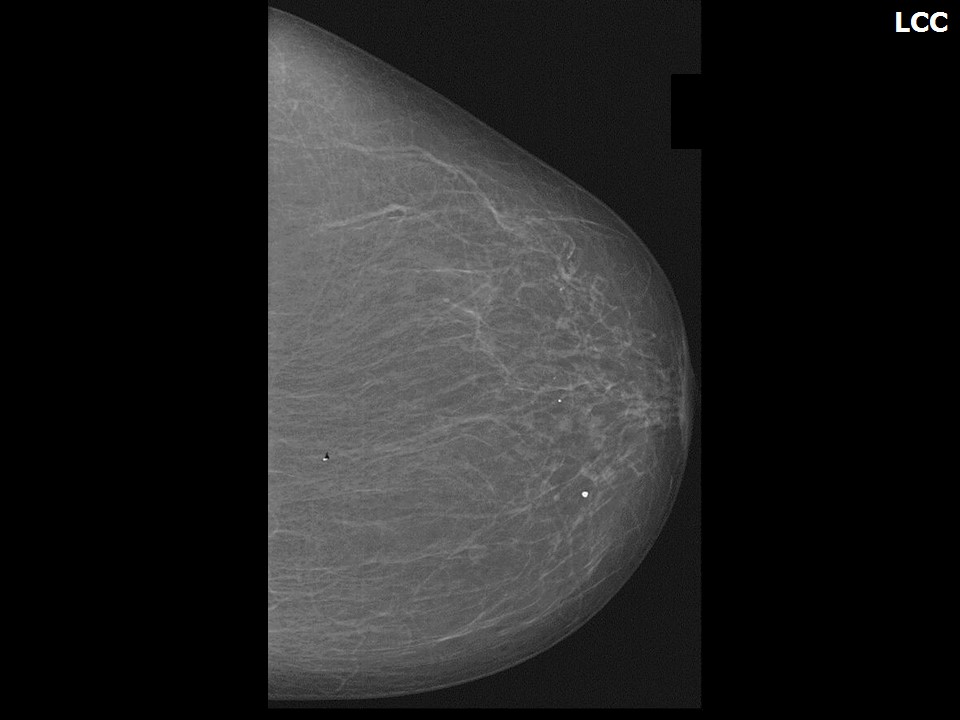 | 2016: 4C High suspicion for malignancy | ||
Case:120 | 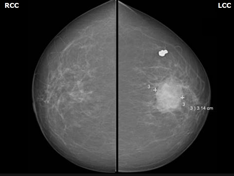 | 2013, left: 5 (Highly Suggestive of Malignancy) 2014, right: 4B Moderate suspicion for malignancy |