


Home / Training / Manuals / Atlas of breast cancer early detection / Cases
Go back to the list of case studies
Total record number: 186
Atlas of breast cancer early detection
Filter by language: English / Русский Go back to the list of case studies
Case:173 |  | |||
Case:172 |  | |||
Case:170 |  | |||
Case:171 |  | |||
Case:174 |  | |||
Case:163 |  | Left BCS, post operative breast: 2 (Benign) | ||
Case:164 |  | |||
Case:175 |  | |||
Case:176 |  | |||
Case:177 |  | |||
Case:178 |  | |||
Case:179 |  | |||
Case:180 |  | |||
Case:181 |  | |||
Case:182 |  | |||
Case:183 |  | right breast: 2 (Benign) | ||
Case:184 |  | |||
Case:185 |  | |||
Case:186 |  | |||
Case:001 |  | |||
Case:089 |  | |||
Case:006 |  | |||
Case:008 |  | |||
Case:061 |  | |||
Case:062 |  | |||
Case:065 |  | |||
Case:069 |  | |||
Case:071 |  | |||
Case:075 |  | |||
Case:059 |  | |||
Case:050 |  | |||
Case:049 |  | |||
Case:047 |  | |||
Case:044 |  | |||
Case:040 |  | |||
Case:035 |  | |||
Case:010 |  | |||
Case:013 |  | |||
Case:015 |  | |||
Case:025 |  | |||
Case:027 |  | |||
Case:031 |  | Follow-up: | ||
Case:032 |  | |||
Case:033 |  | |||
Case:078 |  | |||
Case:080 |  | September 2012: 6 (Known Biopsy-Proven Malignancy) December 2012: 6 (Known Biopsy-Proven Malignancy) | ||
Case:082 |  | Left BCS, post operative breast: 2 (Benign) | ||
Case:085 |  | |||
Case:090 |  | |||
Case:097 |  | 2012: 4C High suspicion for malignancy | ||
Case:107 |  | |||
Case:116 |  | |||
Case:118 |  | |||
Case:123 |  | |||
Case:124 |  | |||
Case:125 |  | |||
Case:029 |  | |||
Case:024 |  | |||
Case:131 |  | |||
Case:133 |  | |||
Case:134 |  | |||
Case:135 |  | |||
Case:138 |  | |||
Case:042 |  | |||
Case:012 |  | |||
Case:019 |  | |||
Case:021 |  | |||
Case:026 |  | |||
Case:028 |  | |||
Case:034 |  | |||
Case:165 |  | |||
Case:166 |  | |||
Case:002 |  | |||
Case:009 |  | |||
Case:060 |  | July 2013: 6 (Known Biopsy-Proven Malignancy) | ||
Case:063 |  | |||
Case:066 |  | |||
Case:067 |  | |||
Case:068 |  | |||
Case:076 |  | |||
Case:056 |  | |||
Case:055 |  | |||
Case:051 |  | |||
Case:048 |  | |||
Case:046 |  | |||
Case:045 |  | |||
Case:043 |  | |||
Case:041 |  | |||
Case:036 |  | |||
Case:005 |  | |||
Case:018 |  | |||
Case:072 |  | |||
Case:074 |  | |||
Case:081 |  | September 2014: 6 (Known Biopsy-Proven Malignancy) | ||
Case:083 | 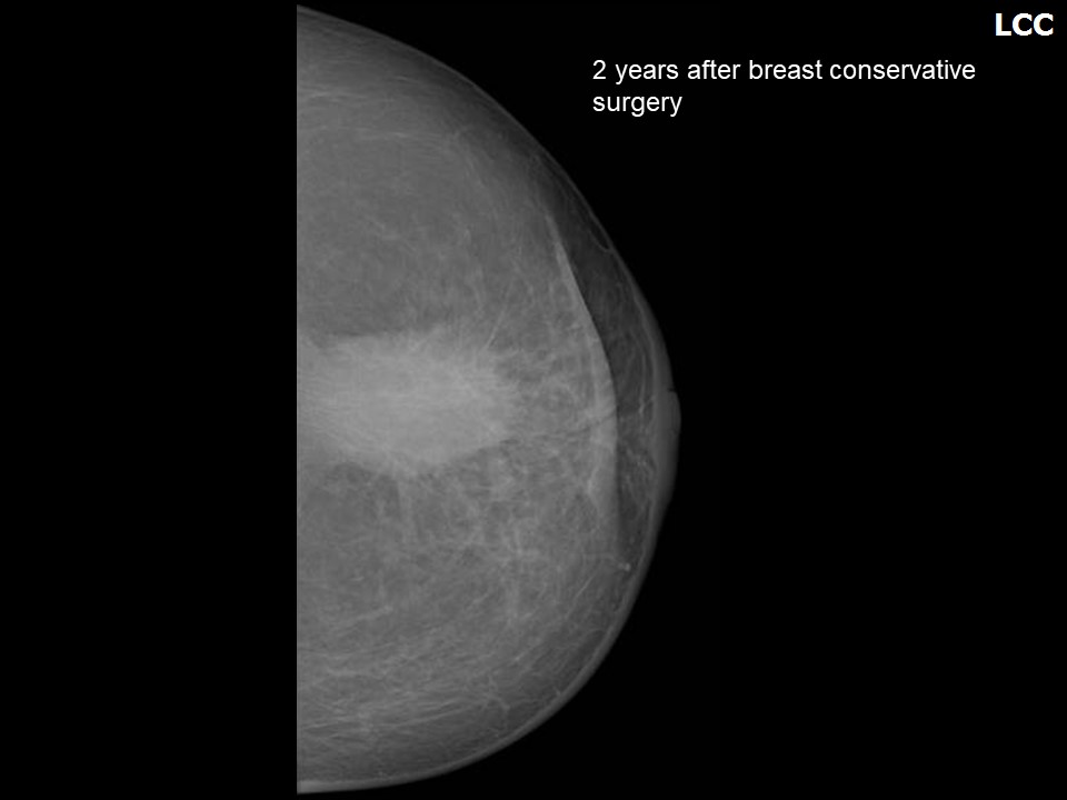 | |||
Case:086 |  | |||
Case:098 |  | 2013: 5 (Highly Suggestive of Malignancy) | ||
Case:108 |  | |||
Case:112 |  | |||
Case:128 |  | |||
Case:130 |  | |||
Case:137 |  | |||
Case:139 |  | |||
Case:091 |  | |||
Case:058 |  | |||
Case:053 |  | |||
Case:014 |  | |||
Case:003 |  | |||
Case:020 |  | |||
Case:147 |  | |||
Case:152 |  | |||
Case:153 |  | |||
Case:157 |  | |||
Case:161 |  | at follow-up (2017): 2 (Benign) at follow-up (2018): 2 (Benign) | ||
Case:169 |  | |||
Case:004 |  | |||
Case:114 |  | |||
Case:121 |  | |||
Case:073 |  | |||
Case:077 |  | |||
Case:057 |  | |||
Case:054 |  | |||
Case:052 |  | |||
Case:039 |  | Right breast: 2 (Benign) | ||
Case:037 |  | |||
Case:079 |  | |||
Case:084 |  | |||
Case:087 |  | |||
Case:092 |  | |||
Case:099 |  | |||
Case:101 |  | |||
Case:111 |  | |||
Case:117 |  | |||
Case:126 |  | 2018: Left BCS, post operative breast: 4C High suspicion for malignancy | ||
Case:127 |  | |||
Case:132 |  | |||
Case:011 |  | |||
Case:016 |  | |||
Case:017 |  | |||
Case:022 |  | |||
Case:154 |  | |||
Case:155 |  | |||
Case:158 |  | |||
Case:162 |  | 2012: 2 (Benign) 2017: 2 (Benign) | ||
Case:007 |  | |||
Case:070 |  | |||
Case:038 |  | |||
Case:093 |  | MRI Bi-RADS: 5 (Highly Suggestive of Malignancy) | ||
Case:095 |  | |||
Case:100 |  | 2014: 5 (Highly Suggestive of Malignancy) | ||
Case:122 |  | |||
Case:136 |  | |||
Case:023 |  | Follow-up: 1 (Negative) | ||
Case:141 |  | |||
Case:143 |  | |||
Case:148 |  | |||
Case:156 |  | |||
Case:167 |  | |||
Case:064 |  | |||
Case:096 |  | |||
Case:102 |  | 2012: 5 (Highly Suggestive of Malignancy) | ||
Case:109 |  | |||
Case:129 |  | |||
Case:030 |  | |||
Case:142 |  | |||
Case:144 |  | |||
Case:149 |  | |||
Case:088 |  | |||
Case:103 |  | 2015: 4C High suspicion for malignancy | ||
Case:110 |  | |||
Case:119 |  | |||
Case:140 |  | |||
Case:150 |  | |||
Case:159 |  | |||
Case:094 |  | |||
Case:104 |  | 2002: 2 (Benign) 2010: 5 (Highly Suggestive of Malignancy) | ||
Case:145 |  | |||
Case:151 |  | |||
Case:160 |  | |||
Case:168 |  | at follow-up (2018): 2 (Benign) | ||
Case:105 |  | 2014: 4B Moderate suspicion for malignancy | ||
Case:106 |  | MRI 2014: 3 (Probably Benign) MRI 2017 and Mammography 2018: 2 (Benign) | ||
Case:146 |  | |||
Case:113 |  | 2016: 4C High suspicion for malignancy | ||
Case:115 |  | 2016: 4C High suspicion for malignancy | ||
Case:120 |  | 2013, left: 5 (Highly Suggestive of Malignancy) 2014, right: 4B Moderate suspicion for malignancy |