


Home / Training / Manuals / Atlas of breast cancer early detection / Cases
Atlas of breast cancer early detection
Filter by language: English / Русский
Go back to the list of case studies
.png) Click on the pictures to magnify and display the legends
Click on the pictures to magnify and display the legends
| Case number: | 171 |
| Age: | 38 |
| Clinical presentation: | Premenopausal woman with average risk of developing breast cancer presented with a painful right breast lump noticed a few days ago. On examination, a mobile palpable lump (3 × 2 cm) with smooth margins was noted in the inferior quadrant of the right breast below the areolar margin. |
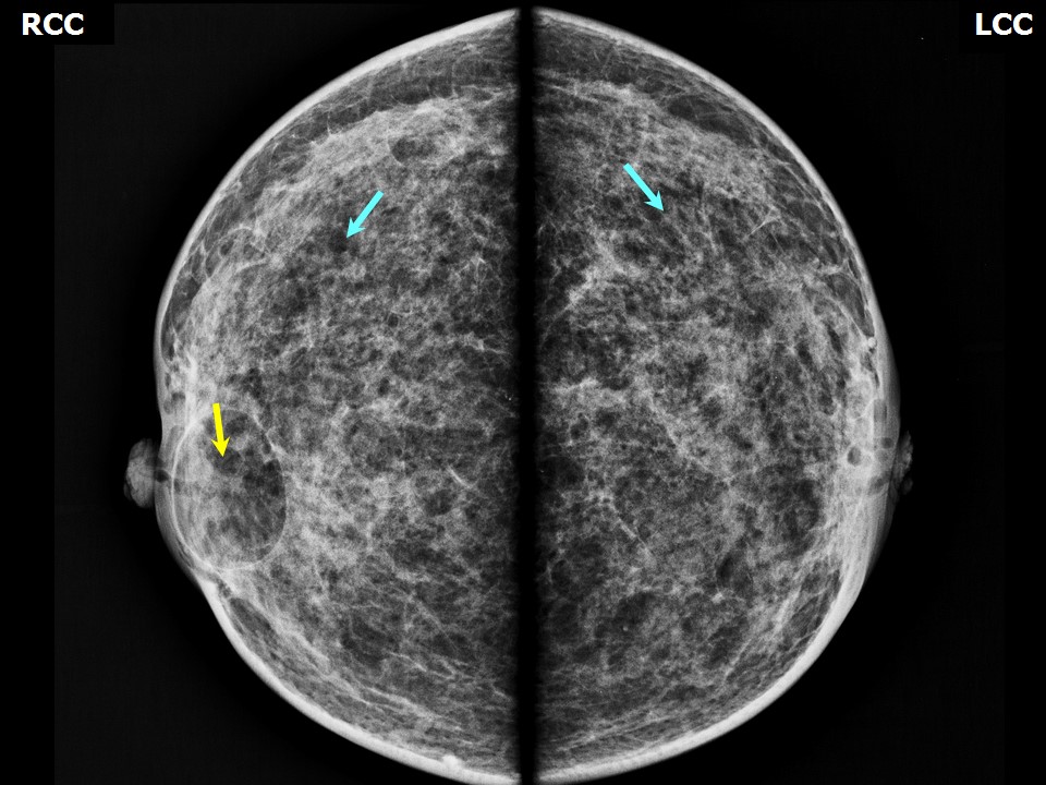
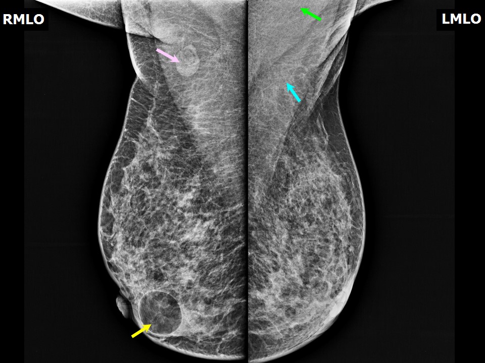
Mammography:
 |  |
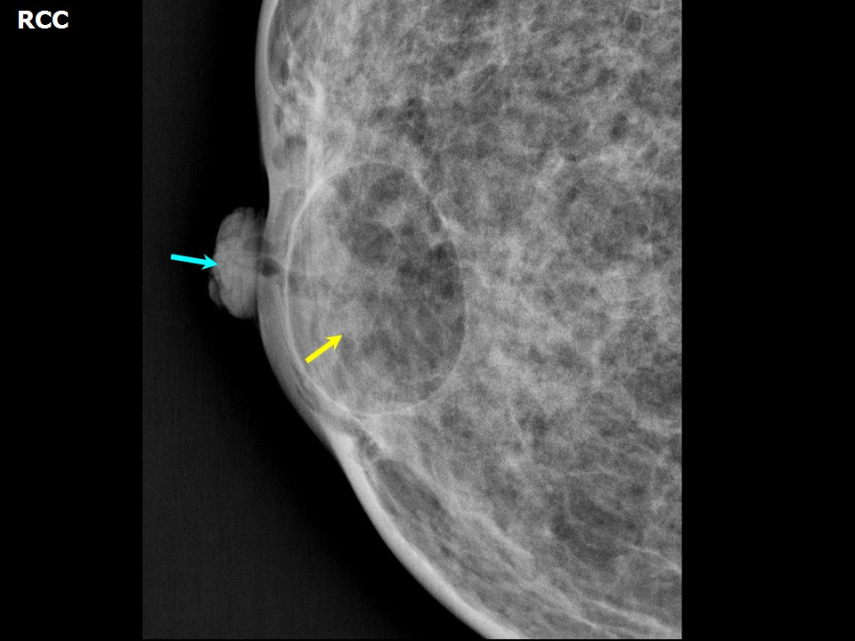 |
| Breast composition: | ACR category c (the breasts are heterogeneously dense, which may obscure small masses) | Mammography features: |
| ‣ Location of the lesion: | Right breast, lower inner quadrant at 5 oclock, subareolar region, anterior third, 0.1 cm from the nipple and at 0.2 cm skin depth |
| ‣ Mass: | |
| • Number: | 1 |
| • Size: | 3.5 × 2.5 cm |
| • Shape: | Oval |
| • Margins: | Circumscribed with perilesional halo |
| • Density: | Fat-containing |
| ‣ Calcifications: | |
| • Typically benign: | None |
| • Suspicious: | None |
| • Distribution: | None |
| ‣ Architectural distortion: | None |
| ‣ Asymmetry: | None |
| ‣ Intramammary node: | None |
| ‣ Skin lesion: | None |
| ‣ Solitary dilated duct: | None |
| ‣ Associated features: | None |
Ultrasound:
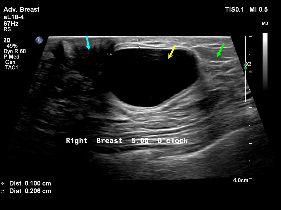 | 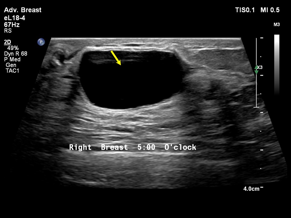 |
| Ultrasound features: Right breast, lower inner quadrant at 5 oclock, subareolar region, 0.1 cm from the nipple and at 0.2 cm skin depth | |
| ‣ Mass | |
| • Location: | Right breast, lower inner quadrant at 5 oclock, subareolar region, 0.1 cm from the nipple and at 0.2 cm skin depth |
| • Number: | 1 |
| • Size: | 3.3 × 1.6 cm |
| • Shape: | Oval |
| • Orientation: | Parallel |
| • Margins: | Circumscribed |
| • Echo pattern: | Anechoic |
| • Posterior features: | No posterior features |
| ‣ Calcifications: | None |
| ‣ Associated features: | None |
| ‣ Special cases: | Fat necrosis |
BI-RADS:
BI-RADS Category: 2 (benign)Further assessment:
Further assessment advised: Referral for cytologyCytology:
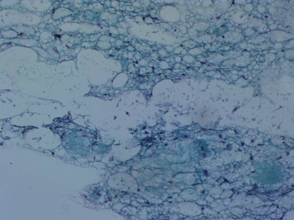 |
| Cytology features: | |
| ‣ Type of sample: | FNAC |
| ‣ Site of biopsy: | |
| • Laterality: | Left |
| • Quadrant: | Subareolar lump |
| • Localization technique: | Palpation |
| • Nature of aspirate: | Scanty, oily fluid |
| ‣ Cytological description: | Smears show adipocytes with bubbly cytoplasm, foamy macrophages and granular debris. Epithelial cells are absent in the smears. A few chronic inflammatory cells are also present. |
| ‣ Reporting category: | Benign |
| ‣ Diagnosis: | Fat necrosis |
| ‣ Comments: | None |
Case summary:
| Premenopausal woman presented with painful right breast lump. Diagnosed as oil cyst, BI-RADS 2 on imaging and as fat necrosis on cytology. |
Learning points:
|