


Home / Training / Manuals / Atlas of breast cancer early detection / Learning
.png)
Click on the pictures to magnify and display the legends

Click on this icon to display a case study
Atlas of breast cancer early detection
Filter by language: English / РусскийBreast imaging Breast ultrasound Ultrasound lexicon Breast masses Posterior features |
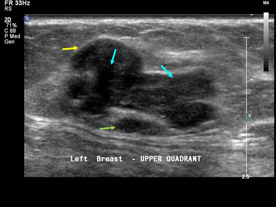
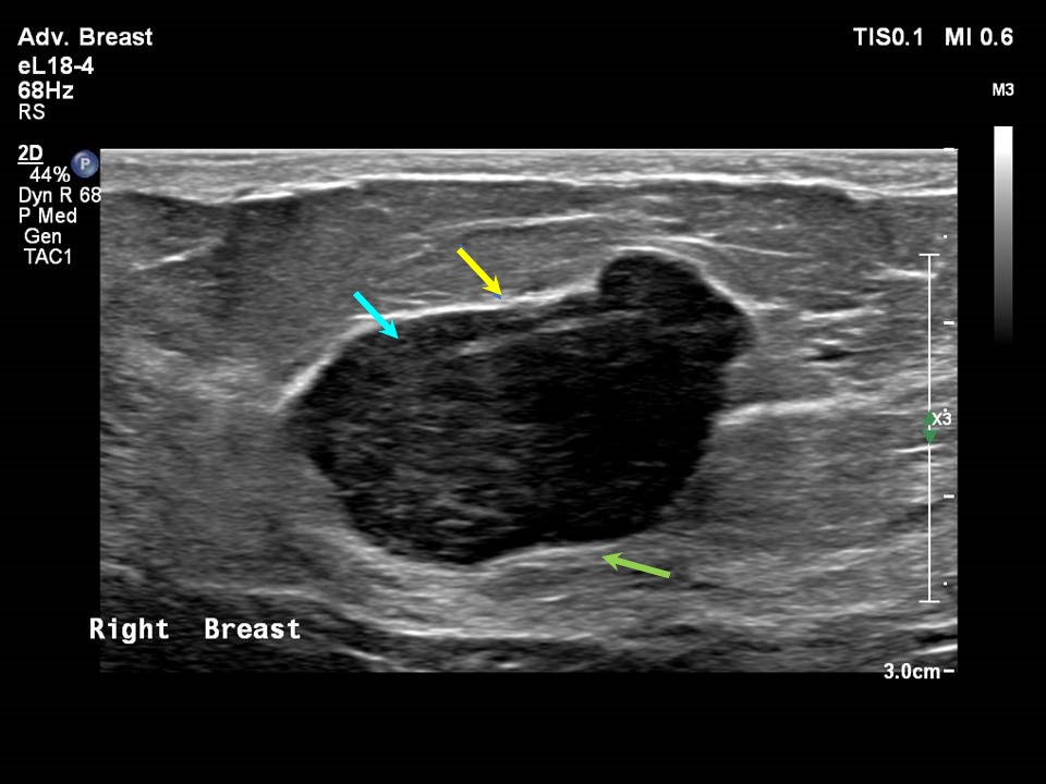
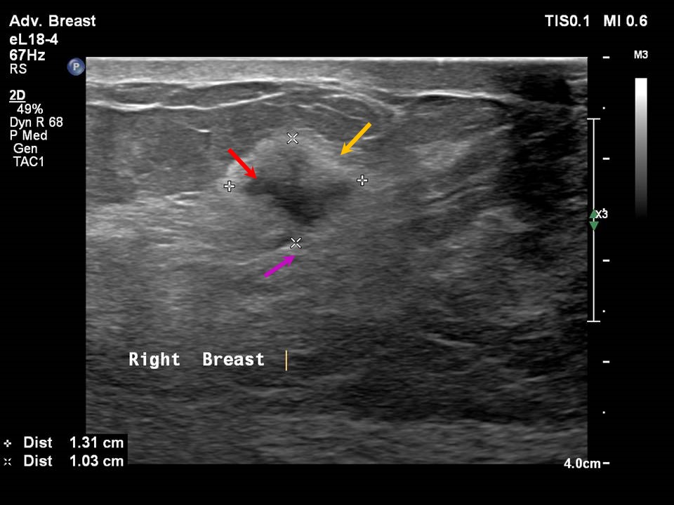
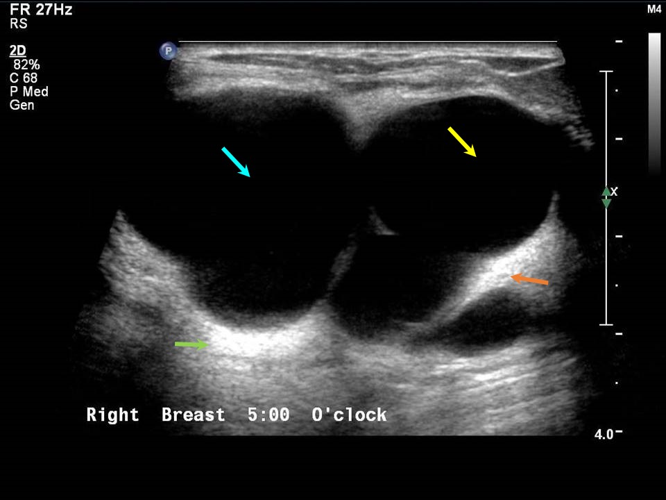
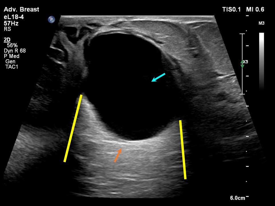
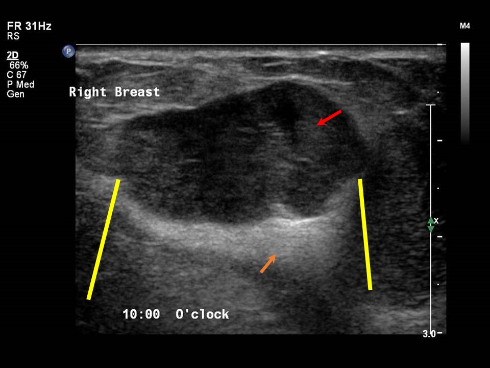
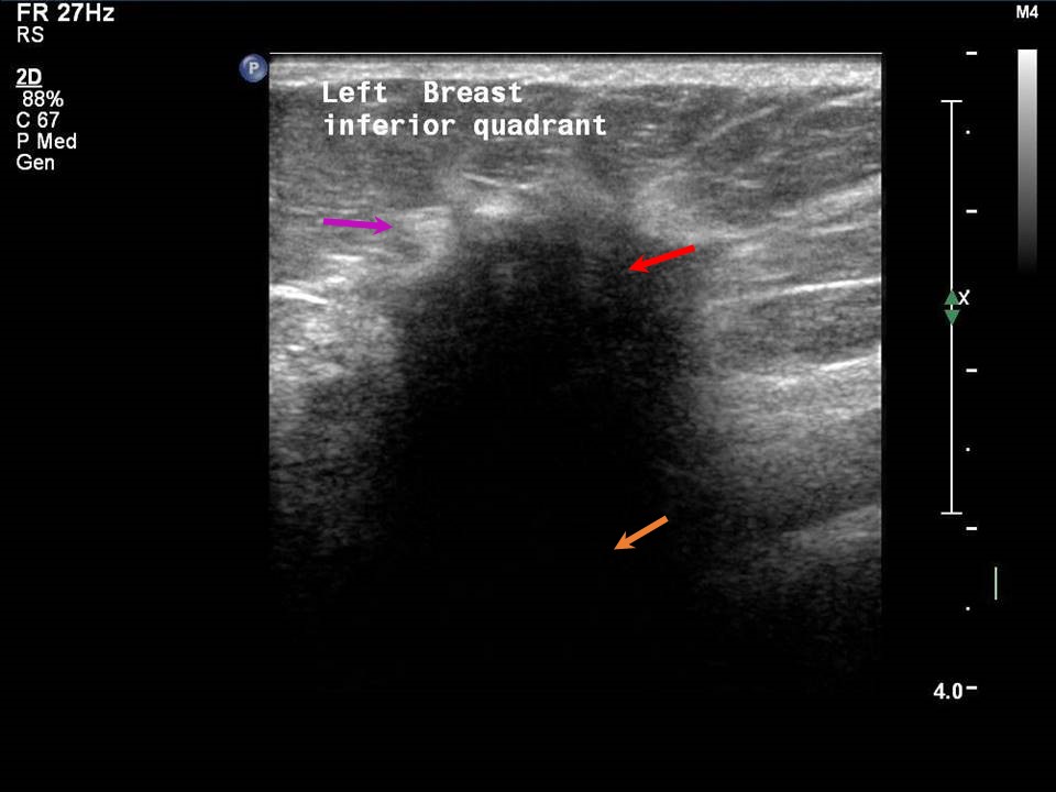
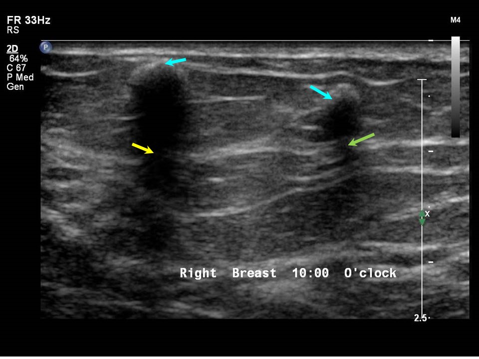
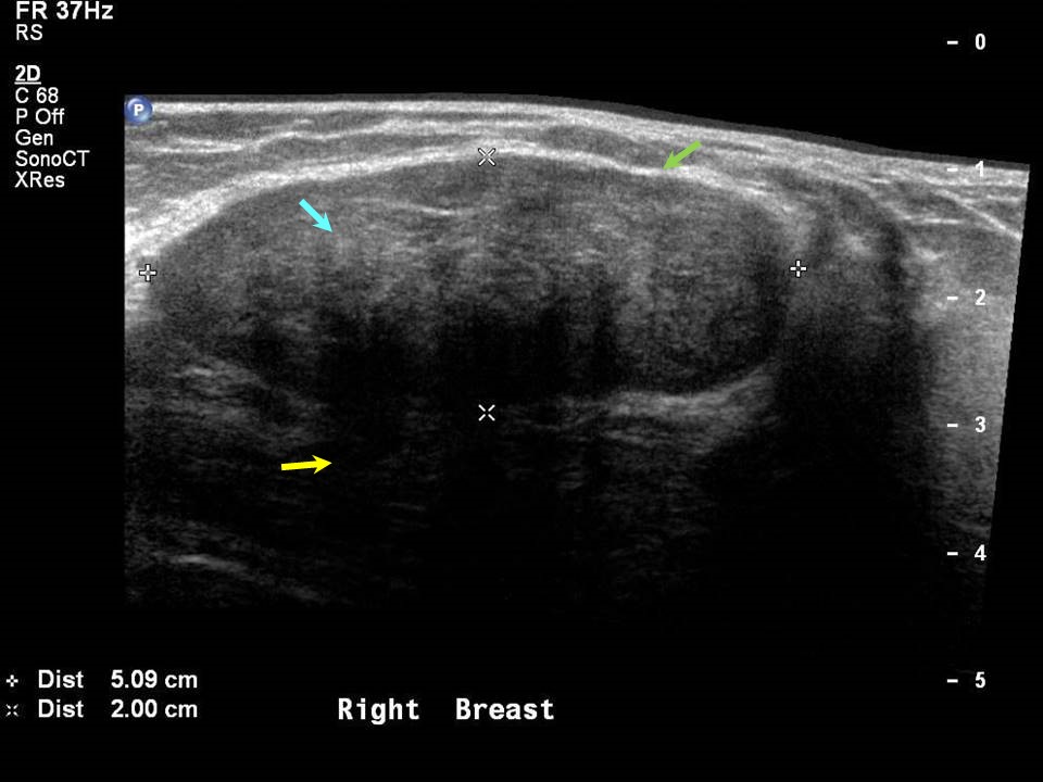
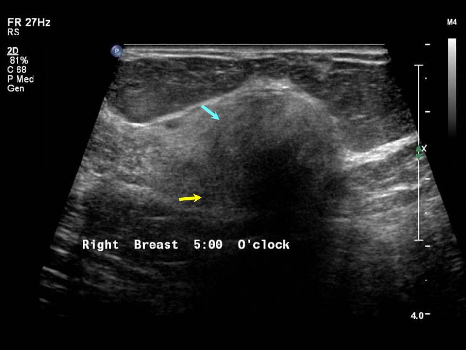
An ultrasound beam may penetrate a lesion with no posterior features or it may be attenuated and scattered, producing posterior shadowing. The density of the tissues through which the sound waves travel determines the posterior features seen. When ultrasound waves pass through a lesion completely, they cause posterior enhancement, as is seen in a clear simple cyst that allows all the sound waves to pass and produces strong posterior enhancement. When ultrasound waves are not able to pass through a mass lesion, either partially or completely, it causes posterior shadowing. Posterior shadowing is seen in malignant masses, postsurgical scarring, calcifications, or fibrotic scarring. No posterior features are seen in benign lesions such as fibroadenomas or phyllodes. Note that no posterior features can also be seen in certain histological types of breast cancer, such as mucinous and medullary carcinoma.
The sound transmitted through a solid mass depends on the amount of the fibrotic tissue in the mass. Thus scirrhous carcinomas, which are gritty to cut, show strong posterior acoustic shadowing on imaging with no transmission of sound waves. Posterior shadowing limits the evaluation of deeper breast structures. No posterior features Posterior enhancement Posterior shadowing Combined posterior features |

Click on the pictures to magnify and display the legends
Click on this icon to display a case study
25 avenue Tony Garnier CS 90627 69366, LYON CEDEX 07 France - Tel: +33 (0)4 72 73 84 85
© IARC 2025 - Terms of use - Privacy Policy.
© IARC 2025 - Terms of use - Privacy Policy.