


Home / Training / Manuals / Atlas of breast cancer early detection / Cases
Atlas of breast cancer early detection
Filter by language: English / Русский
Go back to the list of case studies
.png) Click on the pictures to magnify and display the legends
Click on the pictures to magnify and display the legends
| Case number: | 077 |
| Age: | 65 |
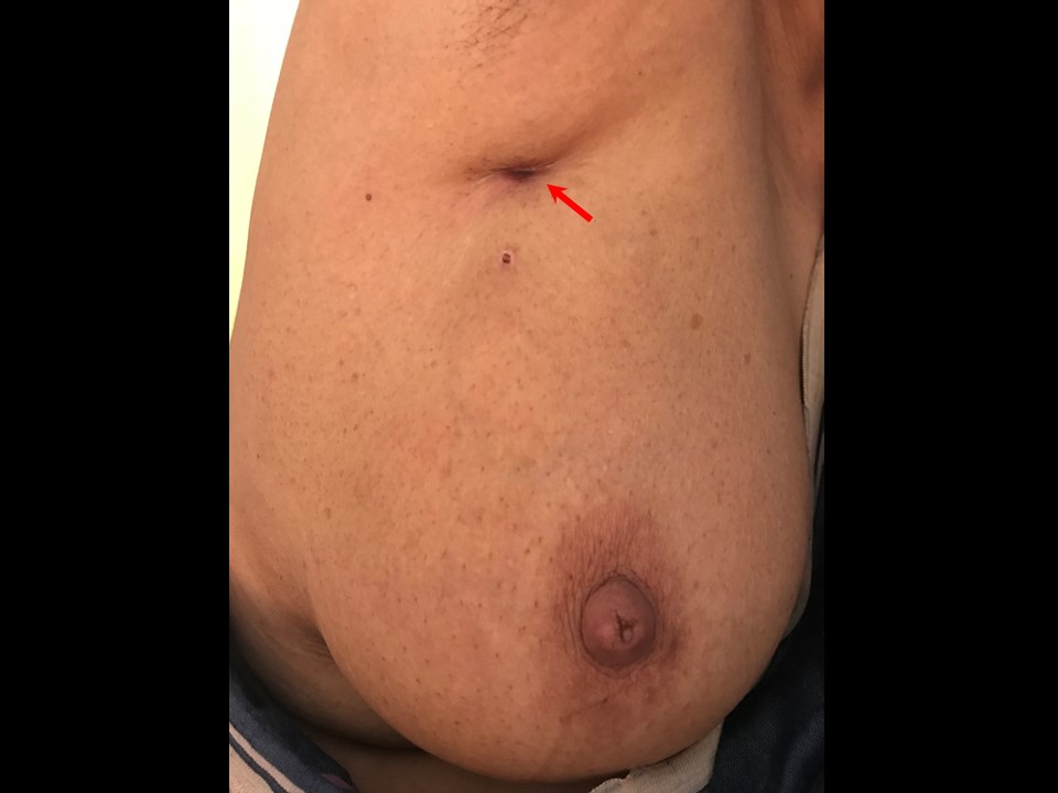
| Clinical presentation: | Postmenopausal woman with average risk of developing breast cancer presented with an ulcer on the upper quadrant of the left breast. Examination revealed a hard irregular lump involving underlying breast and the overlying skin. |
 |
Mammography:
 |  |
 |
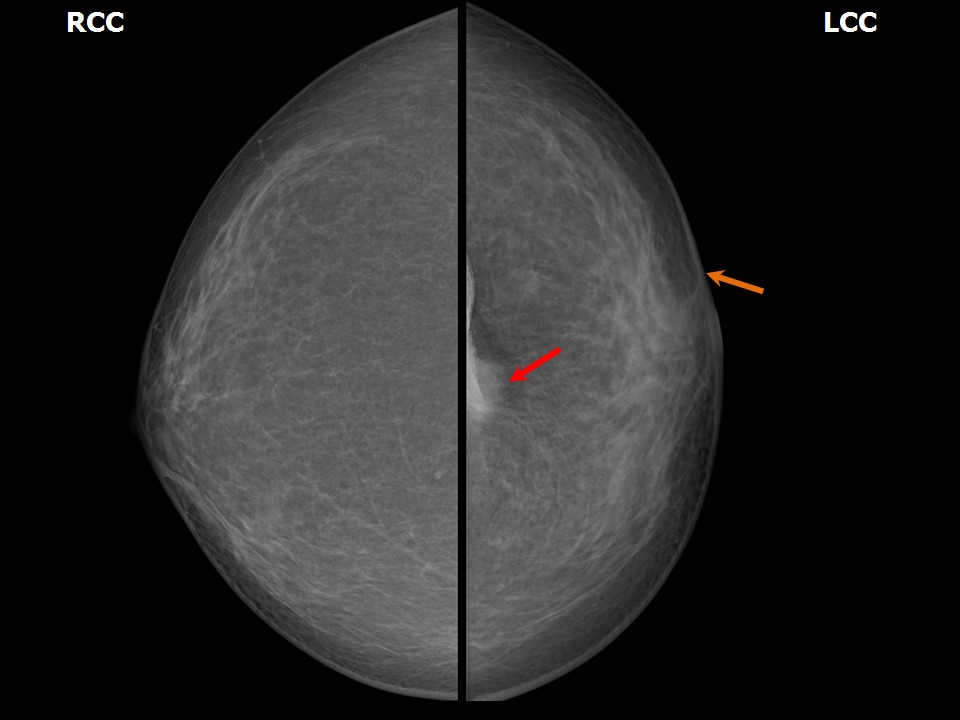
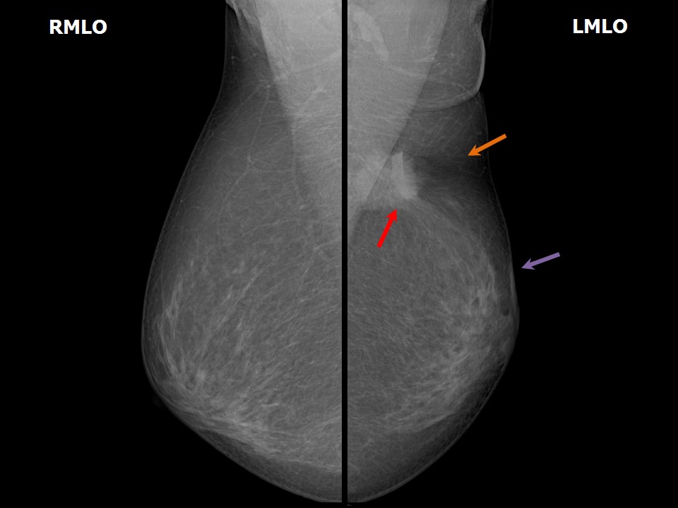
| Breast composition: | ACR category b (there are scattered areas of fibroglandular density) | Mammography features: |
| ‣ Location of the lesion: | Left breast, upper quadrants at 12 oclock, posterior third |
| ‣ Mass: | |
| • Number: | 1 |
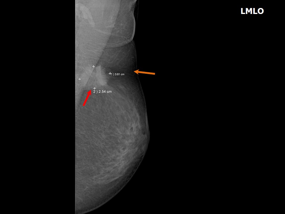
| • Size: | 3.6 × 2.5 cm |
| • Shape: | Irregular |
| • Margins: | Spiculated |
| • Density: | High |
| ‣ Calcifications: | |
| • Typically benign: | None |
| • Suspicious: | None |
| • Distribution: | None |
| ‣ Architectural distortion: | None |
| ‣ Asymmetry: | None |
| ‣ Intramammary node: | None |
| ‣ Skin lesion: | None |
| ‣ Solitary dilated duct: | None |
| ‣ Associated features: | Skin retraction and skin thickening |
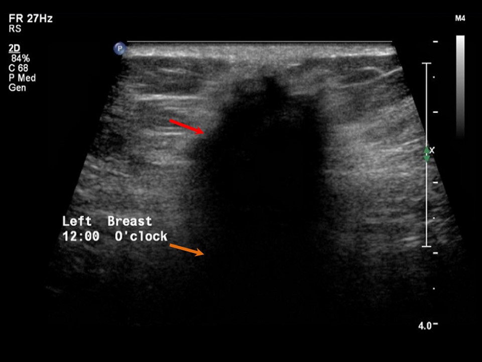
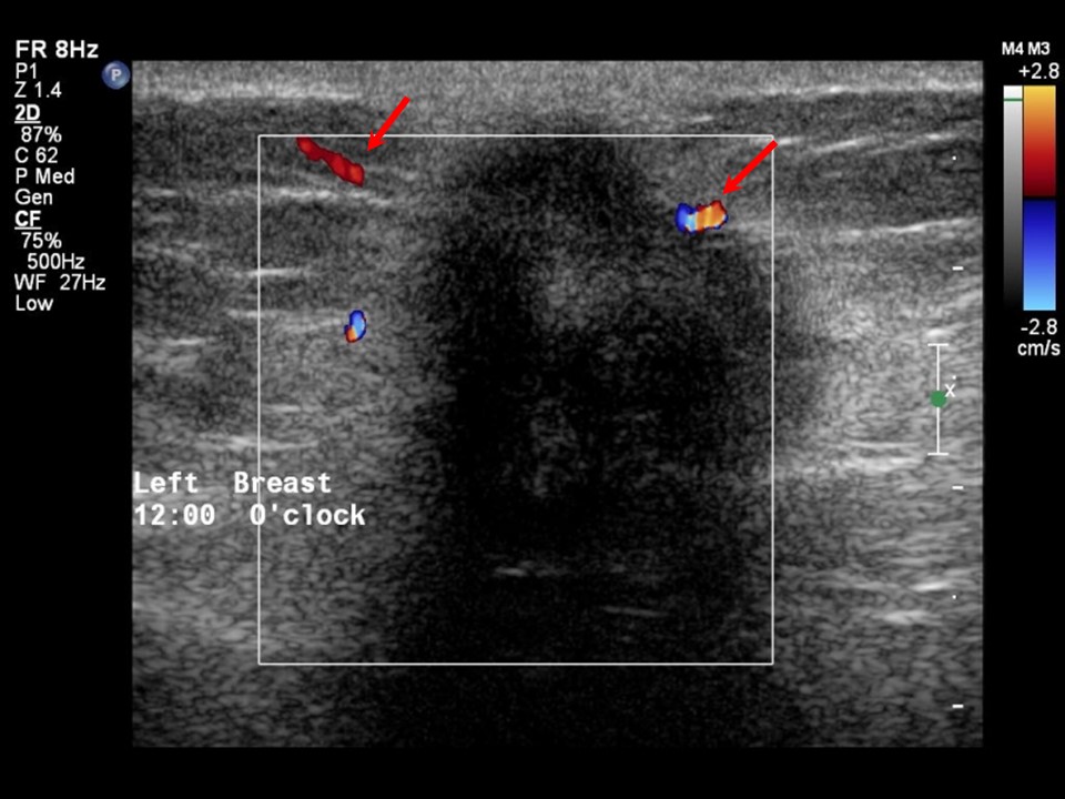
Ultrasound:
 |  |
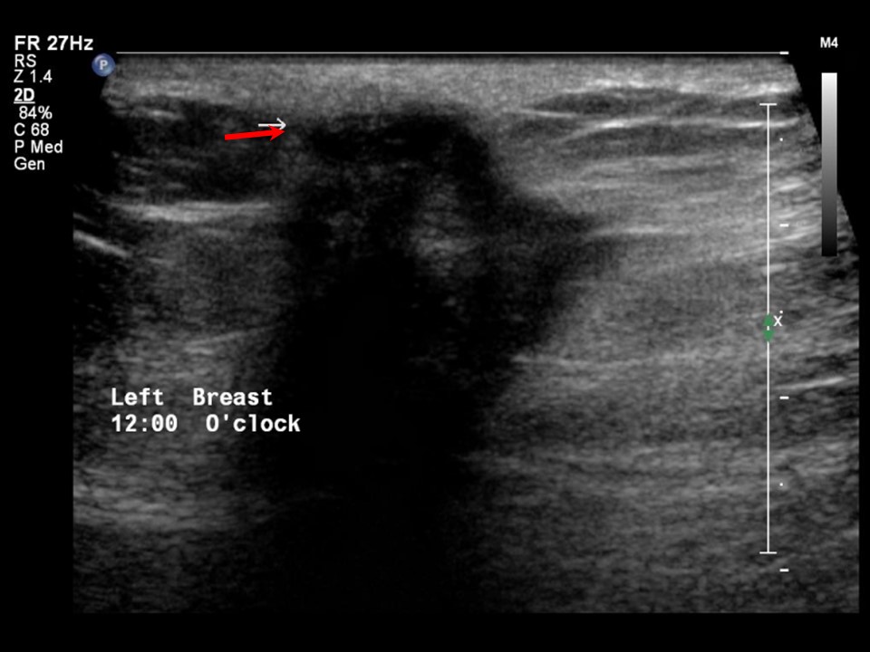 | 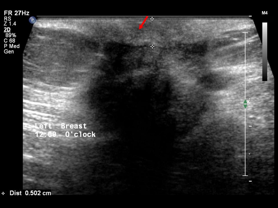 |
| Ultrasound features: Left breast, upper quadrants at 12 oclock | |
| ‣ Mass | |
| • Location: | Left breast, upper quadrants at 12 oclock |
| • Number: | 1 |
| • Size: | 3.5 × 3.0 cm |
| • Shape: | Irregular |
| • Orientation: | Not parallel |
| • Margins: | Spiculated |
| • Echo pattern: | Hypoechoic |
| • Posterior features: | Posterior shadowing |
| ‣ Calcifications: | None |
| ‣ Associated features: | Skin thickening, skin retraction, and vessels in rim |
| ‣ Special cases: | None |
BI-RADS:
BI-RADS Category: 5 (highly suggestive of malignancy)Further assessment:
Further assessment advised: Referral for core biopsyCytology:
 |
| Cytology features: | |
| ‣ Type of sample: | FNAC (solid lesion) |
| ‣ Site of biopsy: | |
| • Laterality: | Left |
| • Quadrant: | Upper at 12 oclock |
| • Localization technique: | Palpation |
| • Nature of aspirate: | Whitish |
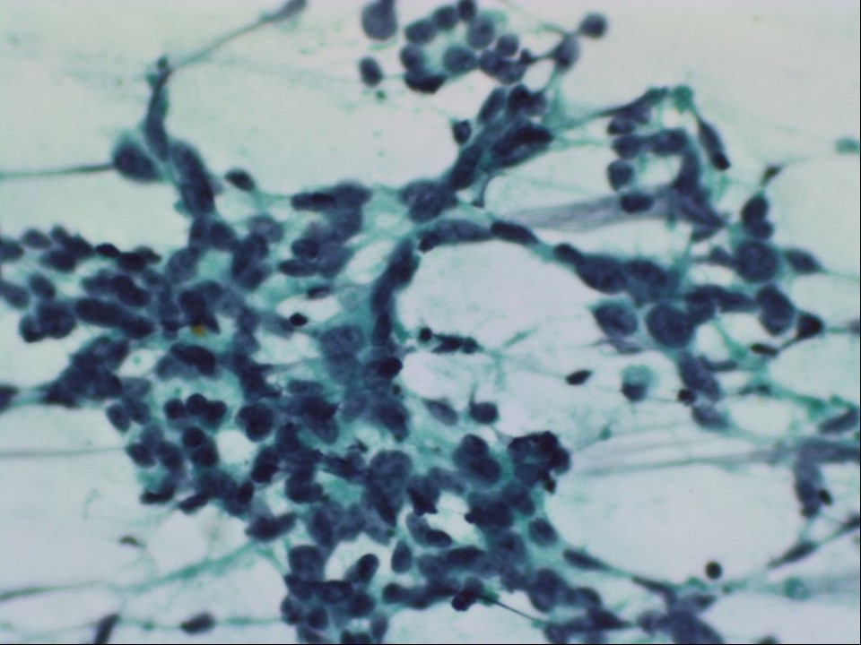
| ‣ Cytological description: | Loosely cohesive malignant cell clusters with many scattered single isolated cells |
| ‣ Reporting category: | Malignant |
| ‣ Diagnosis: | Carcinoma |
| ‣ Comments: | None |
Histopathology:
Breast-conserving surgery
 |  |
| Histopathology features: | |
| ‣ Specimen type: | Breast-conserving surgery |
| ‣ Laterality: | Left |
| ‣ Macroscopy: | Specimen of left breast lumpectomy 9.5 × 6.0 × 3.5 cm with an elliptical flap of skin 9.2 × 4.5 cm. Skin appears puckered in the centre. Cut surface shows a tumour 3.0 × 2.5 × 2.1 cm. The tumour is firm, greyish white with infiltrating margins. The tumour is flush with the skin, 0.2 cm from the base, 4.0 cm from the superior margin, 3.0 cm from the inferior margin, 1.8 cm from the lateral margin, and 1.3 cm from the medial margin |
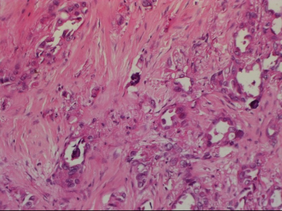
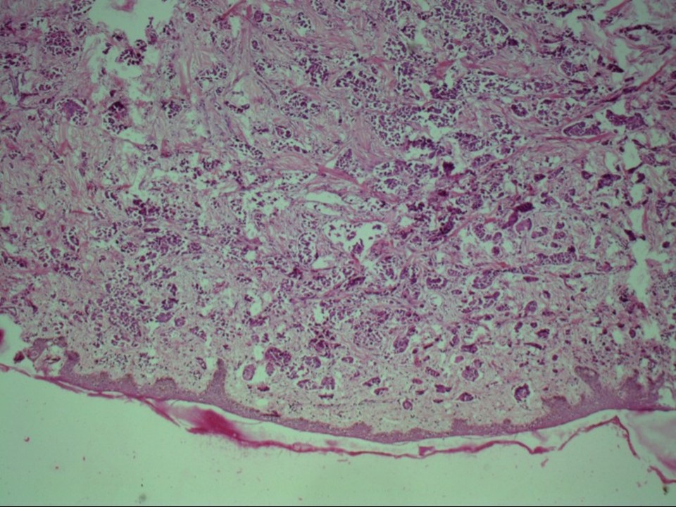
| ‣ Histological type: | Invasive carcinoma of no special type |
| ‣ Histological grade: | Grade 3 (3 + 3 + 2 = 8) |
| ‣ Mitosis: | 15 |
| ‣ Maximum invasive tumour size: | 3 cm in greatest dimension |
| ‣ Lymph node status: | 5/15 |
| ‣ Peritumoural lymphovascular invasion: | Present |
| ‣ DCIS/EIC: | Not identified |
| ‣ Margins: | Posterior margin is free of tumour, involving skin |
| ‣ Pathological stage: | pT4bN2 |
| ‣ Biomarkers: | |
| ‣ Comments: | Foci of calcification seen |
Case summary:
| Postmenopausal woman presented with ulcerated skin in left axilla. Diagnosed as left breast carcinoma with overlying skin retraction and ulceration, BI-RADS 5 on imaging, as left breast carcinoma on cytology, and as invasive breast carcinoma of no special type, pT4bN2 on histopathology. |
Learning points:
|