


Home / Training / Manuals / Atlas of breast cancer early detection / Learning
.png)
Click on the pictures to magnify and display the legends

Click on this icon to display a case study
Atlas of breast cancer early detection
Filter by language: English / РусскийAnatomy of the breast Gross anatomy of the breast |
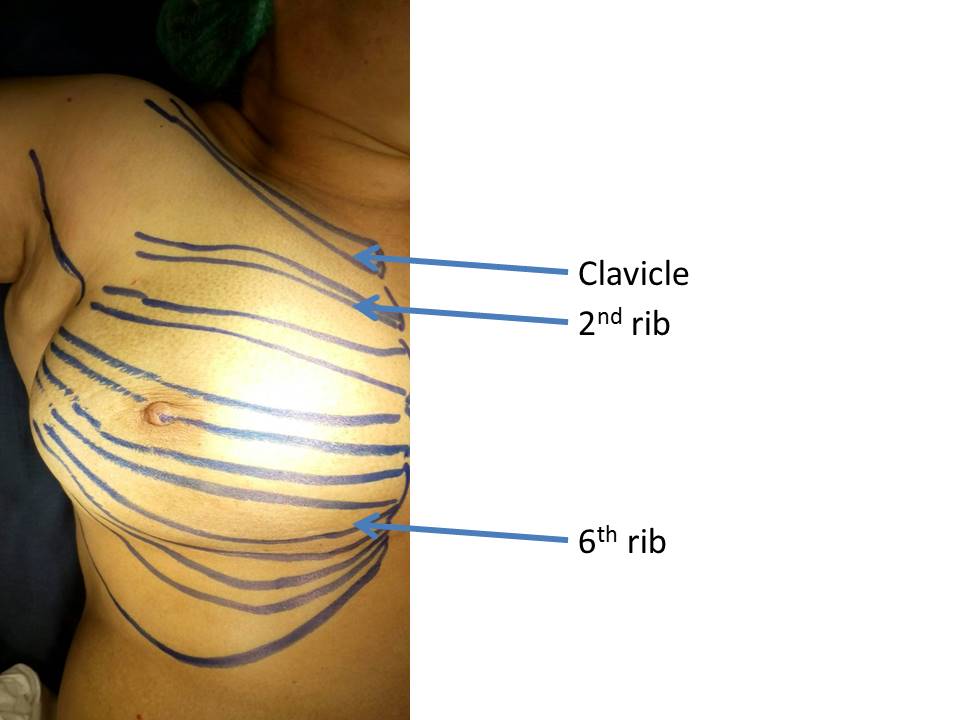
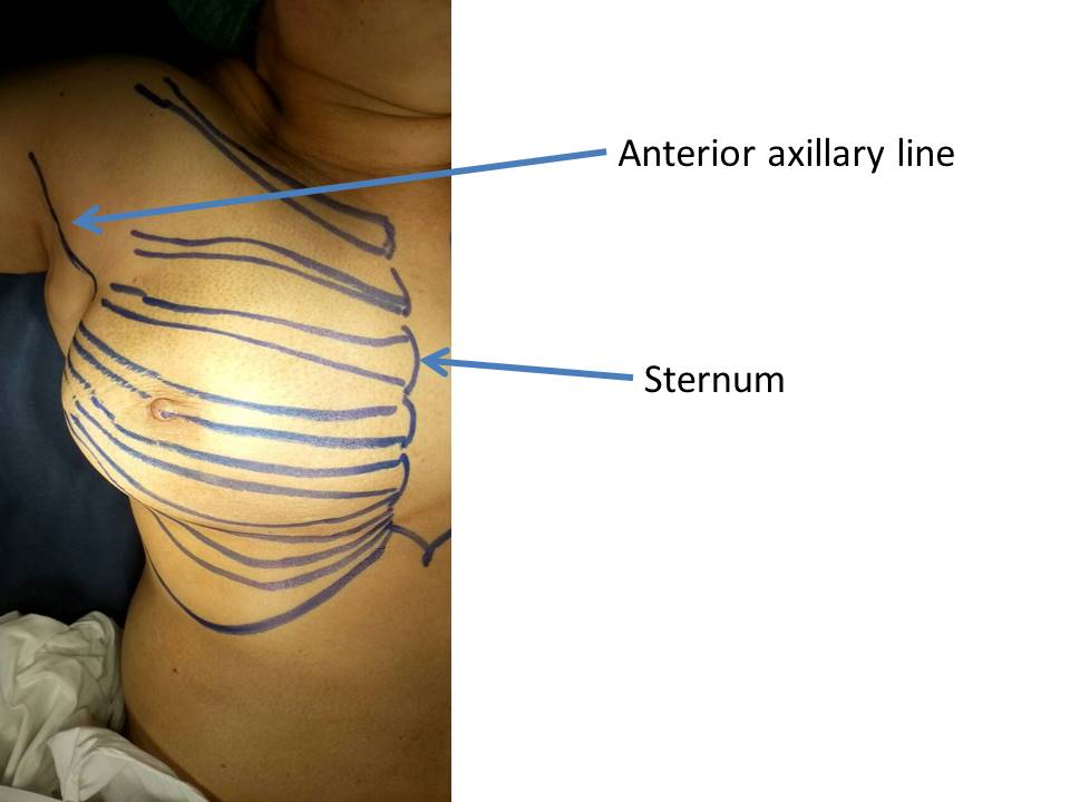
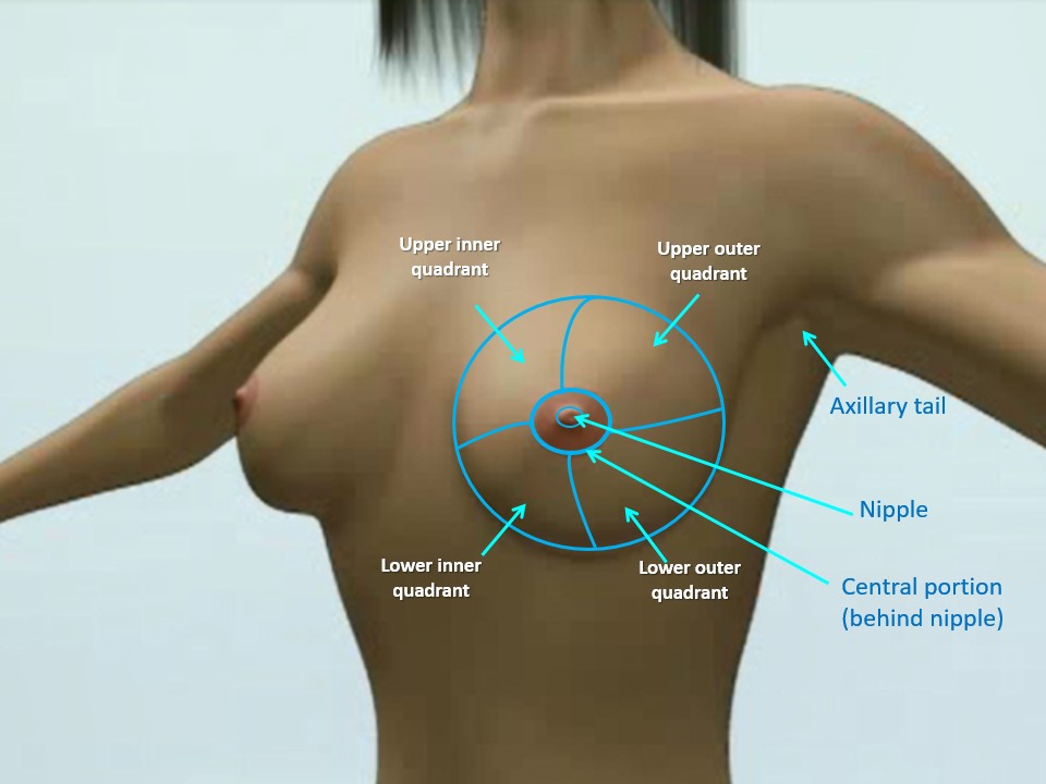
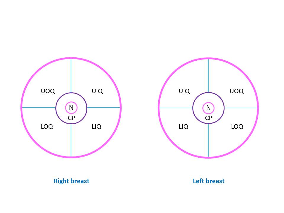
Anatomical relations The adult female breast is situated between the second and the sixth ribs and extends horizontally from the edge of the sternum (the flat bone in the middle of the chest) to the mid-axillary line. The breast overlies the deep fascia covering the pectoralis major muscle (pectoralis fascia), from which it is separated by loose areolar tissue containing small blood vessels and lymphatics. This loose connective tissue in the retromammary space gives rise to a lucency in the mammogram. The lucent area is known as the retromammary bursa or Chassaignac bag. The breast tissue often curves around the lateral free margin of the pectoralis major. The axillary tail (tail of Spence) is a prolongation of the upper and outer quadrants of the breast towards the axilla. This tail passes under the axillary fascia (through the foramen of Langer) and may be mistaken for enlarged lymph nodes. Each breast is divided into four quadrants, with the nipple and areola at the centre:
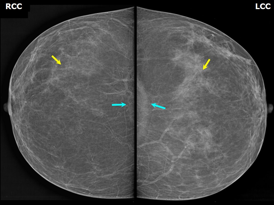
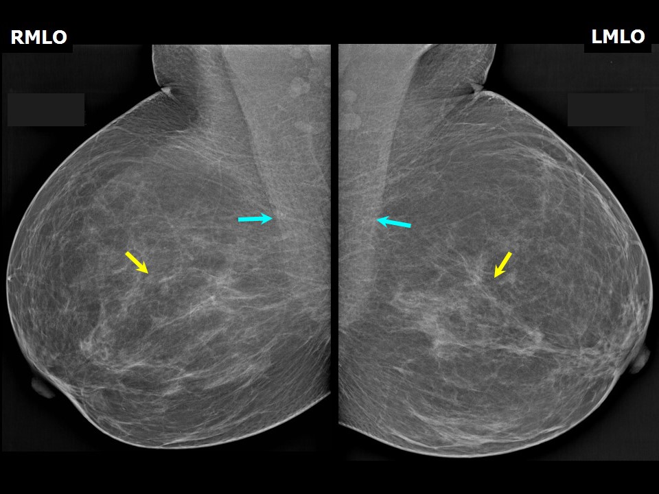
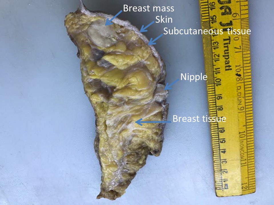
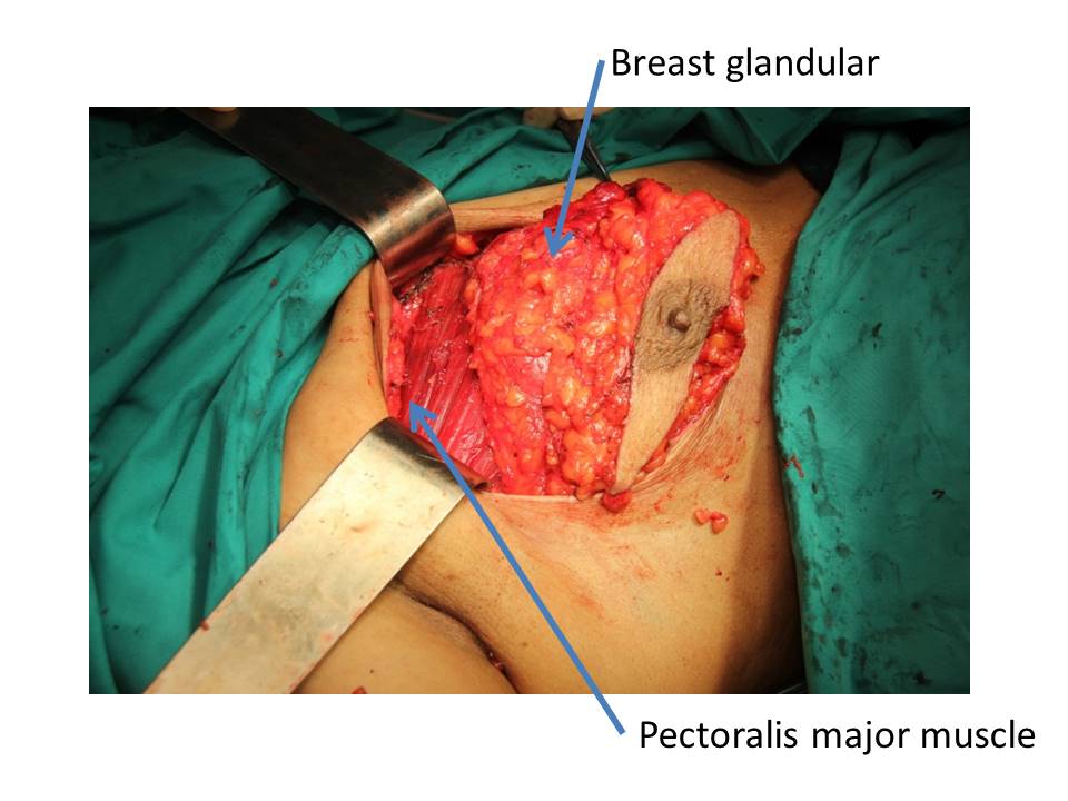
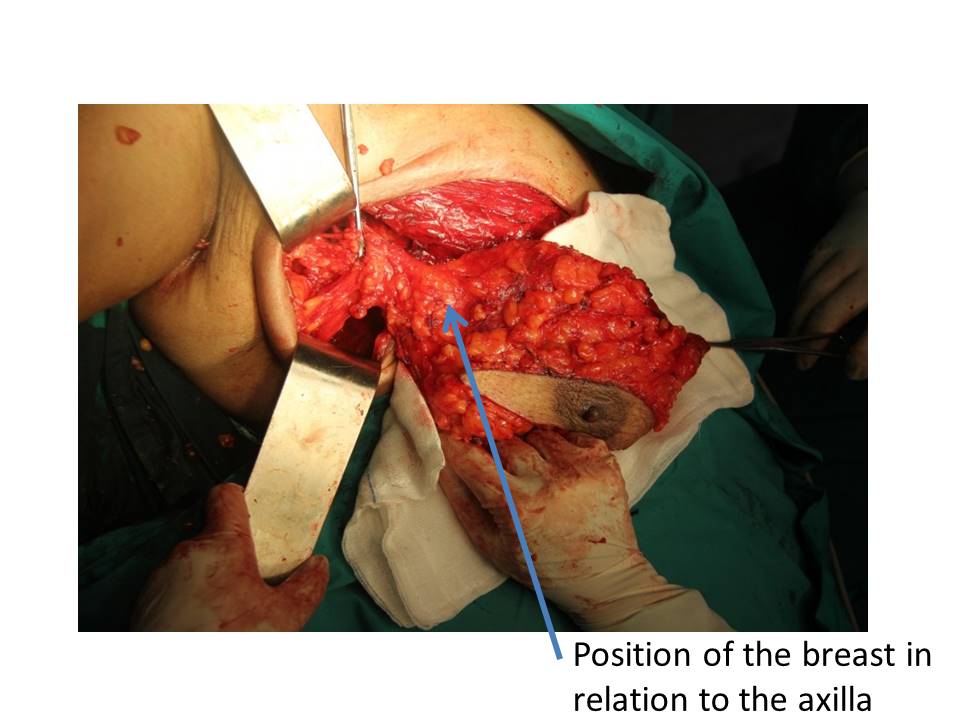
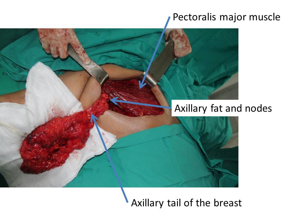
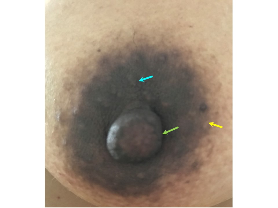
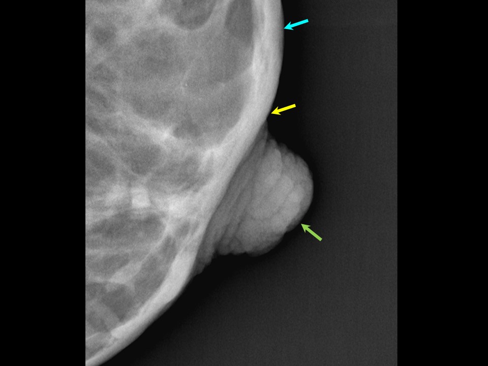
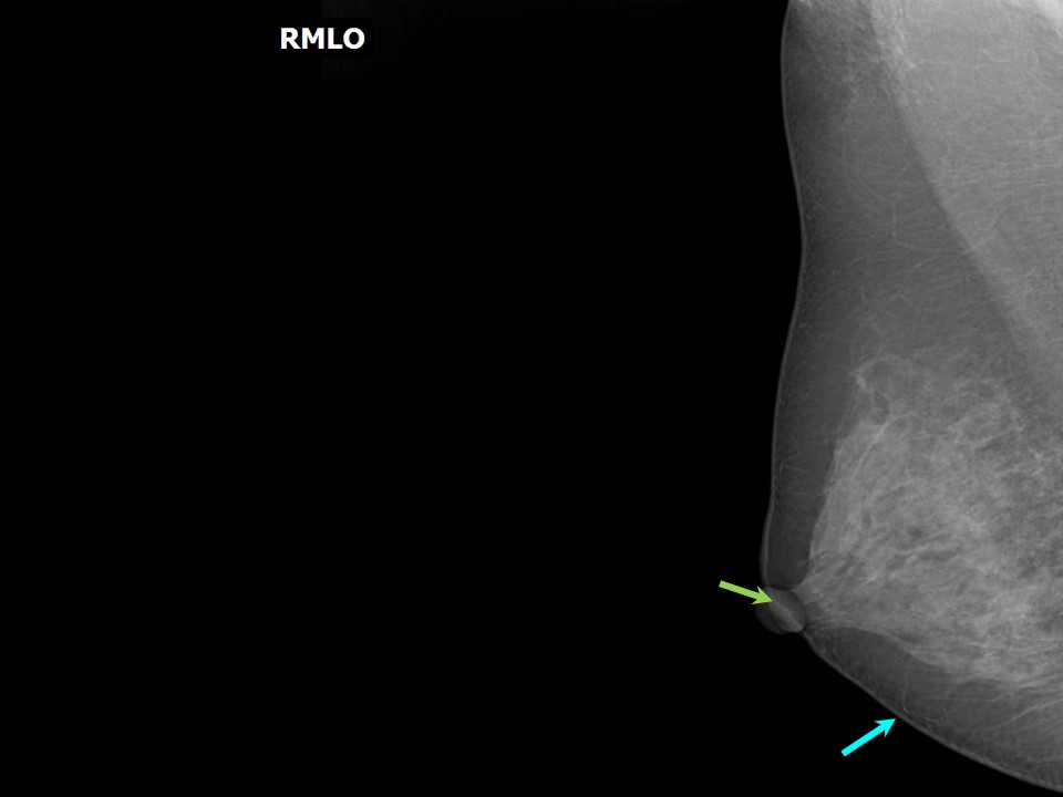
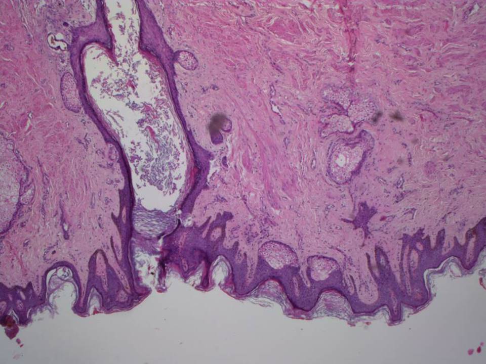
Normal mammogram showing normal breast tissue overlying the pectoralis muscle Mastectomy specimen showing main components of the breast Intraoperative images showing breast anatomy During mastectomy, the anatomical relationships of the breast tissue are clearly seen. In the images below obtained during surgery, the right breast is seen overlying the red fan-shaped pectoralis major muscle. The pectoralis major muscle connects the chest wall to the bones of upper arm. The axillary tail is a prolongation of the upper and outer quadrants of the breast; it passes under the pectoralis major muscle to the axilla. The tail merges with the axillary fat containing the lymph nodes. Skin and subcutaneous tissue The skin of the breast contains the nipple and the areola. The nipple is an erectile structure covered with thick pigmented skin; it also contains muscle. The orifices of the lactiferous ducts are seen near the apex of the nipple. The skin of the areola contains numerous modified sweat glands and sebaceous glands. These glands, also known as Montgomery tubercles, enlarge during pregnancy. The areola contains involuntary muscles in the subcutaneous tissues that are arranged in concentric rings and radially. |
Click on the pictures to magnify and display the legends
Click on this icon to display a case study
25 avenue Tony Garnier CS 90627 69366, LYON CEDEX 07 France - Tel: +33 (0)4 72 73 84 85
© IARC 2025 - Terms of use - Privacy Policy.
© IARC 2025 - Terms of use - Privacy Policy.