


Home / Training / Manuals / Atlas of breast cancer early detection / Learning
.png)
Click on the pictures to magnify and display the legends

Click on this icon to display a case study
Atlas of breast cancer early detection
Filter by language: English / РусскийIntroduction | |
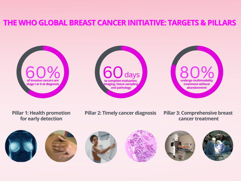
Female breast cancer has surpassed lung cancer as the most commonly diagnosed cancer in the world. The World Health Organizations Global Breast Cancer Initiative (GBCI), which was launched in 2021, sets a target for health systems to complete evaluation, imaging, and tissue diagnosis of women who have symptoms within 60 days. The other targets are to achieve diagnosis of at least 60% of invasive breast cancers at stage I or II and to ensure that at least 80% of patients with breast cancer undergo comprehensive treatment without abandonment. The achievement of these targets requires significant improvement in the capacity to perform triple assessment through clinical breast examination (CBE), breast imaging, and breast pathology. The Atlas of Breast Cancer Early Detection is a step-by-step guide to the procedure of CBE and its interpretation, diagnostic mammography, diagnostic breast ultrasound, image-guided fine-needle aspiration cytology (FNAC), and core biopsy of the breast. The atlas includes nearly 1000 annotated images and videos and 186 case studies. It will be a wonderful resource for surgeons, gynaecologists, radiologists, pathologists, medical students, nurses, and paramedical staff involved in the evaluation of breast abnormalities. The freely downloadable images and videos can be used to train new providers. Readers may start with the section on CBE, the section on Breast imaging, or the section on Breast pathology. The section on CBE describes the normal appearance of the breast and variations in the normal anatomy. It describes various techniques and steps to be followed and their sequence while conducting a comprehensive clinical examination of breast and axilla. It illustrates clinical findings and signs suggestive of and associated with various breast pathologies. The section on Breast imaging discusses the appearance of the normal breast on ultrasound and mammography. It describes the equipment, positioning of the patient, techniques and views, and common pitfalls to be avoided during breast imaging in order to obtain optimal breast images and minimize repetition of procedures. The section describes normal variations and changes in the appearance of the breast and the morphological appearance of the common benign and malignant breast lesions using standardized terminologies. It uses the Breast Imaging Reporting and Data System (BI-RADS) categories appropriately to complete the evaluation of breast conditions. The section on Breast pathology details various methods and steps used to process the tissue sample before its histological examination. The importance of prompt fixation and transport of pathology specimens is explained. The section also discusses the use of cytology as a diagnostic tool in breast lumps, the steps used in processing cytology samples, and the use of standard terminology in cytology in diagnosis. It highlights the diagnostic, prognostic, and predictive findings that should be included in a histopathology report. These are essential to communicate to the multidisciplinary team involved in breast cancer care.
|
Click on the pictures to magnify and display the legends
Click on this icon to display a case study
25 avenue Tony Garnier CS 90627 69366, LYON CEDEX 07 France - Tel: +33 (0)4 72 73 84 85
© IARC 2025 - Terms of use - Privacy Policy.
© IARC 2025 - Terms of use - Privacy Policy.