


Home / Training / Manuals / Atlas of breast cancer early detection / Learning
.png)
Click on the pictures to magnify and display the legends

Click on this icon to display a case study
Atlas of breast cancer early detection
Filter by language: English / РусскийBreast imaging Breast ultrasound Ultrasound Lexicon Special cases Mass in skin |
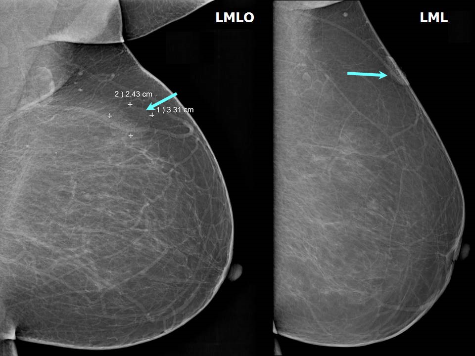
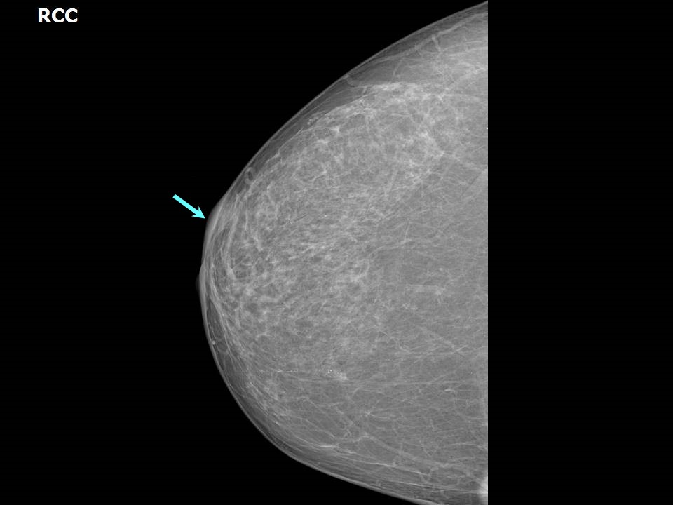
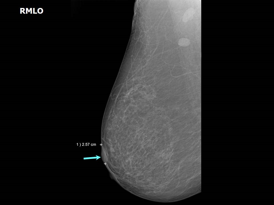
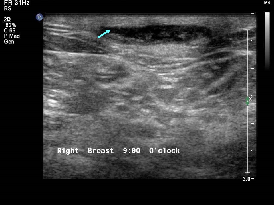
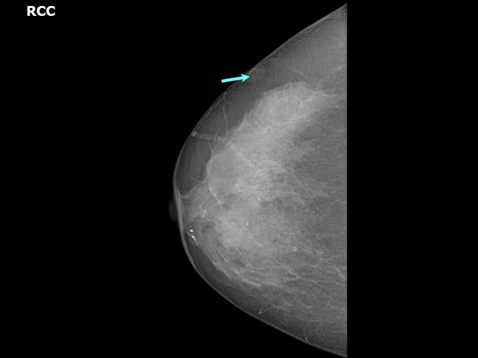
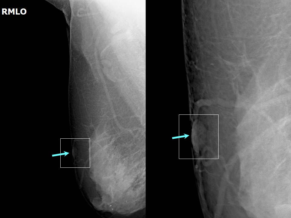
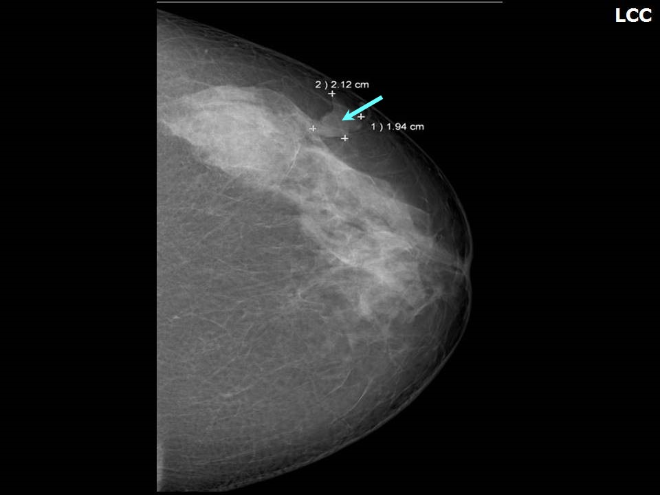
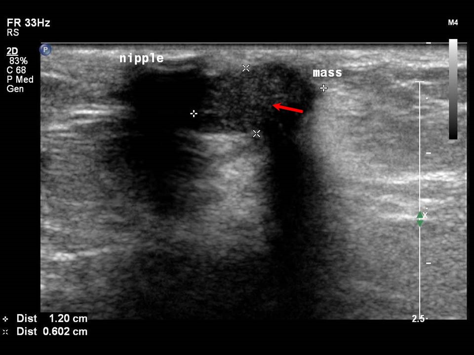
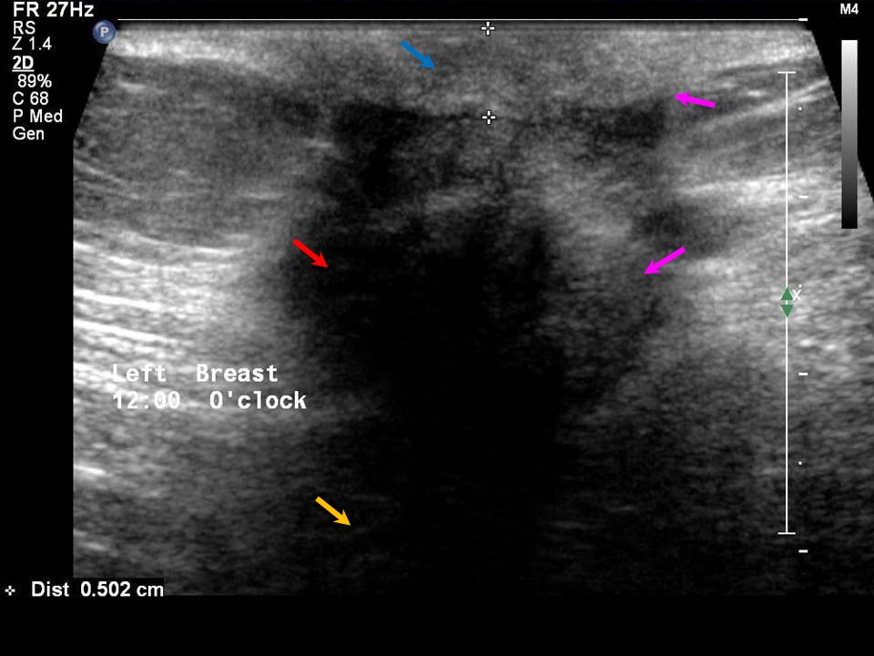
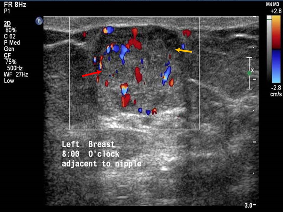
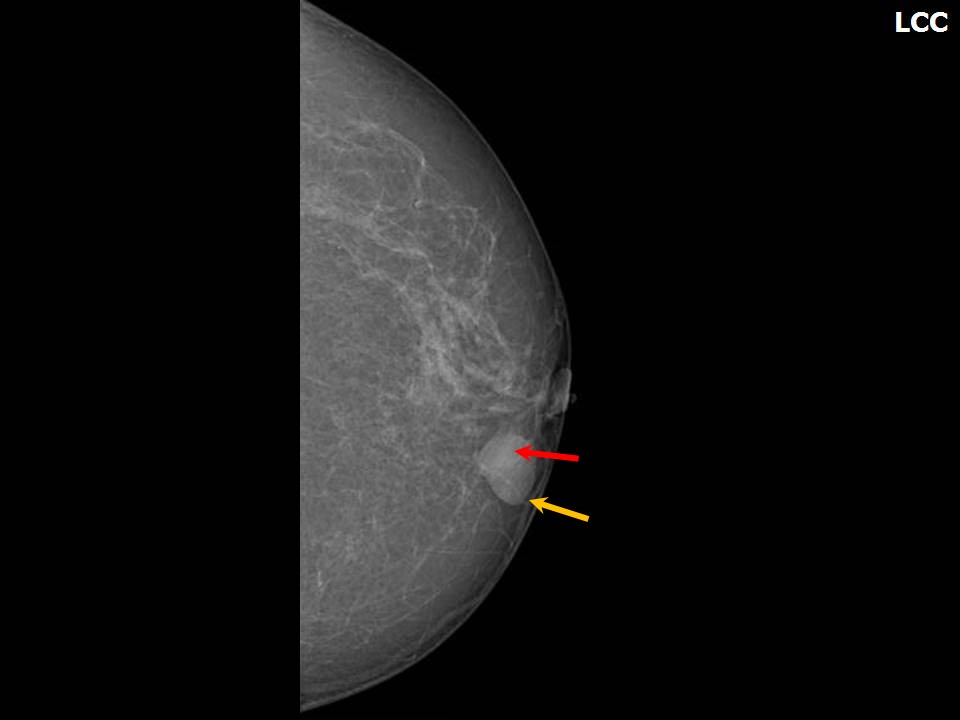
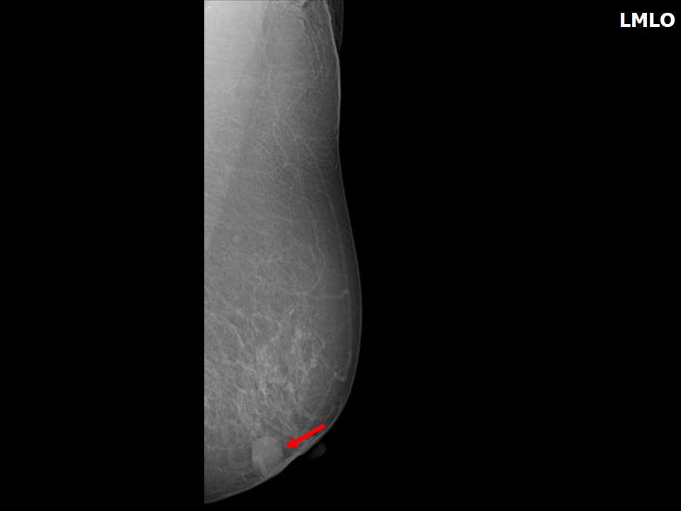
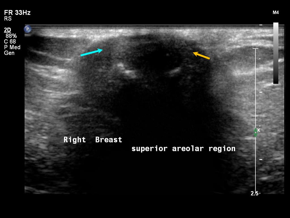
Mass in or on skin is also a special case in the ultrasound lexicon. Breast ultrasound is more accurate than mammography in localizing a lesion in the superficial tissues.
A superficial lesion may originate in:
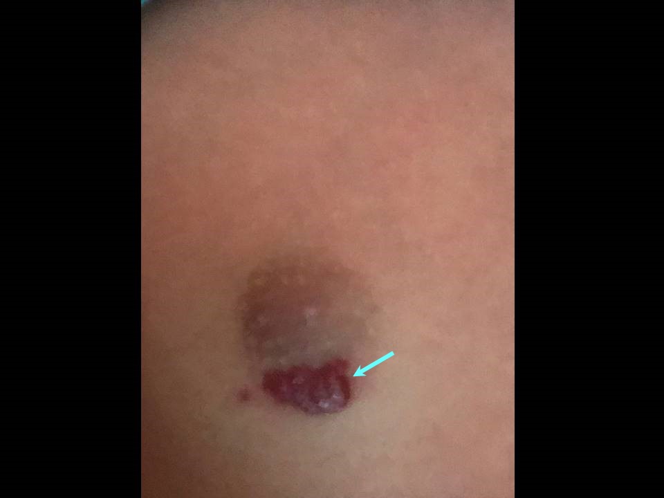
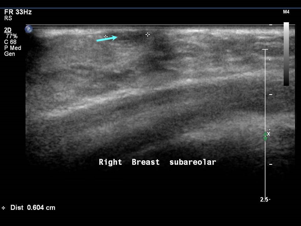
Skin lesions in the dermis A dermal lesion has either some or all of the following diagnostic features on ultrasound:
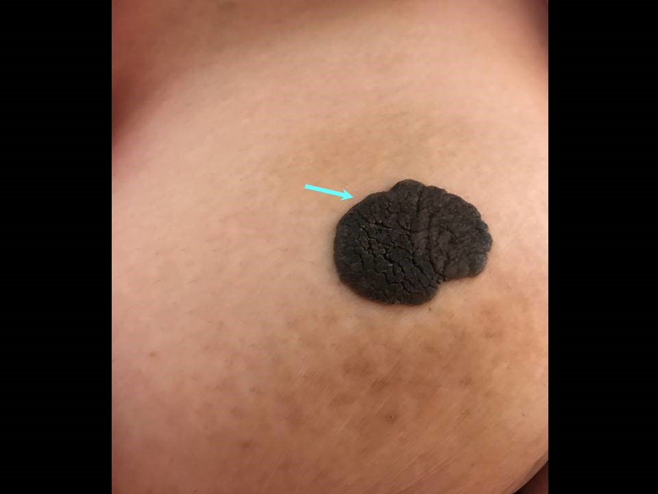
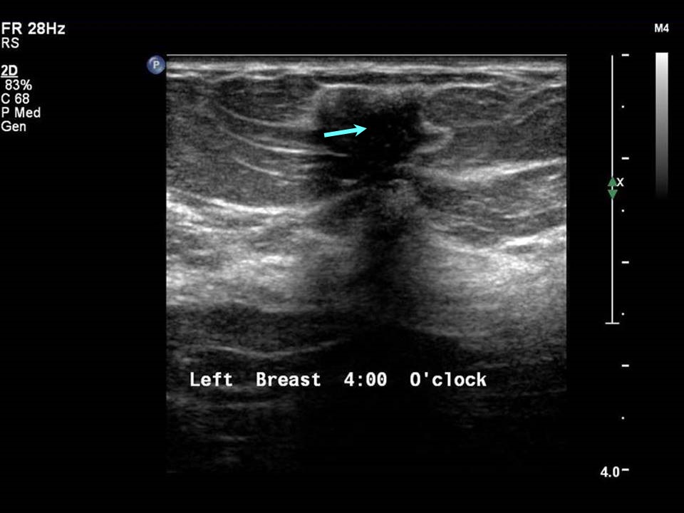
Epidermoid cysts Epidermoid cysts and sebaceous cysts are benign lesions. On ultrasound, they show similar morphology, but epidermoid cysts contain dead cells and sebaceous cysts have oily contents. Other examples of dermal lesions of the breast with a pasted-on-skin appearance are seborrhoeic keratosis, naevus, warts, and dermatofibrosarcoma protuberans.
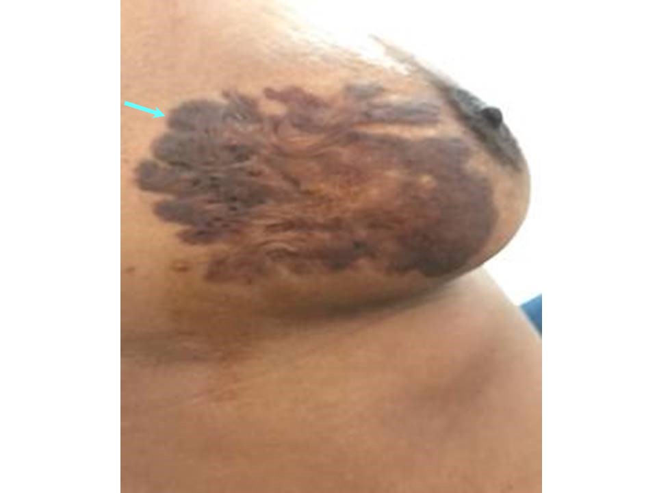
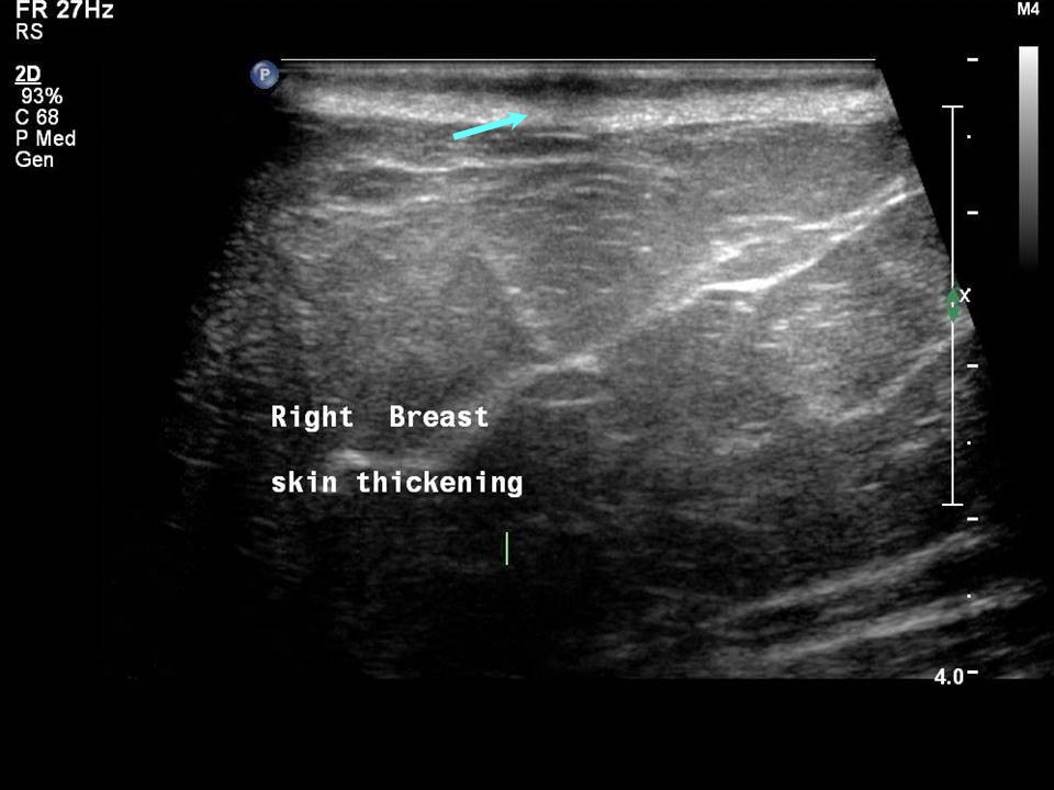
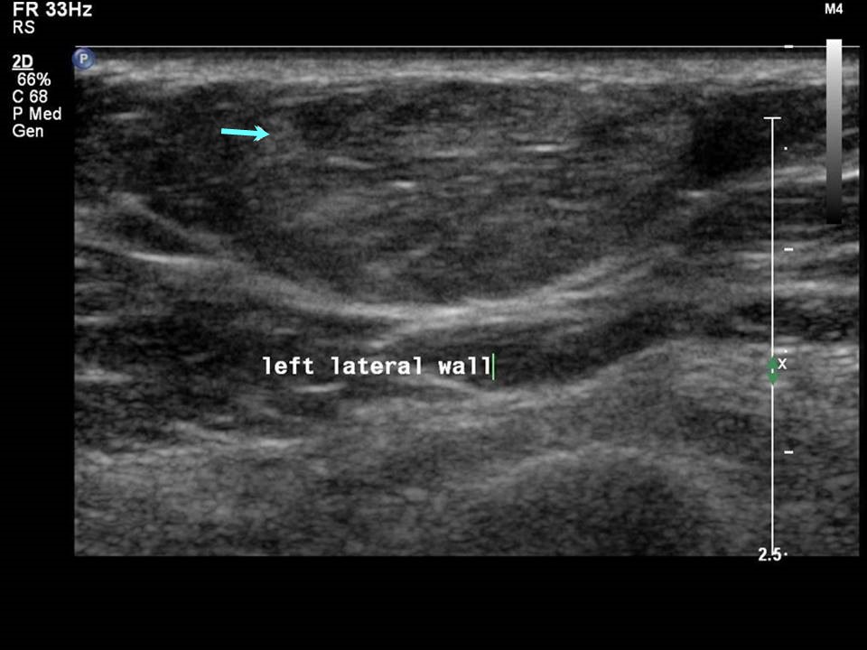
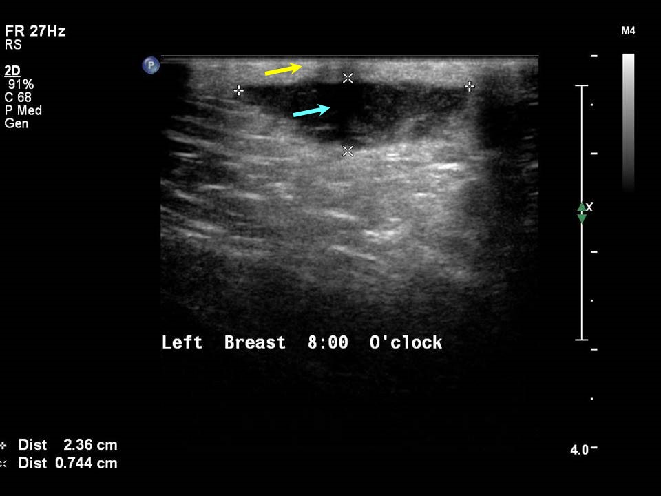
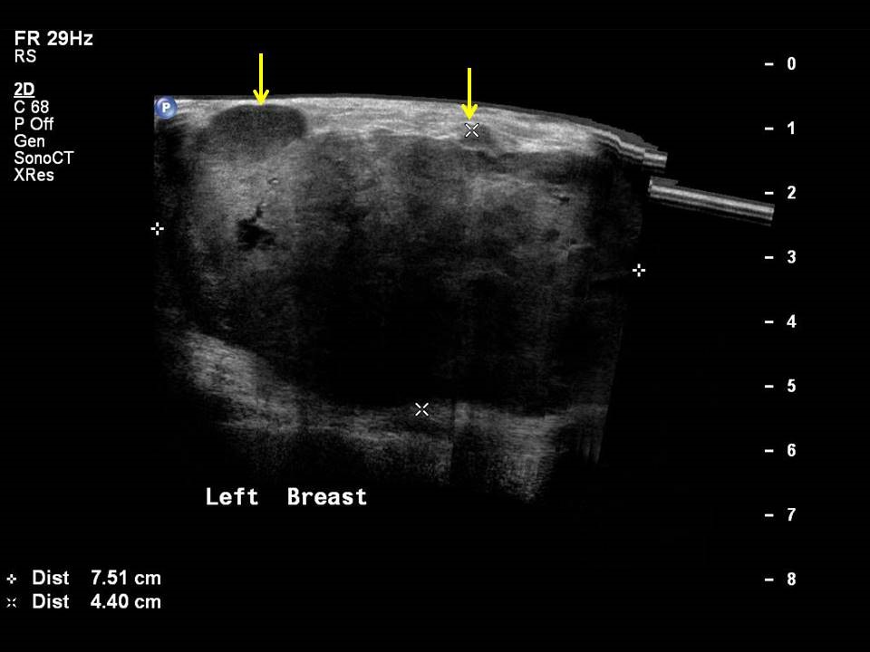
Seborrhoeic keratosis Seborrhoeic keratosis is a benign lesion. It should be distinguished from melanoma. Naevus A naevus is a pigmented chronic dermal lesion, commonly a birth identification mark, also known as a mole. Warts Warts are caused by infection of the skin by human papillomavirus (HPV). Dermatofibrosarcoma protuberans Dermatofibrosarcoma protuberans is a low-grade dermal malignant lesion with a high rate of local recurrence. Skin lesions in the hypodermis Skin lesions in the hypodermis include subcutaneous fat lesions and lesions in the anterior TDLU. Differential diagnoses include fat-containing lesions, haemangiomas, and lesions of neurogenic origin. Fat-containing lesions Lipomas, angiolipomas, and fat necrosis are all examples of fat-containing lesions. Vascular malformations: Haemangiomas and thrombosed vessels Lesions of neurogenic origin Anterior TDLU Anterior TDLU lesions in the subcutaneous tissues include papilloma, adenosis, fibroadenoma, and breast cancer. Breast cancer seen in the hypodermis with skin thickening and infiltrations Papilloma (peripheral) Fibroadenoma Accurate anatomical localization and categorization of the superficial mass by lexicon terminology is important to differentiate the probable superficial cancers and benign lesions. Lexicon features including shape, margins, echotexture, associated features. Biopsy may be warranted to complete the triple assessment of the mass. |


Click on the pictures to magnify and display the legends
Click on this icon to display a case study
25 avenue Tony Garnier CS 90627 69366, LYON CEDEX 07 France - Tel: +33 (0)4 72 73 84 85
© IARC 2025 - Terms of use - Privacy Policy.
© IARC 2025 - Terms of use - Privacy Policy.