


Home / Training / Manuals / Atlas of breast cancer early detection / Learning
.png)
Click on the pictures to magnify and display the legends

Click on this icon to display a case study
Atlas of breast cancer early detection
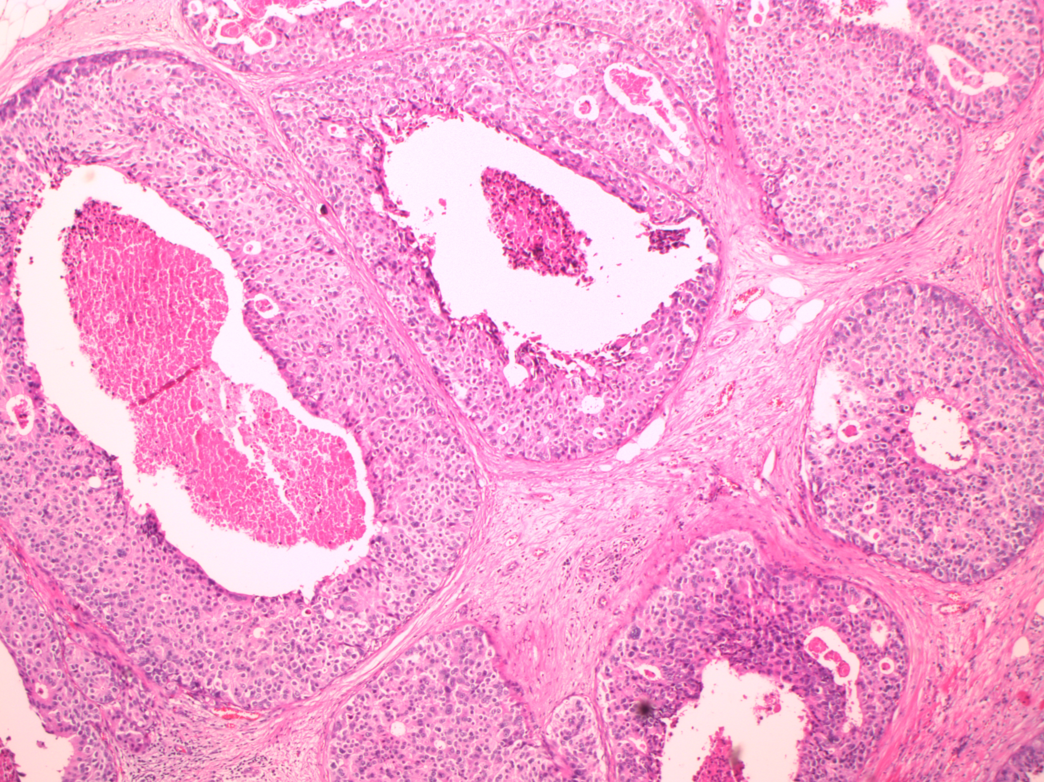
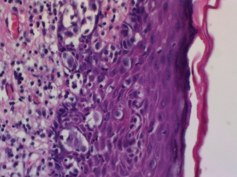
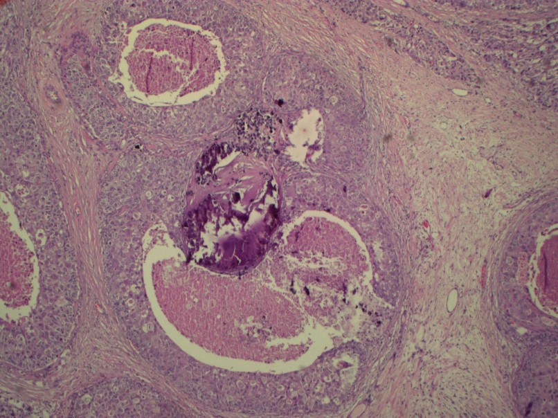
Filter by language: English / РусскийBreast pathology Histopathology of the breast Description and significance of the components of the report Features of DCIS |
DCIS associated with invasive carcinoma increases the risk of local recurrence for women undergoing breast-conserving surgery. It is more important to report the features of DCIS when in situ disease is predominant (e.g. cases of DCIS with microinvasion or extensive DCIS associated with T1a carcinoma). If DCIS is a minimal component of the invasive carcinoma, the features of the DCIS have less clinical relevance.
The pathology report should specify whether extensive DCIS (EIC) is present. EIC-positive carcinomas are defined by the following criteria:
Features of DCIS to be documented for the surgical pathology report
|

.jpg)
.jpg)
.jpg)
.jpg)
Click on the pictures to magnify and display the legends
Click on this icon to display a case study
25 avenue Tony Garnier CS 90627 69366, LYON CEDEX 07 France - Tel: +33 (0)4 72 73 84 85
© IARC 2025 - Terms of use - Privacy Policy.
© IARC 2025 - Terms of use - Privacy Policy.