


Home / Training / Manuals / Atlas of breast cancer early detection / Cases
Atlas of breast cancer early detection
Filter by language: English / Русский
Go back to the list of case studies
.png) Click on the pictures to magnify and display the legends
Click on the pictures to magnify and display the legends
| Case number: | 010 |
| Age: | 78 |
| Clinical presentation: | Postmenopausal woman with average risk of breast cancer underwent CT scan of the chest for retrosternal enlargement of thyroid. The scan revealed an incidental presence of right breast lump. Examination revealed lumpish nodularity in both breasts. |
Mammography:
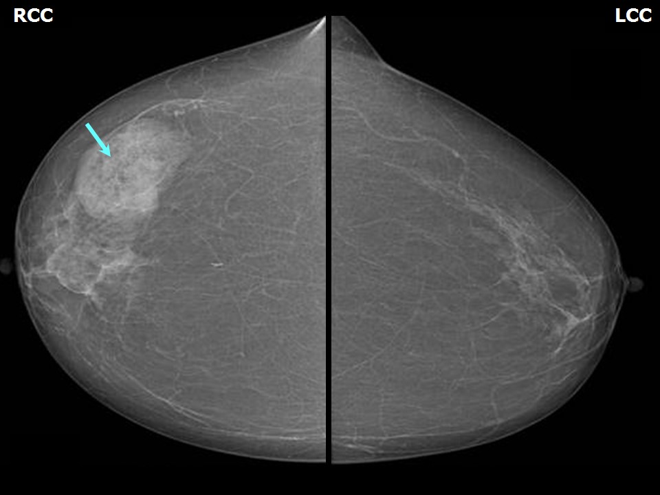 | 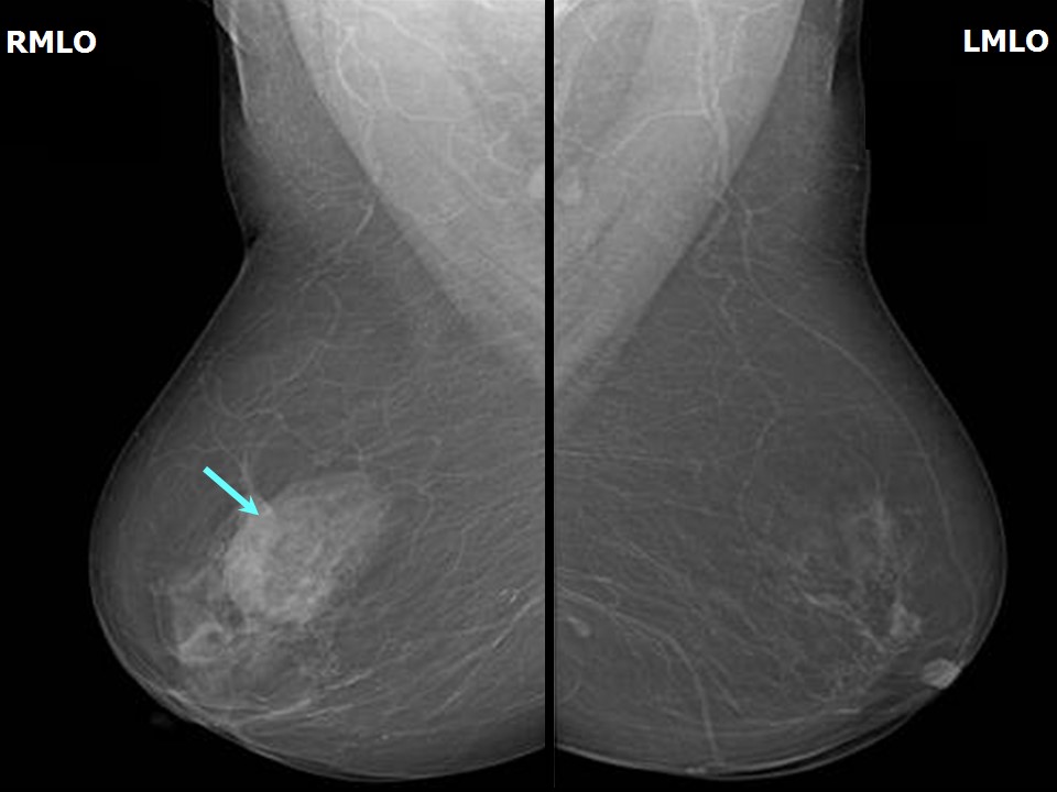 |
| Breast composition: | ACR category a (the breasts are almost entirely fatty) | Mammography features: |
| ‣ Location of the lesion: | Right breast, upper outer quadrant at 10 oclock, middle third |
| ‣ Mass: | |
| • Number: | 1 |
| • Size: | 4.0 × 3.0 cm |
| • Shape: | Oval |
| • Margins: | Circumscribed |
| • Density: | Equal with fat-containing areas |
| ‣ Calcifications: | |
| • Typically benign: | None |
| • Suspicious: | None |
| • Distribution: | None |
| ‣ Architectural distortion: | None |
| ‣ Asymmetry: | None |
| ‣ Intramammary node: | None |
| ‣ Skin lesion: | None |
| ‣ Solitary dilated duct: | None |
| ‣ Associated features: | None |
Ultrasound:
 |  |
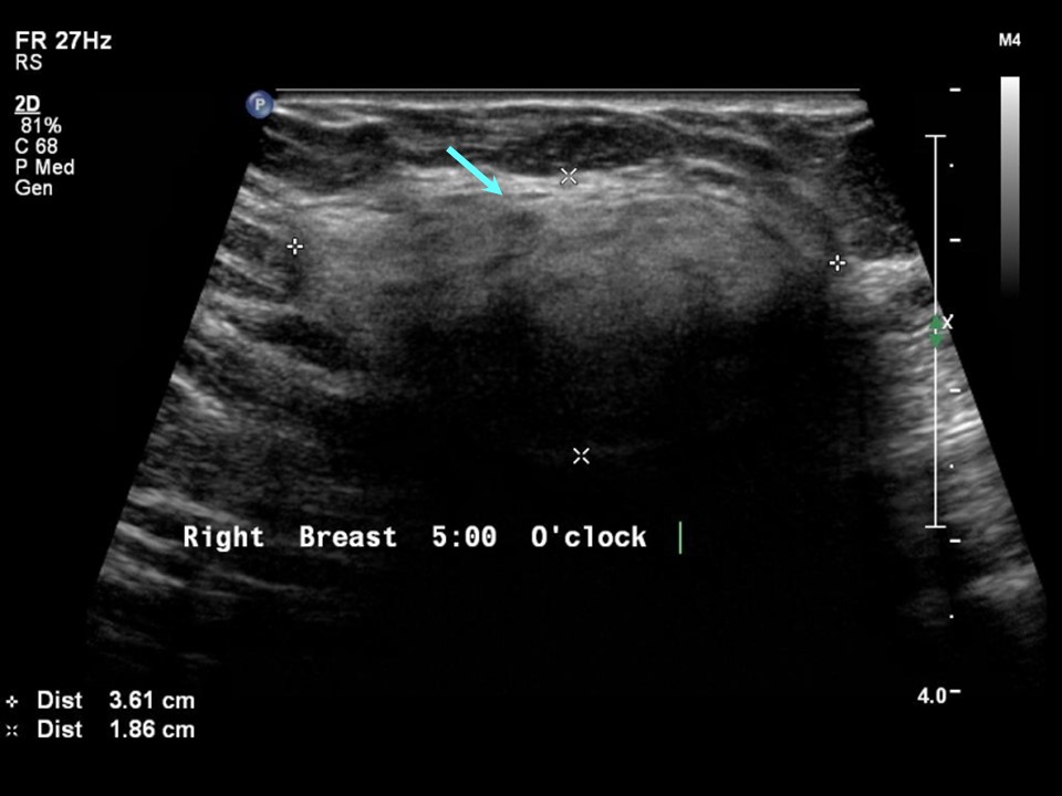 |
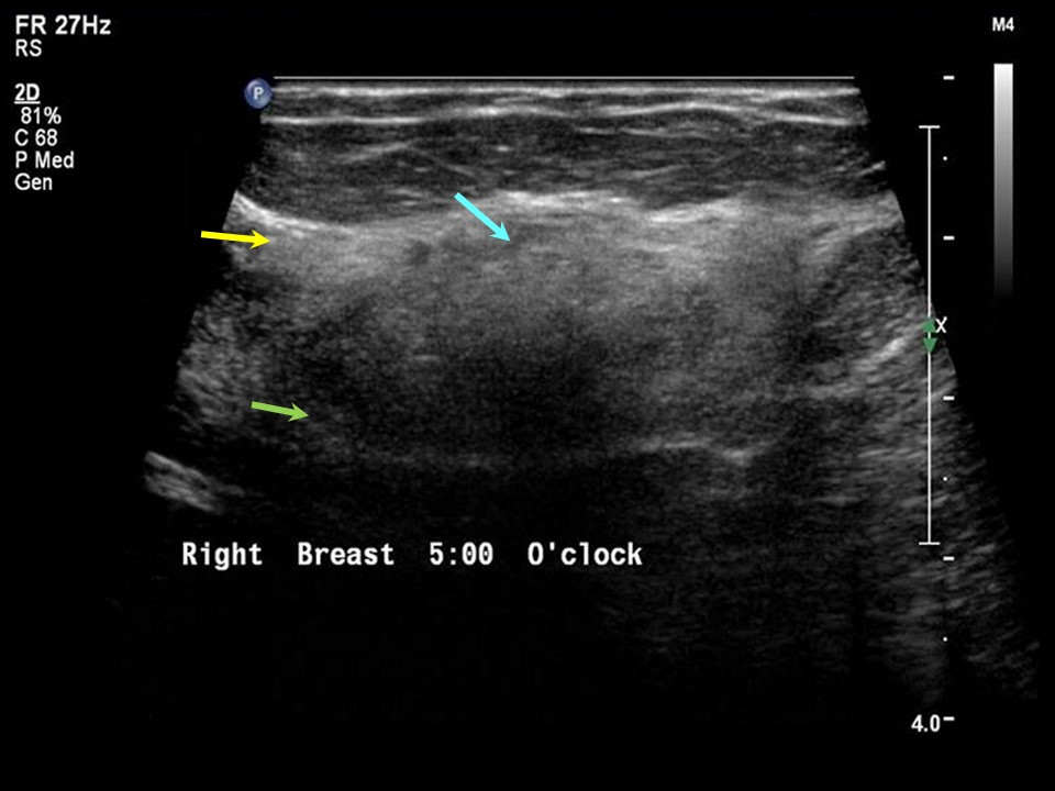
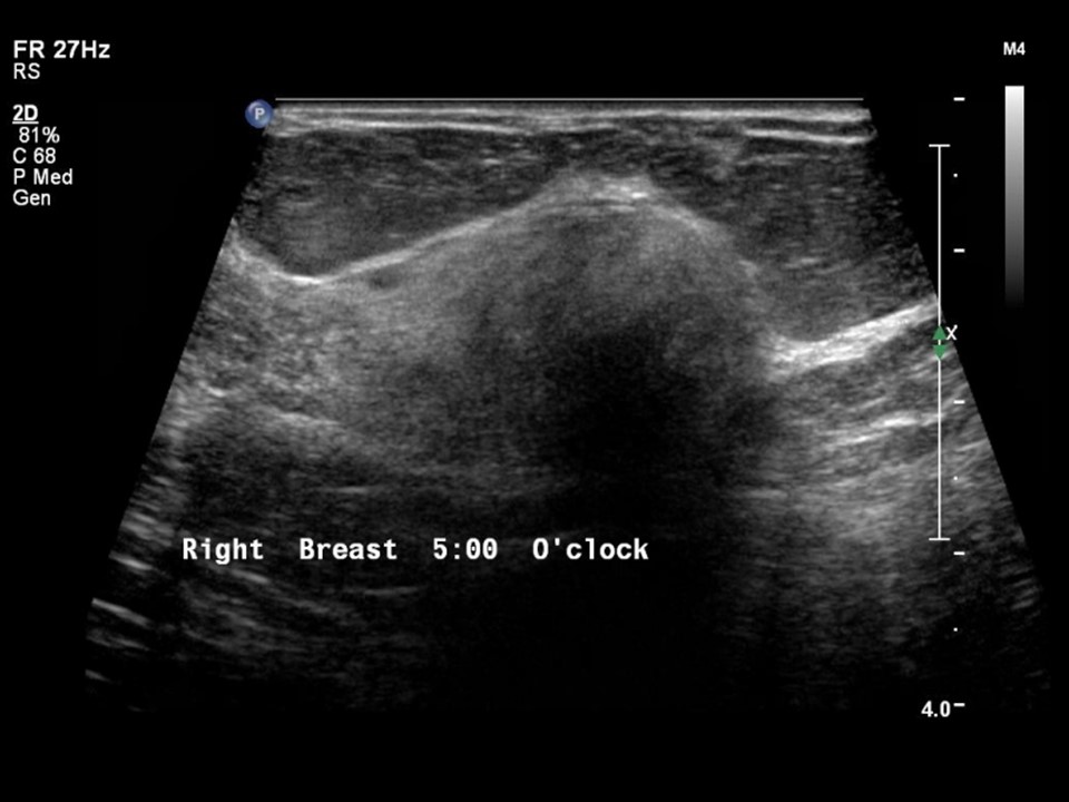
| Ultrasound features: Right breast, upper outer quadrant at 10 oclock | |
| ‣ Mass | |
| • Location: | Right breast, upper outer quadrant at 10 oclock |
| • Number: | 1 |
| • Size: | 3.6 × 2.0 cm |
| • Shape: | Oval |
| • Orientation: | Parallel |
| • Margins: | Circumscribed |
| • Echo pattern: | Heteroechoic |
| • Posterior features: | No posterior features |
| ‣ Calcifications: | None |
| ‣ Associated features: | None |
| ‣ Special cases: | None |
BI-RADS:
BI-RADS Category: 2 (benign)Further assessment:
Further assessment advised: Referral for cytologyCytology:
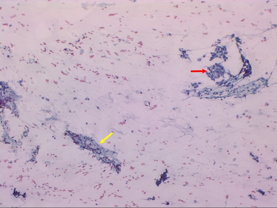 |
| Cytology features: | |
| ‣ Type of sample: | FNAC |
| ‣ Site of biopsy: | |
| • Laterality: | Right |
| • Quadrant: | Upper outer |
| • Localization technique: | Ultrasound-guided, because non-palpable |
| • Nature of aspirate: | Yellowish fatty, greasy material |
| ‣ Cytological description: | Overall low cell yield. Bimodal population of cohesive epithelial cells in small groups and single bipolar nuclei of non-neoplastic glandular breast tissue. Many adipose tissue fragments seen in the background |
| ‣ Reporting category: | Benign |
| ‣ Diagnosis: | Non-neoplastic glandular breast tissue |
| ‣ Comments: | None |
Case summary:
| Postmenopausal woman presented with bilateral breast nodularity. Diagnosed as hamartoma (fibroadenolipoma), BI-RADS 2 on imaging and as non-neoplastic glandular breast tissue on cytology. |
Learning points:
|