


Home / Training / Manuals / Atlas of breast cancer early detection / Cases
Atlas of breast cancer early detection
Filter by language: English / Русский
Go back to the list of case studies
.png) Click on the pictures to magnify and display the legends
Click on the pictures to magnify and display the legends
| Case number: | 026 |
| Age: | 42 |
| Clinical presentation: | Premenopausal woman with average risk of developing breast cancer presented with a right breast lump she had noticed many months ago. It had gradually increased in size. Examination revealed a large firm mobile lump (8.09.0 cm in greatest dimension) in the upper quadrant of the right breast. |
Mammography:
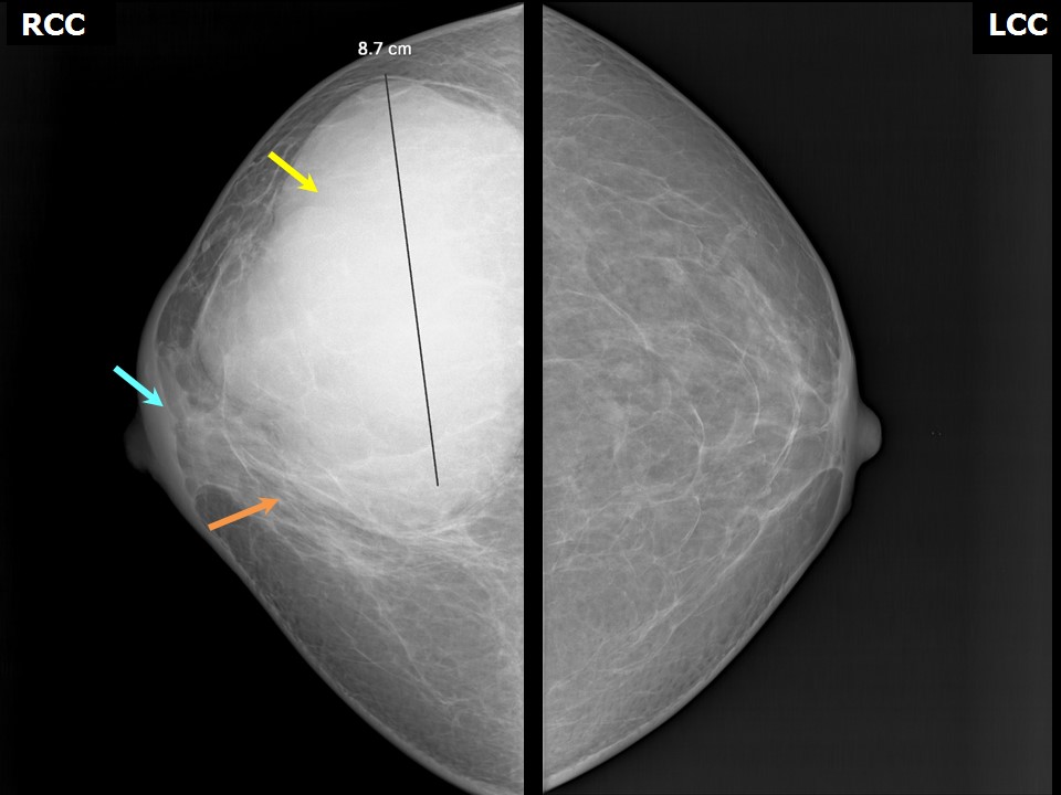 | 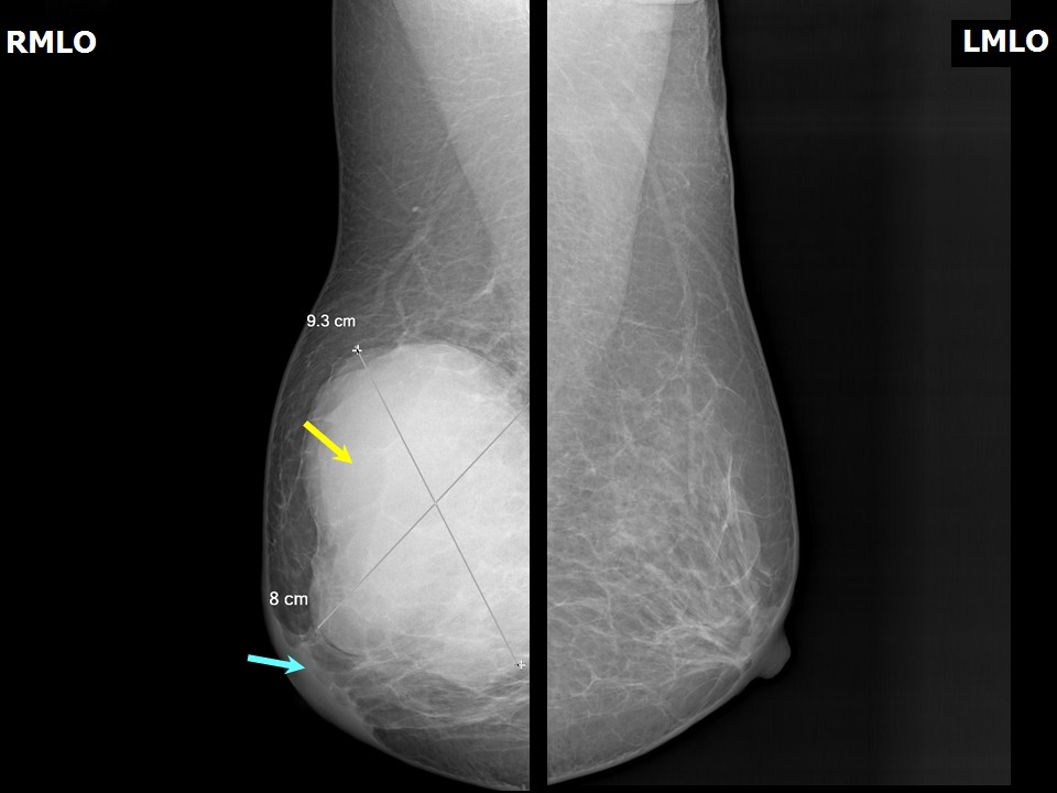 |
| Breast composition: | ACR category b (there are scattered areas of fibroglandular density) | Mammography features: |
| ‣ Location of the lesion: | Right breast, upper outer quadrant at 912 oclock, anterior, middle, and posterior thirds |
| ‣ Mass: | |
| • Number: | 1 |
| • Size: | 9.3 × 8.0 × 8.7 cm |
| • Shape: | Oval |
| • Margins: | Circumscribed with perilesional halo and splaying of adjacent fibroglandular tissues |
| • Density: | High |
| ‣ Calcifications: | |
| • Typically benign: | None |
| • Suspicious: | None |
| • Distribution: | None |
| ‣ Architectural distortion: | None |
| ‣ Asymmetry: | None |
| ‣ Intramammary node: | None |
| ‣ Skin lesion: | None |
| ‣ Solitary dilated duct: | None |
| ‣ Associated features: | None |
Ultrasound:
 |
| Ultrasound features: Right breast, outer quadrants | |
| ‣ Mass | |
| • Location: | Right breast, outer quadrants |
| • Number: | 1 |
| • Size: | 7.6 × 3.6 cm |
| • Shape: | Oval |
| • Orientation: | Parallel |
| • Margins: | Circumscribed |
| • Echo pattern: | Hypoechoic |
| • Posterior features: | No posterior features |
| ‣ Calcifications: | None |
| ‣ Associated features: | Internal vascularity, central areas of necrosis, and liquefaction |
| ‣ Special cases: | None |
BI-RADS:
BI-RADS Category: 4A (low level of suspicion for malignancy)Further assessment:
Further assessment advised: Referral for excision biopsyCytology:
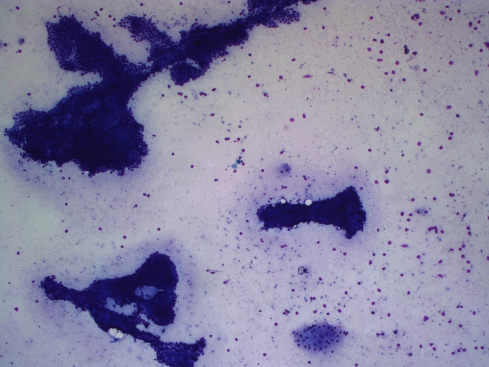 | 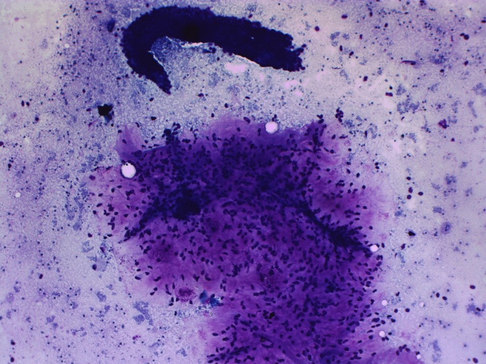 |
| Cytology features: | |
| ‣ Type of sample: | FNAC |
| ‣ Site of biopsy: | |
| • Laterality: | Right |
| • Quadrant: | Large lump involving all quadrants |
| • Localization technique: | Palpation |
| • Nature of aspirate: | Blood-mixed whitish aspirate |
| ‣ Cytological description: | Smears are very cellular and reveal many tightly cohesive sheets of benign ductal epithelial cells. A few bare nuclei are seen. Cellular stromal fragments are seen on the smear. A few clusters of ductal cells with apocrine change are also seen |
| ‣ Reporting category: | Benign |
| ‣ Diagnosis: | To consider phyllodes tumour in view of the cellular stromal fragments |
| ‣ Comments: | None |
Histopathology:
Core needle biopsy
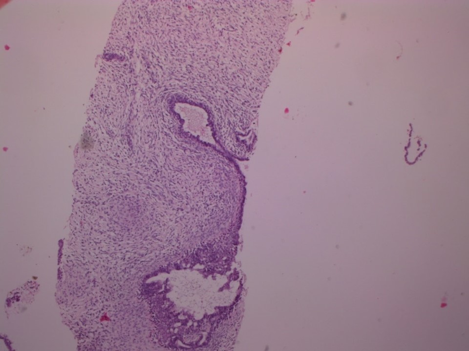 |
| Histopathology features: | |
| ‣ Specimen type: | Core needle biopsy |
| ‣ Laterality: | Right |
| ‣ Macroscopy: | Three whitish soft to firm tissue cores, 15, 12, and 10 mm in length |
| ‣ Histological type: | Sections from all three cores show a very cellular stromal tissue with a few interspersed ductules. The lining ductal epithelium is normal. The stromal cells do not show any nuclear atypia, mitotic figures 5 per 10 hpf |
| ‣ Histological grade: | |
| ‣ Mitosis: | |
| ‣ Maximum invasive tumour size: | |
| ‣ Lymph node status: | |
| ‣ Peritumoural lymphovascular invasion: | |
| ‣ DCIS/EIC: | |
| ‣ Margins: | |
| ‣ Pathological stage: | |
| ‣ Biomarkers: | |
| ‣ Comments: | Benign phyllodes tumour, wide excision advised |
Wide excision
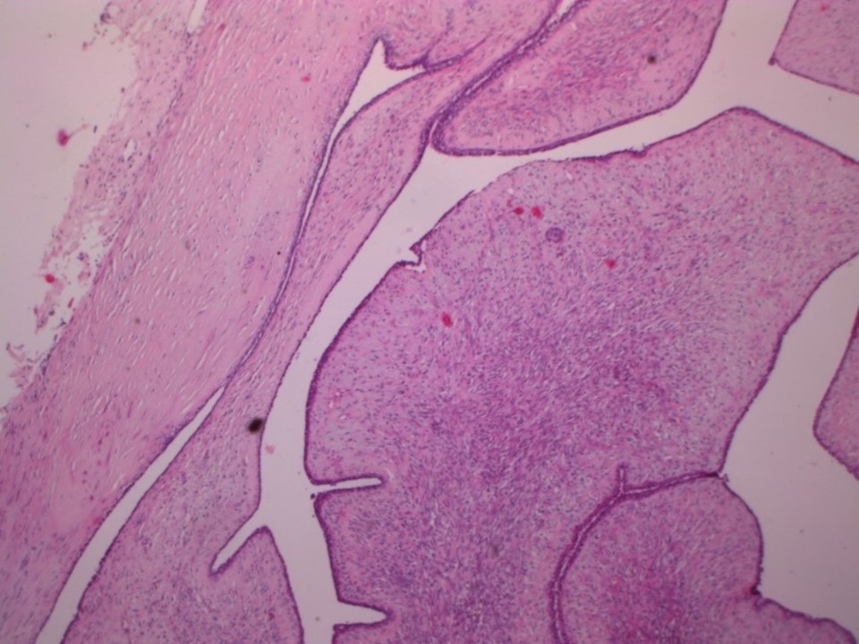 |
| Histopathology features: | |
| ‣ Specimen type: | Wide excision |
| ‣ Laterality: | Right |
| ‣ Macroscopy: | Specimen of wide excision of right breast lump (14.0 × 12.0 × 9.0 cm). Skin flap with nipple and areola (10.8 × 10.0 cm). Externally, the nipple and areola are unremarkable. Serial sectioning of the specimen reveals a whitish leaf-like growth (9.0 × 7.5 × 5.5 cm). The tumour is 2.5 cm from the superior margin, 3.3 cm from the lateral margin, 4.0 cm from the medial margin, 2.5 cm from the inferior margin, 0.3 cm from the base, and 1.8 cm from the skin. On cut surface, small, fluid-filled cystic changes are seen. Myxoid areas that protrude into the cystic cavity are also noted. The cut surface is reddish with fatty areas. Frozen section was reported as negative |
| ‣ Histological type: | Sections from the tumour show features of a benign phyllodes tumour. Sections from cystic areas show hyaline changes with proteinaceous material and many neutrophils. A few of the ductules show epithelial hyperplasia. The very cellular areas of stroma show mitotic figures of 5 per 10 hpf. Nuclear pleomorphism is not significant. Surgical resection margins superior, inferior, lateral, medial, and overlying skin are uninvolved by tumour. The posterior resection margin (base) shows skeletal muscle fibres and is free of tumour |
| ‣ Histological grade: | |
| ‣ Mitosis: | |
| ‣ Maximum invasive tumour size: | |
| ‣ Lymph node status: | |
| ‣ Peritumoural lymphovascular invasion: | |
| ‣ DCIS/EIC: | |
| ‣ Margins: | |
| ‣ Pathological stage: | |
| ‣ Biomarkers: | |
| ‣ Comments: | Right breast benign phyllodes tumour |
Case summary:
| Premenopausal woman presented with a right breast lump of long duration. Diagnosed as large circumscribed mass in the right breast splaying the adjacent fibroglandular tissues, probably phyllodes, BI-RADS 4A on imaging and as benign phyllodes tumour on cytology and histopathology. |
Learning points:
|