


Home / Training / Manuals / Atlas of breast cancer early detection / Cases
Atlas of breast cancer early detection
Go back to the list of case studies
.png) Click on the pictures to magnify and display the legends
Click on the pictures to magnify and display the legends
BI-RADS Category (2014): 4B (moderate suspicion of malignancy)
| Case number: | 105 |
| Age: | 76 |
| Clinical presentation: | Postmenopausal woman with increased risk of developing breast cancer because of family history of breast cancer presented for follow-up. Examination revealed a small palpable nodule in the retroareolar region of the right breast. |
Mammography:
 |
| Breast composition: | ACR category d (the breasts are extremely dense, which lowers the sensitivity of mammography) | Mammography features: |
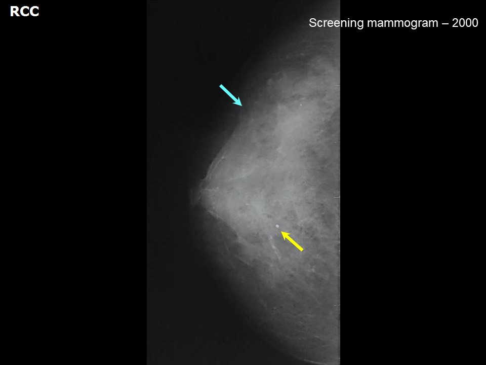
| ‣ Location of the lesion: | 2000: Right breast, CC view |
| ‣ Mass: | |
| • Number: | 0 |
| • Size: | None |
| • Shape: | None |
| • Margins: | None |
| • Density: | None |
| ‣ Calcifications: | |
| • Typically benign: | Typically benign, round |
| • Suspicious: | None |
| • Distribution: | None |
| ‣ Architectural distortion: | None |
| ‣ Asymmetry: | None |
| ‣ Intramammary node: | None |
| ‣ Skin lesion: | None |
| ‣ Solitary dilated duct: | None |
| ‣ Associated features: | None |
 |  |
 |  |
| Breast composition: | ACR category c (the breasts are heterogeneously dense, which may obscure small masses) | Mammography features: |
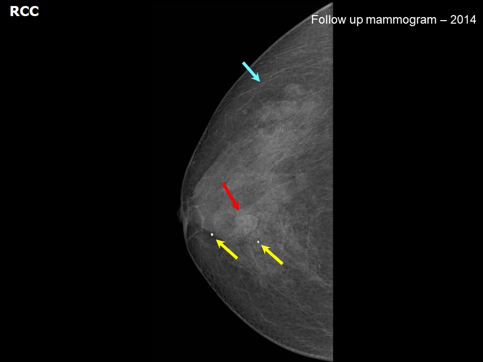
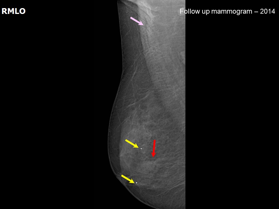
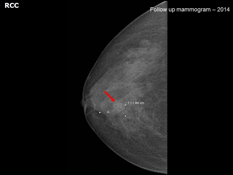
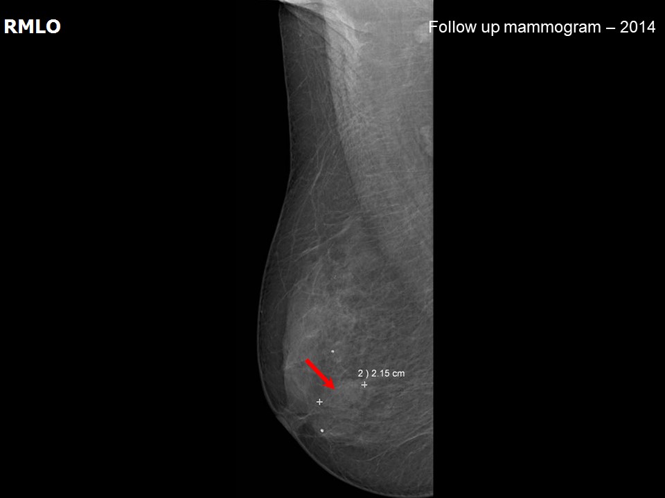
| ‣ Location of the lesion: | 2014: Right breast, central portion of the breast at 12 oclock, middle third |
| ‣ Mass: | |
| • Number: | 1 |
| • Size: | 2.2 × 1.8 cm |
| • Shape: | Oval |
| • Margins: | Indistinct |
| • Density: | Equal |
| ‣ Calcifications: | |
| • Typically benign: | Round |
| • Suspicious: | None |
| • Distribution: | None |
| ‣ Architectural distortion: | None |
| ‣ Asymmetry: | None |
| ‣ Intramammary node: | None |
| ‣ Skin lesion: | None |
| ‣ Solitary dilated duct: | None |
| ‣ Associated features: | None |
Ultrasound:
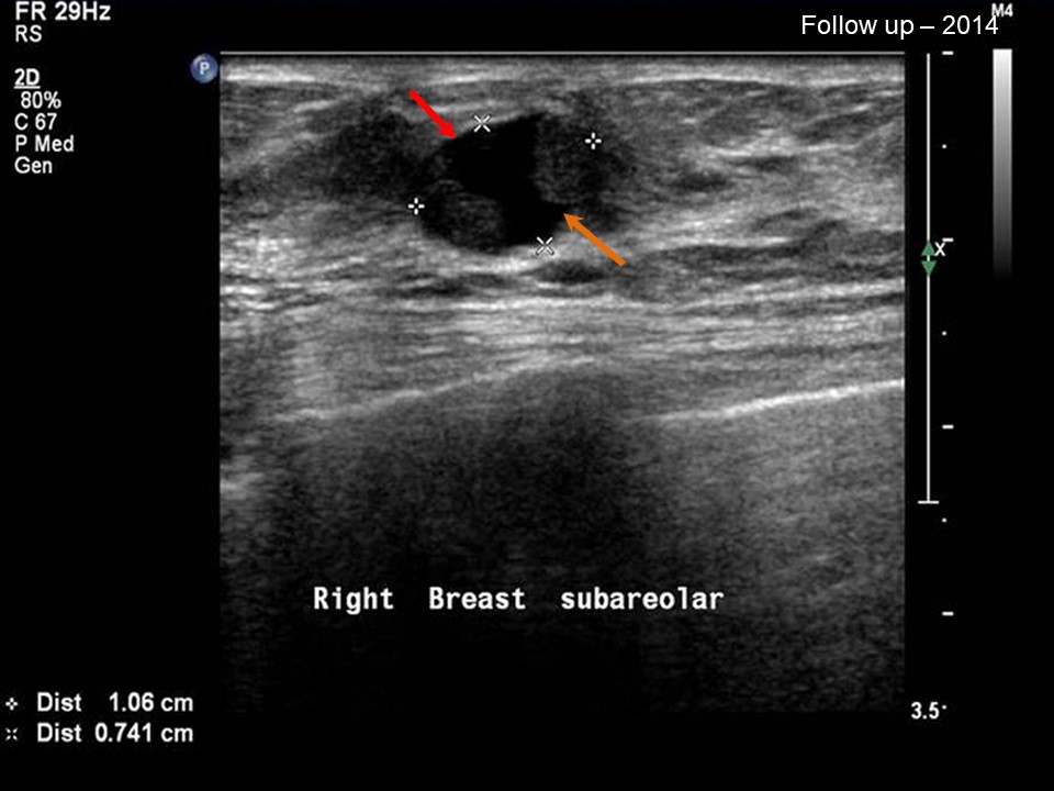 | 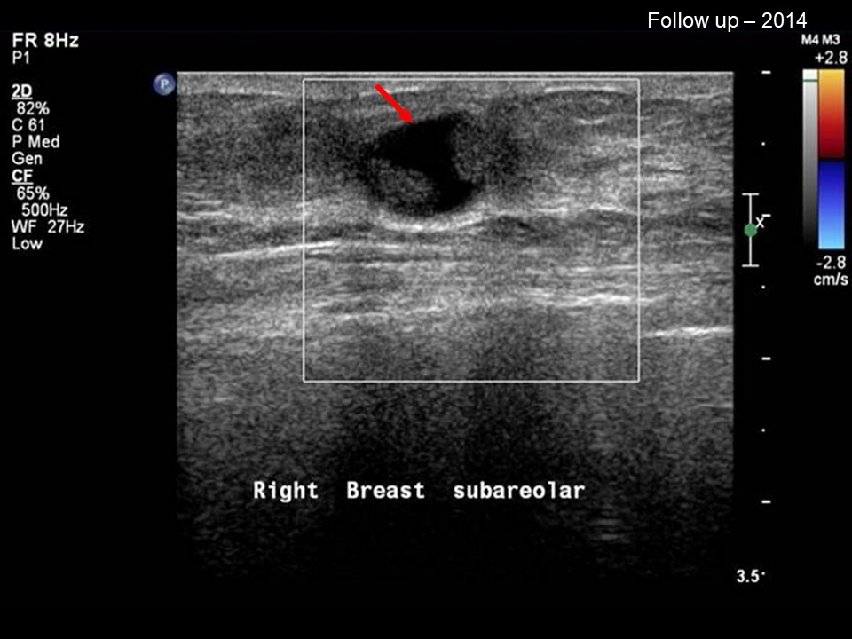 |
| Ultrasound features: Right breast, central portion of the breast | |
| ‣ Mass | |
| • Location: | Right breast, central portion of the breast |
| • Number: | 1 |
| • Size: | 1.0 × 0.7 cm |
| • Shape: | Oval |
| • Orientation: | Parallel |
| • Margins: | Circumscribed |
| • Echo pattern: | Complex cystic and solid |
| • Posterior features: | No posterior features |
| ‣ Calcifications: | None |
| ‣ Associated features: | None |
| ‣ Special cases: | None |
BI-RADS:
BI-RADS Category (2000): 2 (benign)BI-RADS Category (2014): 4B (moderate suspicion of malignancy)
Further assessment:
Further assessment advised: Referral for cytologyCytology:
 |  |
| Cytology features: | |
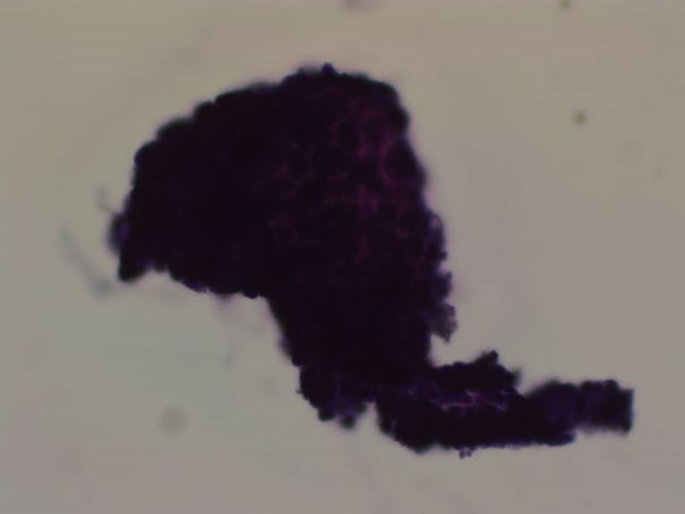
| ‣ Type of sample: | FNAC |
| ‣ Site of biopsy: | |
| • Laterality: | Right |
| • Quadrant: | Subareolar region at 12 oclock |
| • Localization technique: | Ultrasound-guided FNAC |
| • Nature of aspirate: | Scant whitish fluid, < 0.2 mL |
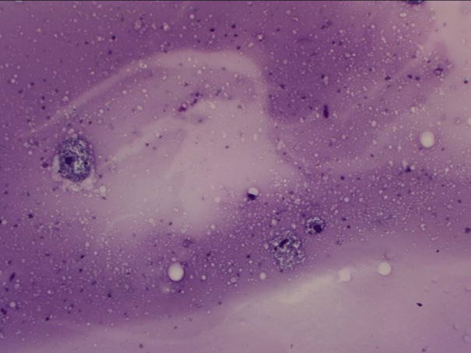
| ‣ Cytological description: | Smears are sparsely cellular. A few three-dimensional clusters of ductal epithelial cells are seen. One cluster shows a papillary configuration. Nuclei are hyperchromatic and cytoplasm is scanty. Nucleoli are not seen. Many foamy macrophages are also seen and a few are haemosiderin-laden. Background shows thick proteinaceous material |
| ‣ Reporting category: | Atypical, probably benign |
| ‣ Diagnosis: | Suggestive of a papillary neoplasm, atypia seen, probably benign. Excision biopsy advised |
| ‣ Comments: | None |
Histopathology:
Surgical excision
 |
| Histopathology features: | |
| ‣ Specimen type: | Surgical excision |
| ‣ Laterality: | Right |
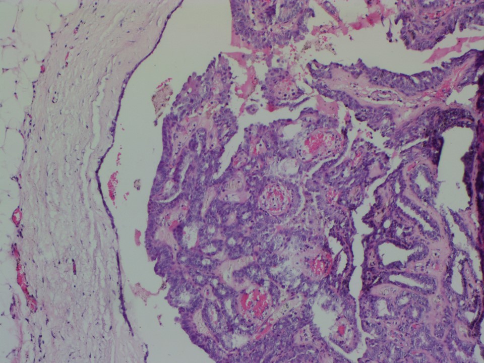
| ‣ Macroscopy: | Fibroadipose tissue (2.5 × 2.0 × 2.0 cm). On cut section a well-circumscribed cystic lesion (1.2 × 1.2 × 1.0 cm) showing papillary excrescences is seen |
| ‣ Histological type: | Breast papilloma |
| ‣ Histological grade: | |
| ‣ Mitosis: | |
| ‣ Maximum invasive tumour size: | |
| ‣ Lymph node status: | |
| ‣ Peritumoural lymphovascular invasion: | |
| ‣ DCIS/EIC: | |
| ‣ Margins: | |
| ‣ Pathological stage: | |
| ‣ Biomarkers: | |
| ‣ Comments: |
Case summary:
| Postmenopausal woman with increased risk of developing breast cancer presented with a small palpable nodule in the retroareolar region of the right breast. Mammography and breast ultrasound dated 2014 reveal a new complex cystic mass with solid lesions along the walls of the cyst in the retroareolar region of the right breast. Compared with a previous mammogram dated 2000, follow-up reveals developing asymmetry in the right breast. Interval change is seen. Diagnosed as BI-RADS 4B on imaging, as papillary neoplasm, atypia seen, probably benign on cytology, and as breast papilloma on histopathology. |
Learning points:
|