


Home / Training / Manuals / Atlas of breast cancer early detection / Cases
Atlas of breast cancer early detection
Go back to the list of case studies
.png) Click on the pictures to magnify and display the legends
Click on the pictures to magnify and display the legends
| Case number: | 152 |
| Age: | 33 |
| Clinical presentation: | Premenopausal woman with average risk of developing breast cancer presented with a right breast lump with severe pain and tenderness first noticed 2 weeks ago. |
 |
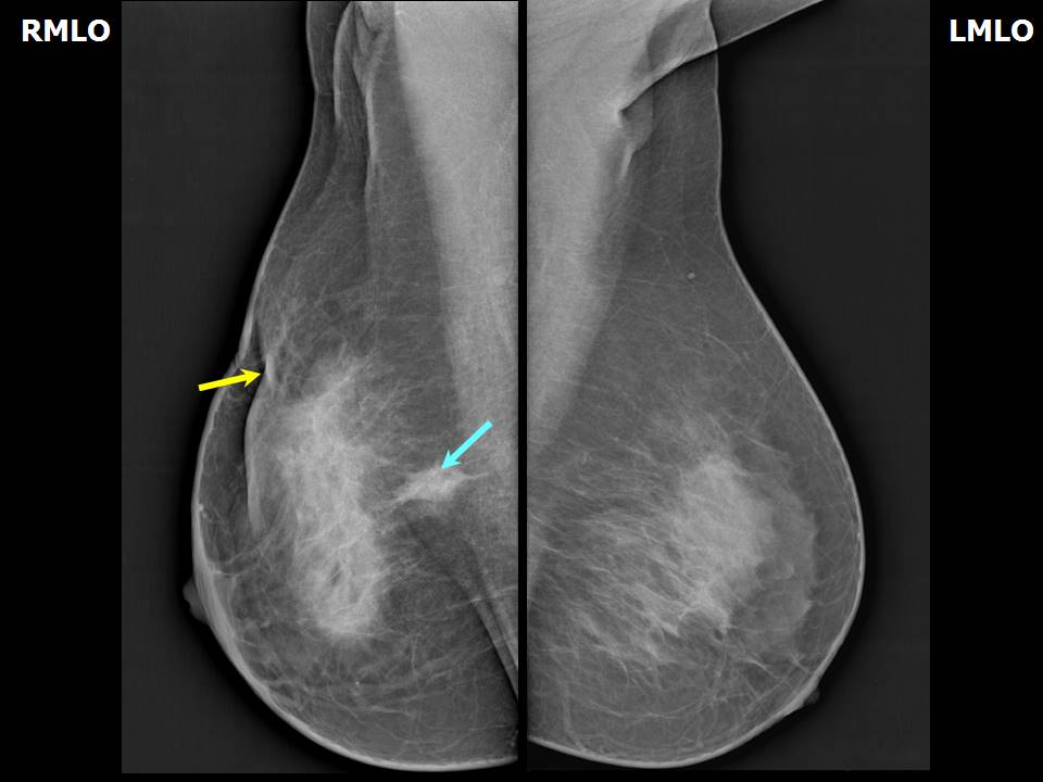
Mammography:
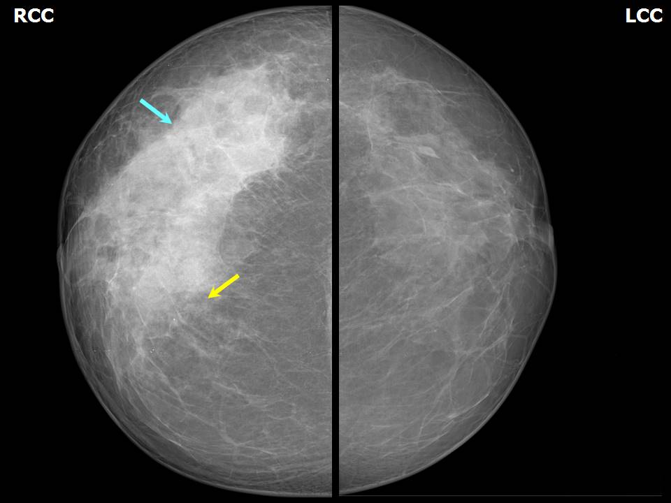 |  |
| Breast composition: | ACR category c (the breasts are heterogeneously dense, which may obscure small masses) | Mammography features: |
| ‣ Location of the lesion: | Right breast, upper outer quadrant at 1012 oclock, anterior and middle thirds |
| ‣ Mass: | |
| • Number: | 0 |
| • Size: | None |
| • Shape: | None |
| • Margins: | None |
| • Density: | None |
| ‣ Calcifications: | |
| • Typically benign: | None |
| • Suspicious: | None |
| • Distribution: | None |
| ‣ Architectural distortion: | Present |
| ‣ Asymmetry: | Present, involving the right upper outer quadrant with prominent glandular tissue |
| ‣ Intramammary node: | None |
| ‣ Skin lesion: | None |
| ‣ Solitary dilated duct: | None |
| ‣ Associated features: | None |
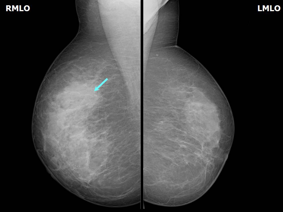
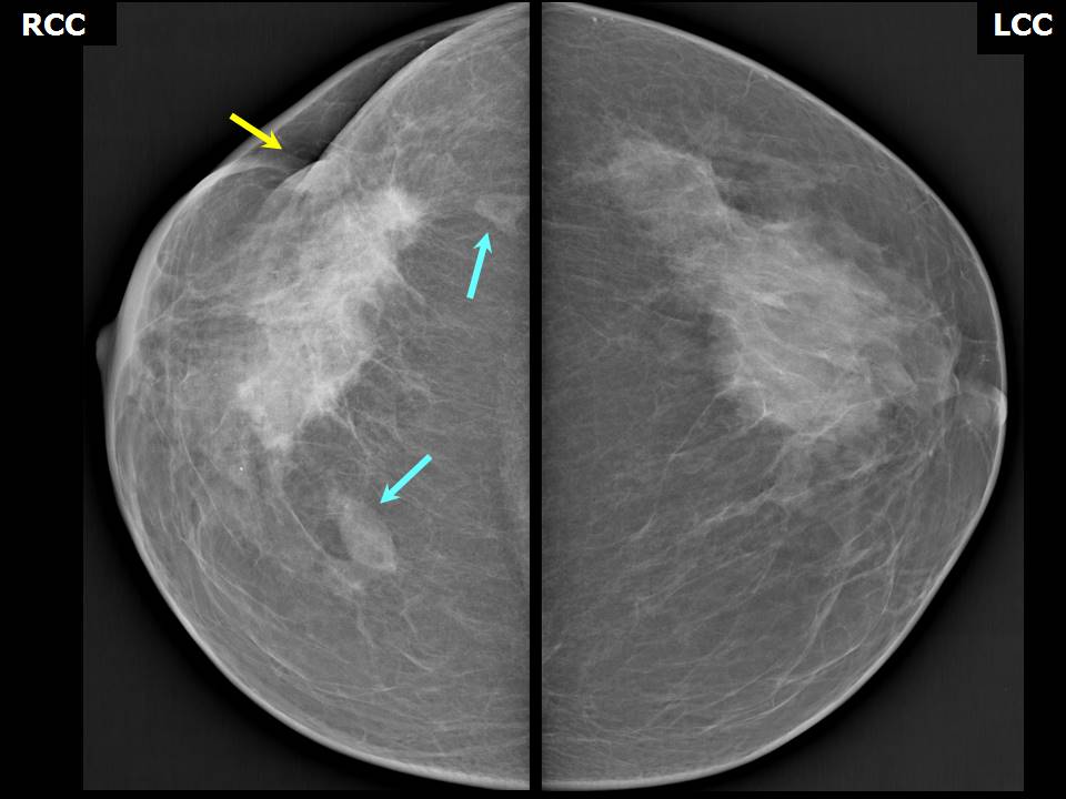 |  |
| Breast composition: | ACR category c (the breasts are heterogeneously dense, which may obscure small masses) | Mammography features: |
| ‣ Location of the lesion: | Follow-up 6 months later after lumpectomy: Right breast, upper outer quadrant at 1012 oclock, anterior and middle thirds |
| ‣ Mass: | |
| • Number: | Multiple |
| • Size: | Largest 1.4 cm in greatest dimension |
| • Shape: | Oval |
| • Margins: | Indistinct |
| • Density: | Equal |
| ‣ Calcifications: | |
| • Typically benign: | None |
| • Suspicious: | None |
| • Distribution: | None |
| ‣ Architectural distortion: | Present |
| ‣ Asymmetry: | Present |
| ‣ Intramammary node: | None |
| ‣ Skin lesion: | None |
| ‣ Solitary dilated duct: | None |
| ‣ Associated features: | Surgical scar skin retraction, skin thickening, and trabecular thickening |
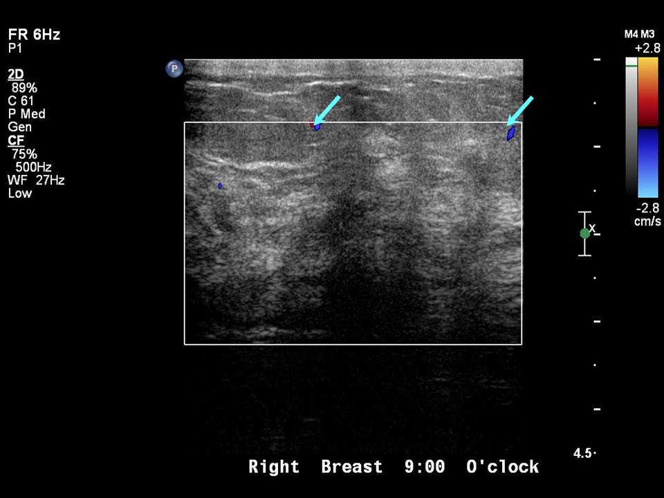
Ultrasound:
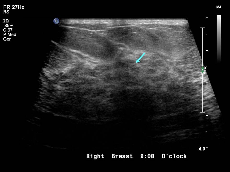 |  |
| Ultrasound features: Right breast, upper outer quadrant at 910 oclock, 2.5 cm from the nipple and at 1.8 cm skin depth | |
| ‣ Mass | |
| • Location: | Right breast, upper outer quadrant at 910 oclock, 2.5 cm from the nipple and at 1.8 cm skin depth |
| • Number: | 1 |
| • Size: | 3.2 × 1.5 cm |
| • Shape: | Irregular |
| • Orientation: | Not parallel |
| • Margins: | Indistinct |
| • Echo pattern: | Heteroechoic |
| • Posterior features: | No posterior features |
| ‣ Calcifications: | None |
| ‣ Associated features: | Vascularity: vessels in rim |
| ‣ Special cases: | None |
 |
| Ultrasound features: Right breast, upper outer quadrant at 910 oclock, 2.5 cm from the nipple and at 1.8 cm skin depth | |
| ‣ Mass | |
| • Location: | Right breast, upper outer quadrant at 910 oclock, 2.5 cm from the nipple and at 1.8 cm skin depth |
| • Number: | Multiple |
| • Size: | 1.2 cm in greatest dimension |
| • Shape: | Irregular |
| • Orientation: | Not parallel |
| • Margins: | Indistinct |
| • Echo pattern: | Heteroechoic |
| • Posterior features: | No posterior features |
| ‣ Calcifications: | None |
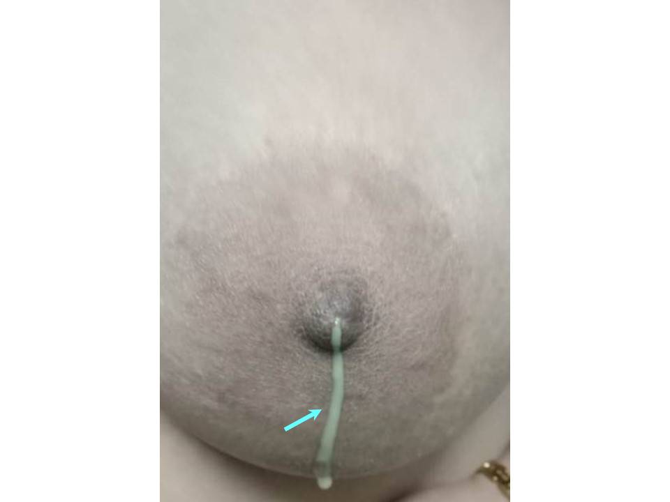
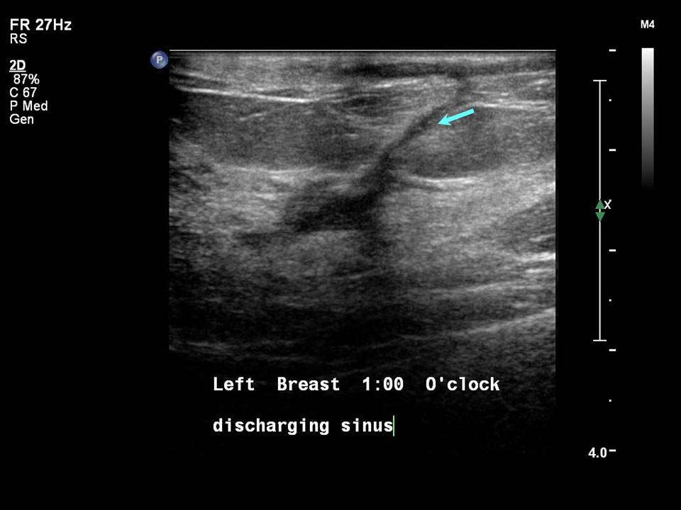
| ‣ Associated features: | Skin thickening and discharging sinus |
| ‣ Special cases: | None |
BI-RADS:
BI-RADS Category: 2 (benign)Further assessment:
Further assessment advised: Referral for cytologyCytology:
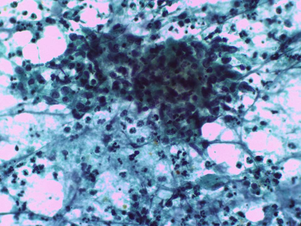 |
| Cytology features: | |
| ‣ Type of sample: | FNAC |
| ‣ Site of biopsy: | |
| • Laterality: | Right |
| • Quadrant: | Upper outer |
| • Localization technique: | Palpation |
| • Nature of aspirate: | Scant whitish purulent material |
| ‣ Cytological description: | Smears reveal many neutrophils admixed with lymphocytes, histiocytes, and plasma cells. Many histiocytes have an epithelioid appearance. Many multinucleated giant cells are also noted |
| ‣ Reporting category: | Benign |
| ‣ Diagnosis: | Granulomatous mastitis |
| ‣ Comments: | None |
Histopathology:
Excision of lump in breast
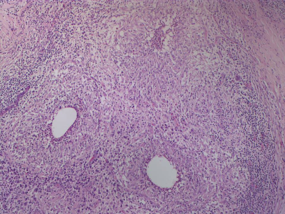 | 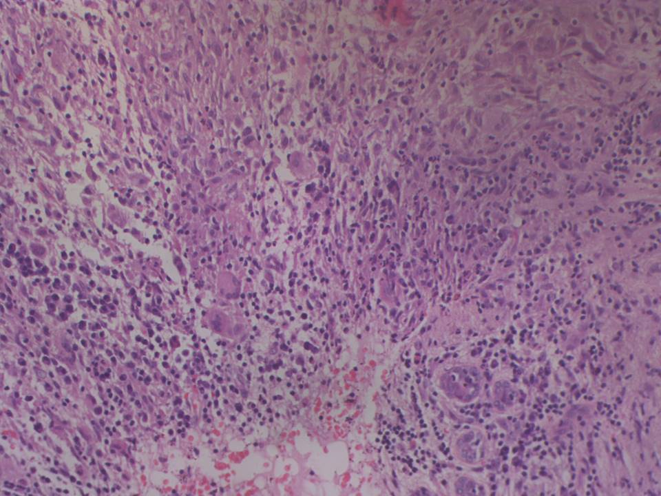 |
| Histopathology features: | |
| ‣ Specimen type: | Excision of lump in breast |
| ‣ Laterality: | Right |
| ‣ Macroscopy: | Single fibrofatty tissue bit (4.0 × 3.0 × 1.5 cm). Cut section firm and whitish |
| ‣ Histological type: | Sections reveal breast tissue showing aggregates of lymphoplasmacytoid cells and abscesses replacing and infiltrating the ducts. Granulomas composed of epithelioid cells, lymphocytes, and multinucleated giant cells are present. Some of these granulomas shows necrosis and neutrophilic infiltration. ZiehlNeelsen stain for acid-fast bacteria negative. Routine bacterial culture and culture for mycobacteria were negative. PCR test for Mycobacterium tuberculosis was negative. Impression: Granulomatous mastitis with acute inflammation |
| ‣ Histological grade: | |
| ‣ Mitosis: | |
| ‣ Maximum invasive tumour size: | |
| ‣ Lymph node status: | |
| ‣ Peritumoural lymphovascular invasion: | |
| ‣ DCIS/EIC: | |
| ‣ Margins: | |
| ‣ Pathological stage: | |
| ‣ Biomarkers: | |
| ‣ Comments: |
Case summary:
| Premenopausal woman presented with a right breast lump with severe pain and tenderness. Diagnosed as BI-RADS 2 on imaging and as granulomatous mastitis on cytology and histopathology. |
Learning points:
|