


Home / Training / Manuals / Atlas of breast cancer early detection / Cases
Atlas of breast cancer early detection
Go back to the list of case studies
.png) Click on the pictures to magnify and display the legends
Click on the pictures to magnify and display the legends
| Case number: | 108 |
| Age: | 52 |
| Clinical presentation: | Premenopausal woman presented for mammography screening. She had an increased risk of breast cancer because of family history, and was scheduled for mammographic surveillance. |
Mammography:
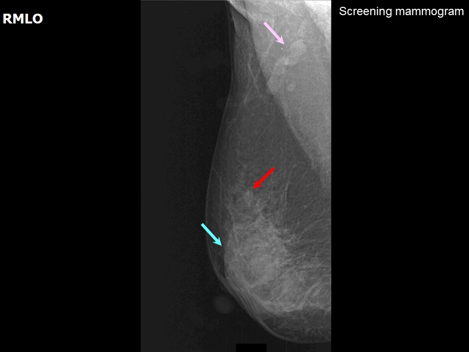 |
| Breast composition: | ACR category b (there are scattered areas of fibroglandular density) | Mammography features: |
| ‣ Location of the lesion: | Right breast, upper outer quadrant at 10 oclock, middle third |
| ‣ Mass: | |
| • Number: | 1 |
| • Size: | 0.7 cm in greatest dimension |
| • Shape: | Irregular |
| • Margins: | Indistinct |
| • Density: | Equal |
| ‣ Calcifications: | |
| • Typically benign: | None |
| • Suspicious: | None |
| • Distribution: | None |
| ‣ Architectural distortion: | None |
| ‣ Asymmetry: | None |
| ‣ Intramammary node: | None |
| ‣ Skin lesion: | None |
| ‣ Solitary dilated duct: | None |
| ‣ Associated features: | None |
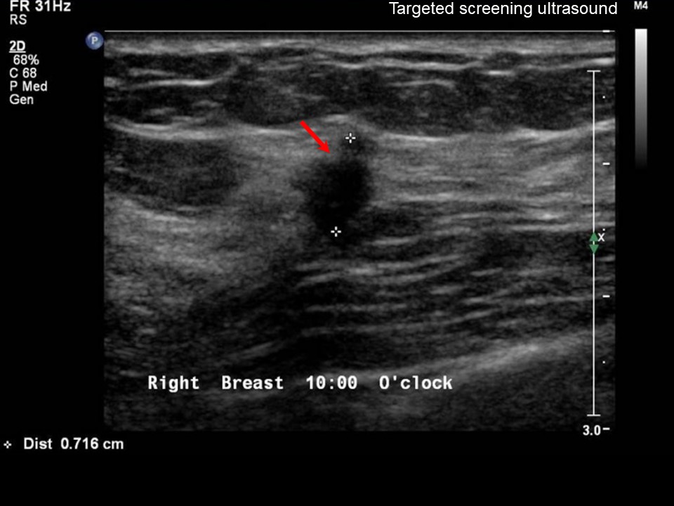
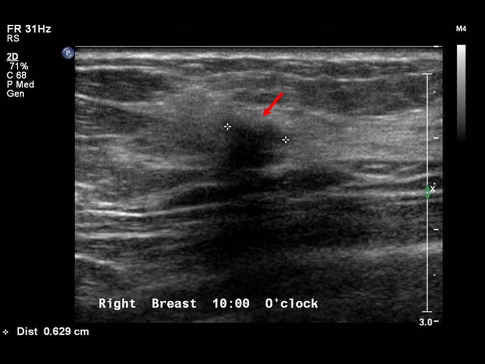
Ultrasound:
 |  |
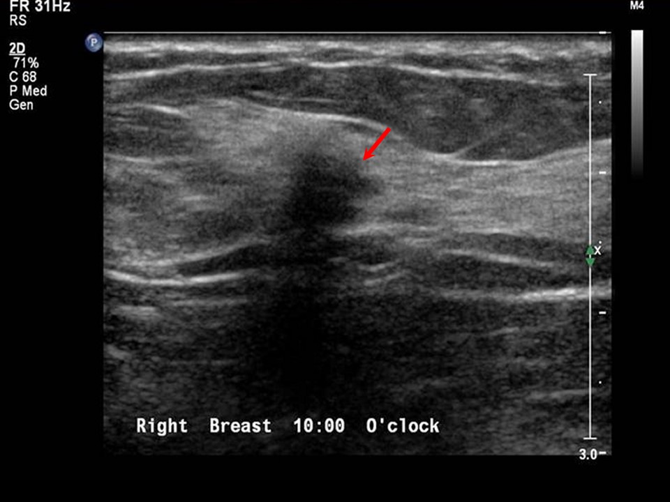 | 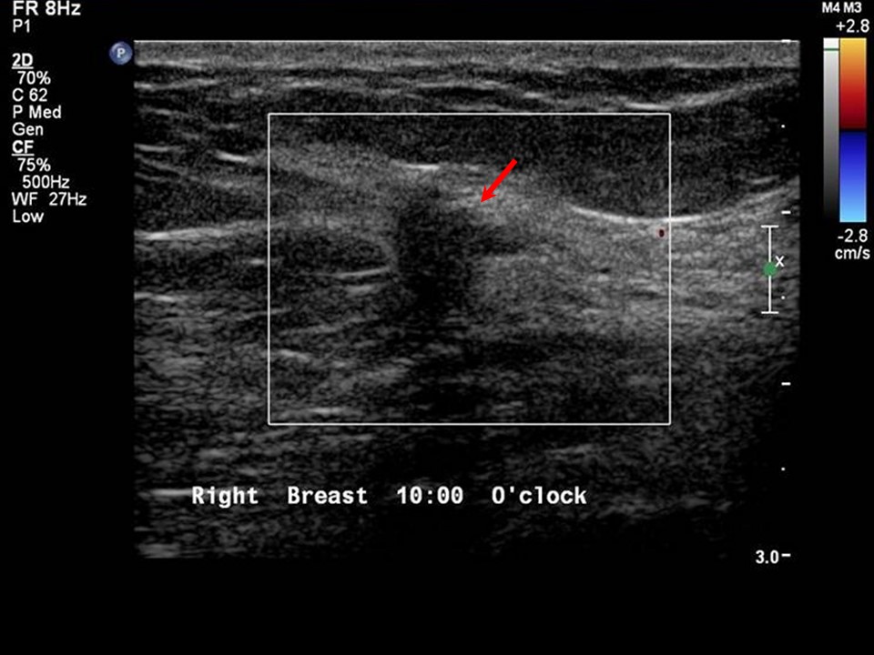 |
| Ultrasound features: Right breast, upper outer quadrant at 10 oclock | |
| ‣ Mass | |
| • Location: | Right breast, upper outer quadrant at 10 oclock |
| • Number: | 1 |
| • Size: | 0.7 cm in greatest dimension |
| • Shape: | Irregular |
| • Orientation: | Not parallel |
| • Margins: | Spiculated |
| • Echo pattern: | Hypoechoic |
| • Posterior features: | Posterior shadowing |
| ‣ Calcifications: | None |
| ‣ Associated features: | None |
| ‣ Special cases: | None |
BI-RADS:
BI-RADS Category: 5 (highly suggestive of malignancy)Further assessment:
Further assessment advised: Further imaging with breast MRIMRI:
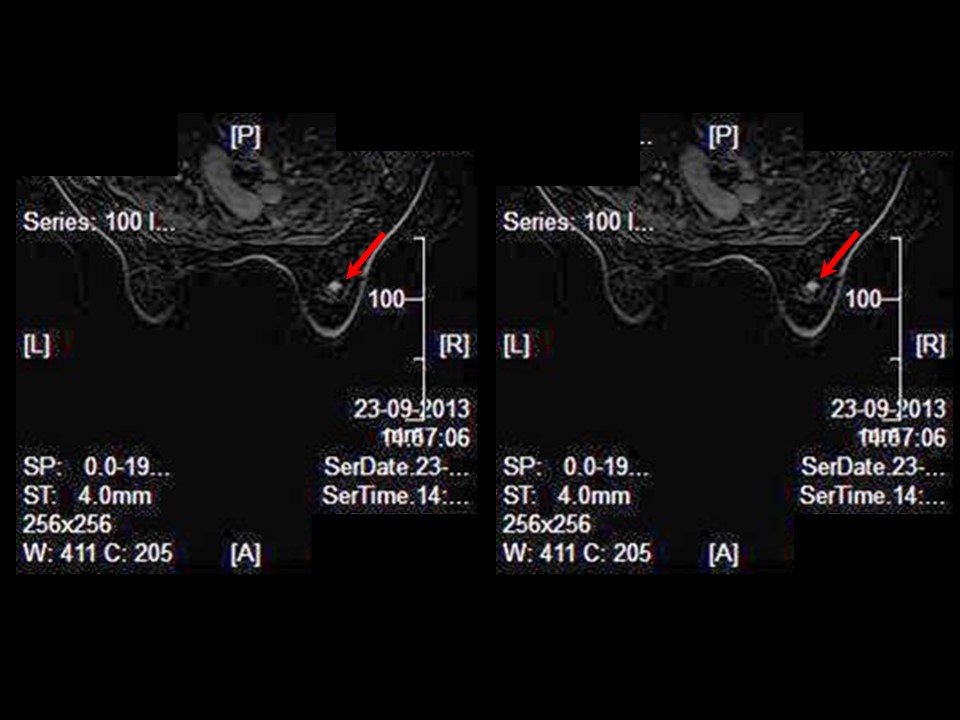 | 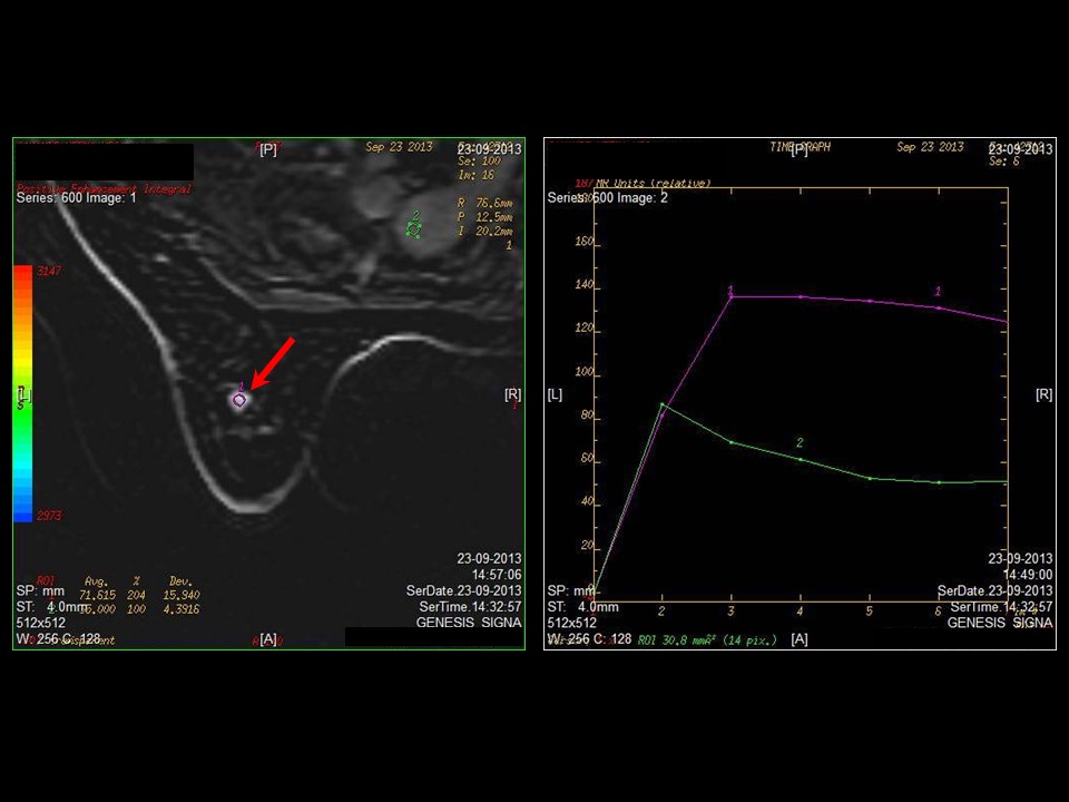 |
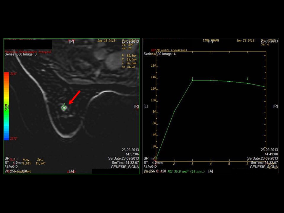 |  |
| MRI features: | ||
| ‣ MRI features: | Amount of fibroglandular tissue: category b (there are scattered areas of fibroglandular density). Background parenchymal enhancement: Mild (2550%), symmetrical | |
| ‣ Location: | Right breast, upper outer quadrant | |
| ‣ Focus: | No | |
| ‣ Mass: | ||
| • Shape: | Round | |
| • Margin: | Spiculated | |
| • Internal enhancement: | Intense homogeneous | |
| • Kinetic curve: | Type 3 | |
| ‣ Non-mass enhancement: | ||
| • Distribution: | No | |
| • Internal enhancement: | No | |
| ‣ Non-enhancing findings: | No | |
| ‣ Associated features: | No | |
| ‣ Axillary nodes: | No | |
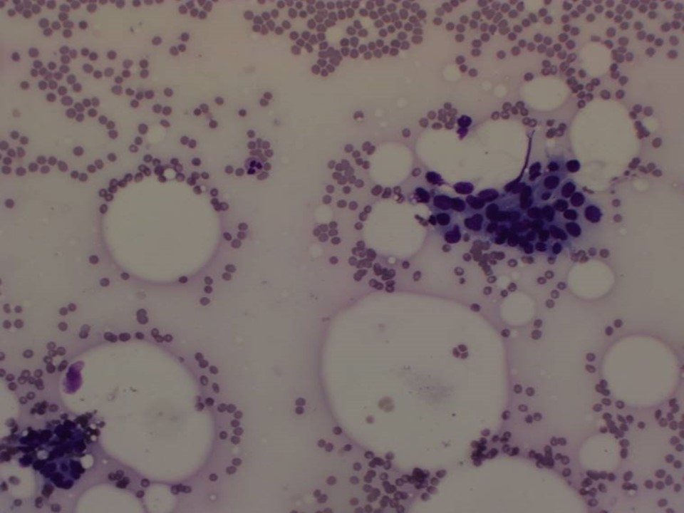
Cytology:
 |
| Cytology features: | |
| ‣ Type of sample: | FNAC |
| ‣ Site of biopsy: | |
| • Laterality: | Right |
| • Quadrant: | Upper outer |
| • Localization technique: | Palpation |
| • Nature of aspirate: | Whitish |
| ‣ Cytological description: | Smears reveal many sheets of tightly cohesive, benign ductal epithelial cells and bare nuclei. A few sheets of ductal epithelial cells show anisonucleosis, enlarged, mildly hyperchromatic nuclei and moderate amounts of cytoplasm |
| ‣ Reporting category: | Suspicious, probably in situ or invasive carcinoma |
| ‣ Diagnosis: | Predominantly benign cells with a few cells suspicious for malignancy |
| ‣ Comments: | None |
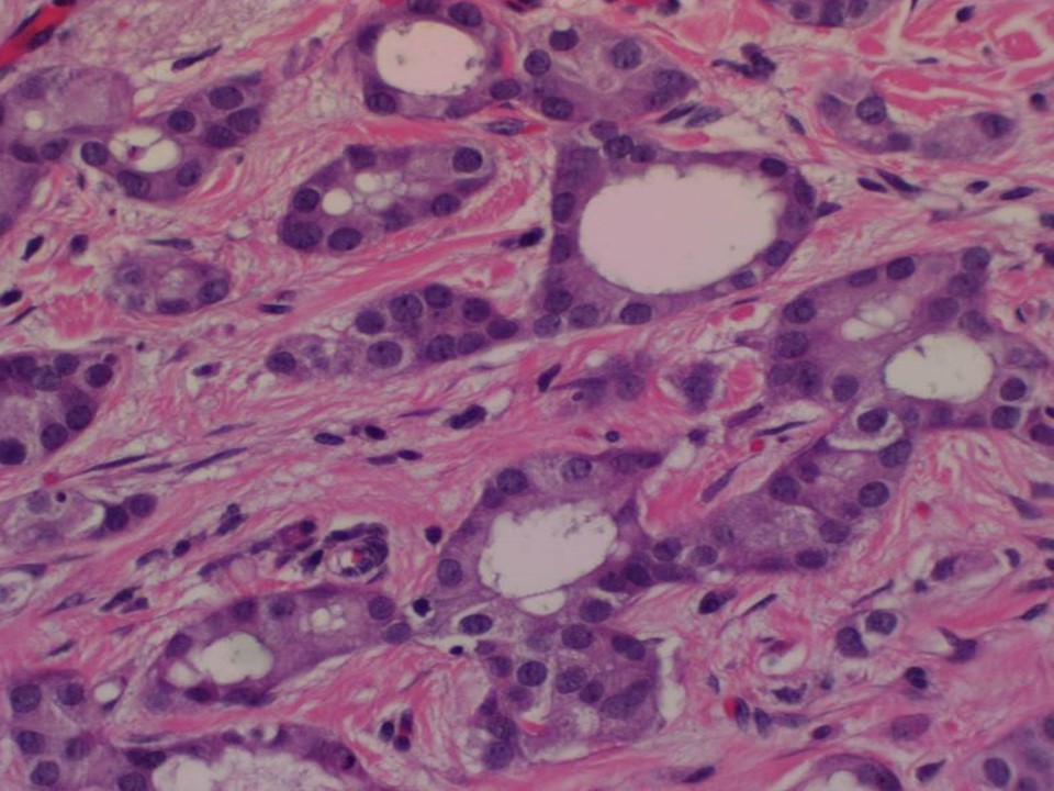
Histopathology:
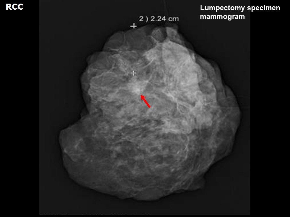
Breast-conserving surgery
 |
| Histopathology features: | |
| ‣ Specimen type: | Breast-conserving surgery |
| ‣ Laterality: | Right |
| ‣ Macroscopy: | On serial sectioning, a small firm greyish white area (0.6 × 0.5 × 0.5 cm) is seen. The closest cut margin is the superior margin, which is 1.5 cm from the tumour |
| ‣ Histological type: | Invasive carcinoma of no special type, a few with tubular carcinoma morphology |
| ‣ Histological grade: | Grade 1 (2 + 1 + 1 = 4) |
| ‣ Mitosis: | 3 |
| ‣ Maximum invasive tumour size: | 0.6 cm in greatest dimension |
| ‣ Lymph node status: | 0/14 |
| ‣ Peritumoural lymphovascular invasion: | Not identified |
| ‣ DCIS/EIC: | Small foci of cribriform type low grade |
| ‣ Margins: | Free of tumour |
| ‣ Pathological stage: | pT1N0 |
| ‣ Biomarkers: | |
| ‣ Comments: | Amount of fibroglandular tissue: category b (scattered fibroglandular tissue). Background: parenchymal enhancement: Mild (2550%), symmetrical. |
Case summary:
| Premenopausal woman with increased risk of developing breast cancer presented for screening. Mammography, breast ultrasound, and breast MRI reveal right breast irregular subcentimeter-size lesion of suspicious morphology, diagnosed as BI-RADS category 4C on imaging, suspicious for malignancy on cytology, and invasive breast carcinoma of no special type, pT1N0 on histopathology. |
Learning points:
|